88.07 Differential expression of Cathepsin L in the coronary arteries of atherosclerotic swine.

P. Gunasekar1, J. Dabestani1, D. K. Agrawal1, J. A. Asensio1  1Creighton University Medical Center,Department Of Surgery, Div. Trauma Surgery, Department Of Clinical & Translational Science,Omaha, NE, USA

Introduction:
Neointimal hyperplasia and restenosis following interventional procedures, including percutaneous transluminal coronary angioplasty (PTCA) and intravascular stenting still remain a significant clinical problem. These interventional procedures cause endothelial denudation and damage to intimal and medial layer which stimulates intimal smooth muscle proliferation and extracellular matrix deposition resulting in intimal hyperplasia (IH) and restenosis. Vascular sterile inflammation has been attributed to the formation of IH. Cathepsin L (CTSL), a member of lysosome protease, is highly associated with diet-induced atherogenesis and IH in animal studies. Vitamin D regulates several proteases and protease inhibitors in different cell types, contributing to its regulatory effects of cell physiology. In our study, we examined the effect of vitamin D on CTSL activity in the coronary arteries of atherosclerotic swine.

Methods:
Yucatan microswine were fed with a high cholesterol atherosclerotic diet. The swine received approximately 500 IU of vitamin D3/per day on the vitamin D-deficient diet, 2,500-3,500 IU of vitamin D3/per day on vitamin D-sufficient diet, and 4,500-5,500 IU of vitamin D3/per day on the vitamin D-supplement diet. After 5-6 months of the experimental diet, PTCA (percutaneous transluminal balloon angioplasty) was performed in the left circumflex coronary artery (LCX) in each swine.  After a year of the diet, angiography and optical coherence tomography (OCT) imaging were performed and swine euthanized. Coronary arteries were embedded in methyl methacrylate or paraffin. Tissue sections were stained with H&E, trichrome, and Movat’s pentachrome.  The expression of Ki67 (proliferation marker), CCR7 (Macrophage marker), Cathepsin L (lysosomal proteases) was evaluated by Immunofluorescence and Immunohistochemistry.

Results:
There was significantly greater density of Ki-67+ cells in post angioplasty LCX in vitamin D-deficient swine compared to vitamin D-sufficient swine and vitamin D-supplemented swine. CCR-7 was found to be higher in vitamin D deficient than vitamin D-sufficient swine and vitamin D-supplemented swine. CTSL expression and its activity were significantly increased in post angioplasty LCX of vitamin D-deficient swine than the supplemented swine.

Conclusion:
Cathepsin L drives IH and macrophage infiltration in coronary arteries after angioplasty in atherosclerotic swine. These findings suggest that vitamin D inhibits CTSL and thus has direct effect on neointimal hyperplasia after coronary intervention.
 

88.02 Vitamin D supplementation Reduces HMGB1, TLRs and RAGE in Post-interventional Coronary Arteries

P. Gunasekar1, J. Dabestani1, D. K. Agrawal1, J. A. Asensio1  1Creighton University Medical Center,Department Of Surgery, Div. Trauma Surgery, Department Of Clinical & Translational Science,Omaha, NE, USA

Introduction:

Coronary angioplasty and stent implantation is a common coronary procedure for patients with coronary artery disease. These interventional procedures stretch and denude the endothelial layer. This promotes local inflammatory response in the injured vessel wall, which is characterized by smooth muscle cell proliferation, migration, neointimal formation. Clinical studies support that plasma vitamin D deficiency is associated with increased risk for coronary artery disease (CAD). It is unclear whether vitamin D status is causally related to CAD or is a marker of health. In this study, we examined the inflammatory profile of coronary arteries in atherosclerotic swine.

Methods:

Yucatan microswine were fed with high cholesterol atherosclerotic diet. The swine received approximately 500 IU of vitamin D3/per day on the vitamin D-deficient diet (VD DEF.), 2,500-3,500 IU of vitamin D3/per day on vitamin D-sufficient diet (VD SUF.), and 4,500-5,500 IU of vitamin D3/per day on the vitamin D-supplement diet (VD SUP.). After 5-6 months of the experimental diet, PTCA (percutaneous transluminal balloon angioplasty) was performed in the left circumflex coronary artery (LCX) and bare mental stent implantation in the left anterior descending coronary artery (LAD) for each swine. Six months following coronary intervention, angiogram and optical coherence tomography (OCT) imaging were performed and swine were euthanized. Coronary arteries were then embedded in methyl methacrylate or paraffin. Tissue sections were stained with H&E. The protein expression of HMGB1 (inflammation and necrosis marker), RAGE (receptor for advanced glycosylation end product), TLR2 and TLR4 (pattern recognition receptors) were evaluated by Immunohistochemistry.

Results:

Optical coherence tomography readings showed the degree of percentage area in-stent restenosis and PTCA was greatest in VD DEF. compared to VD SUF. or VD SUP. swine. We found a greater inflammatory profile in the coronary arteries of VD DEF. compared to VD SUF. or VD SUP. swine, based on histological staining and immunoreactivity to HMGB1, RAGE, TLR2, TLR4, in both LCX and LAD. The ligands for RAGE and receptor for HMGB1 (TLR2 and TLR4) were highly expressed in neointimal cells in stented LAD arteries of VD DEF. swine. This inflammatory profile decreases with increasing the levels of Vitamin D.

Conclusion:

Vitamin D deficiency increases the HMGB1-mediated pathways, resulting in the release of inflammatory cytokines from macrophages and other immune cells; and the recruitment of inflammatory cells through TLR4. Vitamin D supplementation suppresses the cytokine activity and prevents neointimal proliferation and restenosis from damage caused by PTCA in atherosclerotic swine. Vitamin D supplementation could be used as an adjunct therapy to prevent intimal hyperplasia and restenosis following coronary interventions.
 

60.01 Axillary Vessel Injuries: What have we Learned From an Analysis of the National Trauma Data Bank

M. A. Dale1, A. Person1, G. Mendoza1, S. Brown1, D. Keleny1, D. Rigg1, J. Dabestani1, D. Agrawal1, J. Asensio1  1Creighton University Medical Center,Department Of Surgery,Omaha, NE, USA

Introduction: Axillary vessel injuries remain uncommon even in busy urban trauma centers. Most trauma surgeon possess limited experience with these injuries. The proximity of the surrounding anatomic structures account for a large number of associated injuries and mobidity associated with such injuries.

Methods: The National Trauma Data bank was queried for pre-hospital admission data for axillary vessel injuries. Data extracted included demographics, physiologic conditioins, surgical interventions and Functional Independent Measure Score (FIM). Univariate and stepwise logistic regression analysis were used.

Results: 590 cases were identified from 1,466,887 patients in NTDB from 2001-2005. Incidence = 0.004%. Mean age 33 ±  16, mean RTS 6.8 ±  2.3, mean Glasgow Coma Score 13 ±  4.4, mean ISS 14.9 ± 10.3. Mechanism of inury: penetrating n=329 (55.6%), blunt n=259 (43.9%), and non-specified n=2 (0.34%). Mean initial SBP 119.7 ±  37.7, mean in survivors 122.8 ± 24.8, mean in non-survivors 79.7 ±  56.1. Base deficit mean -3.7 ±  10.7, in survivors -3.3 ±  8.5, non-survivors -7.3 ± 19.2. Total number of injuries n=681, of these axillary artery n=455 (67%), axillary vein n=144 (21%), axillary nerve n=68 (9.98%), unspecified n=14 (2%). Sugical procedures for axillary injuries were documented in 238 patients, these included n=54 (15.4%) suture of the artery, n=38 (8.91%) vascular shunt or bypass. Complications n=2637 associated with axillary vessel injuries: pneumonia n=22 (16.05%), wound infection n=16 (11.67%), compartment syndrome n=14 (10.21%), ARDS n=12 (8.76%). Fuctional Independent Measures: dependent n=11 (1.86%), partial help required n=13 (2.2%), independent with device n=28 (4.75%), independent n=210 (35%), not applicable (7.3%), and not documented n=285 (48.3%). Survivors n=548 (92.88%), non-survivors n=42 (7.12%).

Conclusion: Axillary vessel injuries remain an uncommon occurence in the United States as denoted by their low incidence as reported in the NTDB and literature. Initial admitting systolic blood pressure has a higher correlation with morbidity based on mechanism of inury. However, when using stepwise logitistic models, ISS, TRISS, and LOS had the highest correlation with survival. Axillary vessel repairs require complex surgical interventions. Most injuries carry an associated high morbidity, good functional outcomes, and relative low mortality.


49.11 Perioperative Outcomes and Predictors of Mortality Following Surgery for Sigmoid Volvulus.

A. Easterday1, A. Person1, S. Aurit1, R. Driessen1, D. Mukkai Krishnamurty1  1Creighton University Medical Center,Department Of Surgery,Omaha, NE, USA

Introduction: Data on perioperative outcomes following surgery for sigmoid volvulus is limited. The aim of this study is to develop a model to predict need for emergent surgery and post-operative mortality following resection for sigmoid volvulus.

Methods: The NSQIP database was queried from 2012-2016 to identify patients undergoing segmental resection for sigmoid volvulus. Pre-, intra-, and post-operative variables were compared. Primary and secondary outcomes were emergent surgery and risk of mortality, respectively. Chi-square and Fischer’s test for categorical variables and the Mann-Whitney test for continuous variables were used. Significant variables for each outcome were entered into a logistic regression model to predict the outcomes. Statistical significance was set at p<0.05. 

Results: A total of 2,086 patients met inclusion criteria and 51.6 percent underwent an elective resection. Median age was 68 years. Laparoscopic resection (30.5% vs. 9.1%; p<0.001) and mechanical bowel preparation (50% vs. 8.7%; p<0.001) were more commonly used in the elective setting. Patients having emergent resection were more likely to suffer from post-operative superficial surgical site infection (p<0.001), pneumonia (p<0.001), cardiac arrest (p=0.038), septic shock (p=0.001), myocardial infarction (p=0.034), and had a higher need for perioperative transfusion (p<0.001). No difference was seen in ileus, anastomotic leaks, readmission or re-operation rates in patients undergoing emergency vs. elective resection. Overall mortality rate was 6.2% (8.5% in the emergent group vs. 4.1% in the elective group; p<0.001). On multivariate analysis, factors associated with increased risk of emergency surgery included female gender (OR 2.01; p<0.001), relative hematocrit elevation (OR=1.058; p<0.001), relative leukocytosis (OR=1.172; p<0.001), acute kidney injury (OR=6.274; p=0.035), pre-operative sepsis (OR=2.037; p<0.001), functional independence prior to surgery (OR=1.525; p=0.011), and bleeding disorders (OR=1.592; p=0.020). Factors predictive of post-operative mortality included increased age (OR=1.064; p<0.001), systemic sepsis (OR=3.848; p<0.001), and emergent surgery (OR=2.086; p=0.007). Independence prior to illness (OR=0.371; p<0.001), higher albumin levels (OR=0.579; p=0.007), and lower BMI (OR=0.952; p=0.037) were shown to be protective. 

Conclusion: Emergent resection for sigmoid volvulus is independently associated with poor post-operative outcomes and mortality. Pre-operative factors that predict need for emergent resection and post-operative mortality identified in this study can be used to aid in shared decision making and counselling for patients admitted with sigmoid volvulus.

103.11 Brachial Vessel Injuries: An Analysis of the National Trauma Data Bank

D. R. Rigg1, A. Person1, M. Dale1, G. Mendoza1, S. Brown1, D. Keleny1, J. Dabestani1, D. Agrawal1, J. Asensio1  1Creighton University Medical Center,Department Of Trauma Surgery And Surgical Critical Care,Omaha, NE, USA

Introduction:  Brachial vessel injuries remain uncommon, even in busy urban trauma centers, and therefore many trauma surgeons have limited experience with these types of injuries. Despite being a relatively uncommon injury, the brachial artery is the most frequently damaged artery in the upper extremity due to its vulnerability and close proximity to upper extremity bony structures. Therefore, trauma surgeons must be familiar with this injury and its associated outcomes. The objective of this study are: 1) review of the nationally reported experience with these injuries; 2) Identify predictors of outcome; 3) Compare predictors of outcome; 4) Report total charges incurred in the management of these injuries.

Methods:  The National Trauma Data Bank (NTDB) was queried for pre-hospital admission data for brachial vessel injuries. Data extracted included demographics, vital signs on admission, and mechanism of injury. Univariate and stepwise logistics regression statistical analysis was used

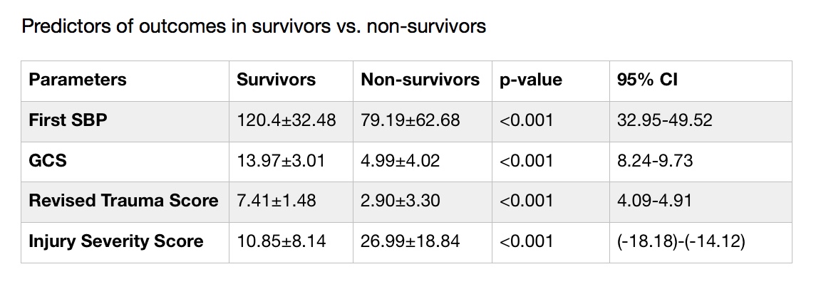
Results: There were 1,799 patients sustaining 1,900 brachial vessel injuries out of 1,466,887 patients registered in the NTDB from 2001 to 2005. Incidence was calculated to be 0.12%. Mean age at injury was 30 ± 14, mean RTS 7.23 ± 1.8, mean Glasgow Coma Scale 13.6 ± 3.5, mean ISS 11.5 ± 9.5. Mechanism of Injury: penetrating n=1,114 (61.9%), blunt n=699 (37.1%), and non-specific n=16 (1%). Mean initial SBP 119 ± 32.5; overall and adjusted survival rates: survivors 94.7%; non-survivors 5.3%. Patients underwent surgical repair n=671. Stratified as primary arteriorrhaphy 369 (55%), RSVG 165 (24.5%), resection of upper limb vessel with replacement 116 (17.3%), PTFE 21 (3.2%). Analysis revealed an increased ISS in the blunt injury group vs penetrating (10.65±8.82 vs 13.09±10.43; p<0.001). In survivors vs non-survivors, initial systolic blood pressure, glasgow coma scale, revised trauma score, and injury severity score were all statistically significant between the two groups (see table).

Conclusion: Brachial vessel injuries remain a low incidence injury as is shown in the NTDB. The injury severity score has a higher correlation with morbidity based on mechanism of injury. The overall cost of all brachial injuries was $22,954,998 in patient with extractable information (n=727).


102.16 Femoral Vessel Injuries: A Review of Cases from the National Trauma Data Bank.

D. J. Keleny1, A. D. Person1, G. Mendoza-Barrera1, S. R. Brown1, D. Rigg1, M. Dale1, J. Dabestani1, D. K. Agrawal1, J. A. Asensio1  1Creighton University Medical Center,Trauma Surgery And Surgical Critical Care,Omaha, NE, USA

Introduction: Femoral vessel injuries are the most frequent vascular injuries seen at Trauma Centers accounting for 70% of all peripheral large blood vessel injuries treated in large volume hospitals. This makes it important to determine parameters contributory to morbidity and mortality with such injuries in an effort to optimize management and predict outcome. Our objective is to review the National Trauma Data Bank with respect to location of femoral vessel injuries with related mortality, to identify predictors of patient outcome, and to report the outcomes of these injuries based on the aforementioned predictive variables including mechanism of injury.


Methods: The National Trauma Data Bank was queried for pre-hospital and admission data for femoral vessel injuries. The primary outcome measured was survival. Statistical analysis included univariate and stepwise logistic regression.


Results: A total of 2,021 patients were identified with a total of 2,693 femoral vessel injuries from 1,466,887 patients in the National Trauma Data Bank. This is an incidence of 0.13%, with the number of survivors being 1,788 (89%). Mean age of patients was 34.1±17.34. The mean revised trauma score (RTS) for survivors was 7.2±1.67 ; for non-survivors 3.57±3.35. The mean GCS for survivors was 13±3.65 ; for non-survivors 6.59±5.11. The mean injury severity score (ISS) for survivors was 16.73±6.32 ; for non-survivors 28.0±13.88. Mechanism of injury was documented in 1,996 patients, with the majority due to penetrating injuries (1,419; 71%). The most commonly injured vessel was the superficial femoral artery (1,044; 39%). The next most commonly injured vessels were the femoral vein (817 ; 30%), the common femoral artery (645; 24%), and the femoral nerve (153; 5.7%). The highest mortality was 19%, in patients with injuries to the common femoral artery.


Conclusion: Although there is a relatively high survival rate for femoral vessel injuries, they incur high complication rates. Initial admission parameters correlate well with morbidity and mortality, including neurological status and hemodynamic stability. When comparing mortality rates by location of injury, common femoral vessel injuries had a significantly higher mortality rate although the most commonly injured vessel is by far the superficial femoral artery. Femoral vein injuries were also more lethal than superficial femoral artery injuries. These correlations should be looked to for optimization of management.

102.14 Pattern of Vascular Injuries From The Colombian Military Conflict

G. E. Mendoza-Barrera1, W. Sanchez2, S. R. Brown1, A. Person1, D. Keleny1, D. Rigg1, M. Dale1, J. Dabestani1, D. K. Agrawal1, J. A. Asensio1  1Creighton University Medical Center,Surgery,Omaha, NE, USA 2Colombian Military Hospital,Nueva Granada Military School Of Medicine,Bogota, BOGOTA, Colombia

Introduction:  Recent military conflicts have changed from more conventional to guerrilla and counter insurgency warfare with the subsequent refinement of newer antipersonnel devices such as improvised explosive devices (IEDs). The Iraq and Afghanistan conflicts have shown newer injury patterns with a subsequent decrease in definitive extremity vascular injury repairs, along with a significant increase in single and multiple limb amputations. Objectives of this study are to analyze the Colombian military experience with combat related extremity vascular injuries. Describe distribution of wounds, mechanisms of injury related to vascular injuries incurred in combat. Identify predictors of outcomes and validate conventional  wounding patterns.

Methods: Retrospective review of 13 year prospective data base. Statistical analysis: Chi square, T test, ANOVA and stepwise logistic regression which included mode of injury (MOI), MOI over time, ISS, RTS, type of vascular injury, time to definitive surgical care, procedures performed, incidence of amputation and mortality.

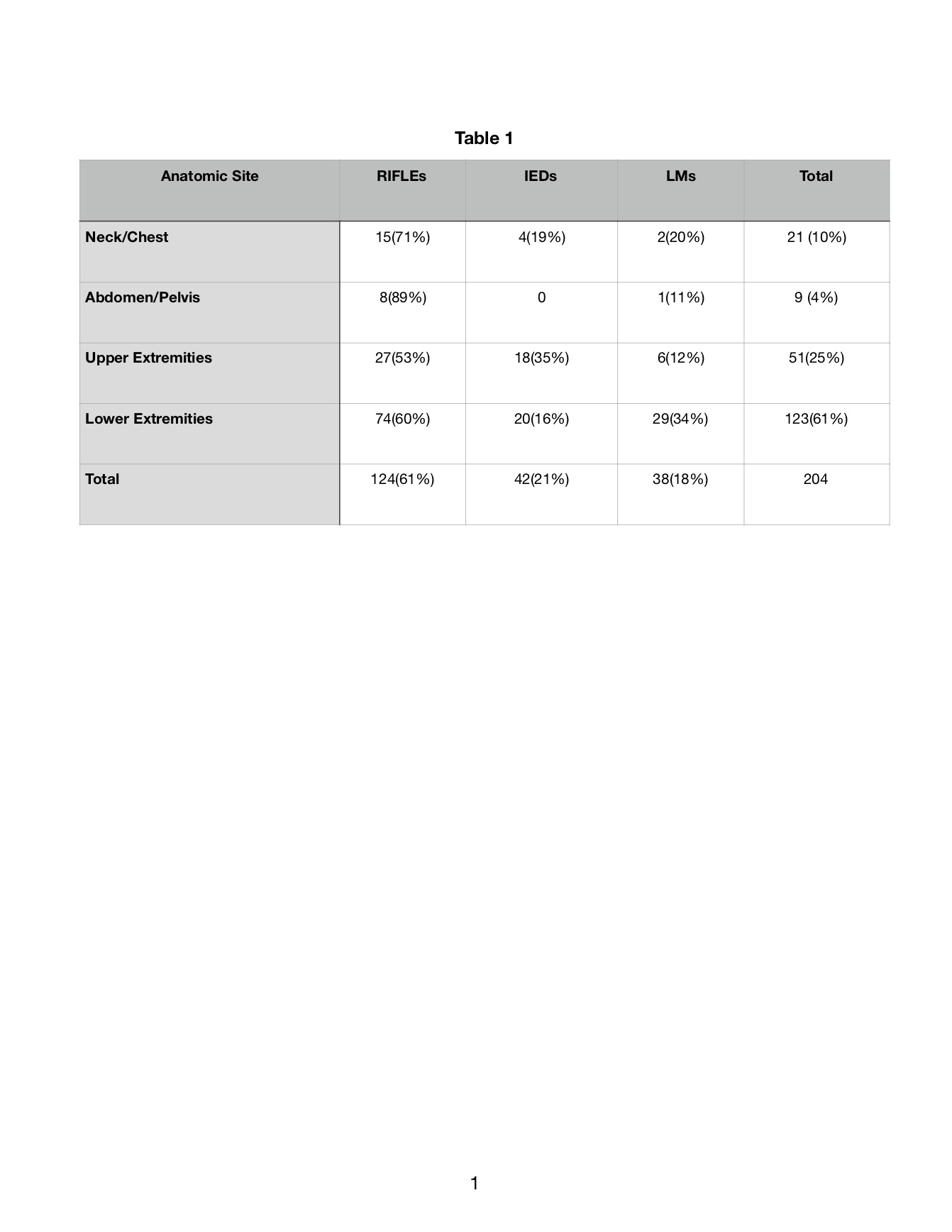
Results: 204 patients sustained 390 vascular injuries 159 (40.8%) arterial and 231(59.2%) . Weapons: Automatic Rifles (RIF) n=124 (60.7%), Improvised Explosive Devices (IEDs) n=42 (20.7%) and Land Mines (LMs) n=38 (18.6%). 70 patients (36.8%) were directly transferred from the field and 134 (63.2%) from MASH units. Mode of injury (MOI) remained unchanged. Extremity vascular injuries accounted for 86%: – 61% upper and 25% lower.  Neck 10% , abdominal injuries 4%. 20 patients (9.8%) required amputation (19 LE and 1 UE). Amputation was independent of MOI – RIF 9.7%, IEDs 13.1% and LM 9.8%, – p < 0.05. Amputation was dependent on presence of combined arterial-venous injuries (AVI), ligation of a named vein and performance of fasciotomy. 11/20 (55%) with lower extremity amputations sustained arterial-venous (AV) injuries, popliteal vessel injury managed with arterial interposition vein graft and popliteal vein ligation. Overall mortality 3.9% (8/204).

Conclusion: For combatants sustaining vascular injuries automatic rifles remain responsible for majority of these injuries. Extremity vascular injuries are as in other wars most prevalent. Strategies to decrease the rate of amputation for popliteal artery injuries: avoid ligation of popliteal vein  and perform complete fasciotomy. Trauma surgeons must remain adept at vascular injuries management.

102.11 Effect of Temporary Shunts on Neurologic Outcomes in Repair of Penetrating Carotid Artery Injuries

S. R. Brown1, A. D. Person1, G. E. Mendoza1, M. Dale1, D. Rigg1, D. Keleny1, J. Dabestani1, D. K. Agrawal1, J. A. Asensio1  1Creighton University Medical Center,Surgery,Omaha, NE, USA

Introduction:  The management of penetrating carotid artery injuries continues to evolve. Early questions regarding repair versus ligation have largely been settled in favor of repair for all but the most devastating injuries. To date, the use of temporary shunts to maintain cerebral perfusion in the repair of carotid artery injuries has not been well studied. Thus far, no single study has had the power to state whether the use of shunts makes any difference in neurological outcomes in the setting of penetrating carotid artery injuries.

Methods: A focused literature search was performed with PubMed, Scopus, and Ovid. PRISMA guidelines were followed. Inclusion criteria were those series reporting surgical repair for penetrating carotid artery injuries. Exclusion criteria were endovascular repair, neurosurgical techniques involving craniotomy, and studies with no reports on preoperative or postoperative neurologic status. Further relevant studies found as references in these papers were also evaluated. A total of 28 papers were identified that met these criteria, ranging from 1970 to 2012. Studies were systematically analyzed to extract cases of surgical repair for which both the utilization of temporary shunts and perioperative neurologic outcome data was reported. Non-parametric data was analyzed with Fisher Exact or Chi-Square tests as applicable. Statistical significance was set to a p-value less than 0.05.

Results: There were a total of 717 carotid artery injuries. 592 were repaired without shunts. 125 were repaired with shunts. There was a trend towards lower overall mortality in repairs with shunts vs. those without shunts (5.6% vs. 9.8%; p=0.17). There was a trend towards increased rates of neurological improvement in repairs with shunts vs. those without shunts (13.6% vs. 10%, p=0.34). Patients undergoing repairs with shunts were more likely to have improved or unchanged neurologic outcomes in comparison to those without shunts (92% vs. 84%, p=0.018).

Conclusion: This focused analysis offers the first large scale evidence that the use of temporary shunts in the surgical repair of penetrating carotid artery injuries results in better neurologic outcomes in comparison to repair without the use of shunts. Based on this evidence, we recommend the routine use of temporary shunts during complex repair of penetrating carotid artery injuries.


101.10 Penetrating Extracranial Vertebral Artery Injuries Collective Review of Rare and Difficult Injuries

J. A. Asensio1, P. J. Dabestani1, C. A. Fernandez1, T. Becker1, R. Bertellotti1, D. Cornell1, T. Kraner1, A. B. Olsen1, D. K. Agrawal1, J. A. Asensio1  1Creighton University Medical Center,Trauma Surgery And Surgical Critical Care,Omaha, NE, USA

Introduction:

Penetrating vertebral artery injuries (VAI) are rare. Their clinical presentation range from asymptomatic to exsanguination. Given their rarity, complex anatomy and difficult surgical exposure, few Trauma Surgeons or Trauma Centers have significant experience with these injuries. The objectives of this study are to review their incidence, clinical presentation, radiologic identification, management – both angiographic and operative, incidence of aneurysms, pseudoaneurysms, arteriovenous fistulas, and outcomes, as well as to review operative approaches for their surgical management.

Methods:

A literature search was conducted on MEDLINE Complete-PubMed. From 1893-2018 all series describing management and outcomes of penetrating VAI's were selected. PRISMA guidelines were employed. Original series and case reports yielded a total 181 studies. They were winnowed to 72 studies, including series and case reports, which form the basis of this collective review. Operative procedures and outcomes were recorded, along with methods of diagnostic imaging, angiographic, and operative management.

Results:

There were a total of 462 patients with penetrating VAI's. Incidence of vertebral artery injury military population ­– 0.2%, civilian population – 3.1%. More complete data was available from 13 collected VAI specific series and 37 case reports for a total of 362 patients. Mechanism of injury data was available for 341 patients (94.2%): GSW’s – 178 patients (52.2%), SW’s – 131 (43.2%), miscellaneous mechanisms of injury – 32 (9.4%). Anatomic site of injury data was available for 177 (49%) patients: 92 (52%) left, 84 (47.5%) right, and bilateral ­– 1 (0.5%). Anatomic segment of injury data was available for 206 patients (57%): 28 (13.6%) V-1, 126 (61.2%) V-2, and 52 (25.2%) V-3. Treatment data was available for 212 patients: Operative management – 89 (38.7%), angiography and angioembolization – 67 (29.1%), combined – 16 (7%) and observation – 58 (25.2%). Stenting and repair were less frequently employed – 10 (4.3%). Incidence of aneurysms/pseudoaneurysms – 61 (16.9%), AVF – 67 (21.6%). Calculated mortality VAI specific series ­– 15.1%, individual case report group –10.5%.

Conclusion:

The majority of VAI's injuries are asymptomatic upon presentation but may present with exsanguinating hemorrhage. CTA is the first line imaging modality to establish diagnosis, if hemodynamically stable. Gunshot wounds account for the majority of these injuries. Most frequently injured segment is V-2. Operative interventions were required in 38.7% and includes: ligation, clipping and packing. Angiography and angioembolization are diagnostic and therapeutic and was required in 29.1%. Trauma surgeons must be adept to surgically expose this vessel, control hemorrhage, prevent exsanguination, and address associated injuries.

100.16 Iliac Vessel Injuries: An Analysis of the National Trauma Data Bank

A. D. Person1, S. Brown1, G. Mendoza1, D. Keleny1, D. Rigg1, M. Dale1, J. Dabestani1, D. K. Agrawal1, J. A. Asensio1  1Creighton University Medical Center,Department Of Trauma Surgery And Surgical Critical Care,Omaha, NE, USA

Background: Iliac vessel injjuries are uncommon, even in high volume trauma centers.  THey incur high morbidity and mortality.  The objectives of this study were to review the National Trauma Data Bank experience with iliac injuries, identify predictors of outcome, and then report the outcomes of these injuries.

Methods: The National Trauma Data Bank was queried for pre-hospital and admission data for all iliac vessel injuries.  The main outcome measured was survival.  Statistical analysis included univariate and stepwise logistic regression.

Results: 1,590 patients were identified from 1,466,887 patients from the National Trauma Data Bank.  Incidence of iliac vessel injury was 0.1%. Mean age of patients with these injuries was 35.7 +/- 18.8. Mean Revised Trauma Score (RTS) was 5.98 +/- 2.8, while the mean Injury Severity Score (ISS) was 26.1 +/- 13.4. The mechanism of injury distribution between penetrating and blunt was 804 (51%) and 777 (49%) respectively. In terms of the vessels injured, the iliac artery was injured, there were 1,174 arterial injuries, 651 venous injuries, and 84 unspecified injuries.  Span data on surgical repairs was available in 380 patients.  Surgical management is as follows: arteriorrhaphy 129 (34%), venorrhaphy 170 (45%), primary repair of artery and/or vein with vein and/or prosthetic patch 52 (14%), and interposition grafts 29 (8%). In total, there were 1,040 survivors (65.4%) and 550 non-survivors (34.6%).

Multiple factors, able to be assessed on presentation demonstrated statistical significance between survivors and non-survivors. Mean Systolic Blood Pressure on presentation was 110.36 vs. 83.45 (p<0.001) between survivors and non-survivors respectively.  Mean Revised Trauma Score was 6.80 vs. 4.41 (p<0.001). Mean Injury Severity Score was lower in survivors at 23.62 vs. 31.17 (p<0.001). Glasgow Coma Scale was 12.5 vs. 7.2 (p=0.002).

Conclusion: Iliac vessel injuries are uncommon in the landscape of American trauma as denoted by their low incidence reported in the National Trauma Data Bank.  Mortality remains high. Predictors of outcome may be identified from the initial admitting data. When stratified to mechanism of injury, this idea remains valid and also appears to correlate well with mortality.