I. Lou1, A. Harrison1, A. Dammalapati1, R. Jaskula-Sztul1,2, H. Chen1,2 1University Of Wisconsin,Madison, WI, USA 2University Of Alabama,Birmingham, Alabama, USA
Introduction:
Medullary thyroid cancer (MTC) is a neuroendocrine tumor that when metastatic portends only a 40-50% overall 5-year survival. MTC commonly metastasizes to the liver, and when diffusely disseminated remains a therapeutic challenge as there are currently no curative options. The aim of this study is to determine the feasibility of an in vivo model of MTC liver metastasis.
Methods:
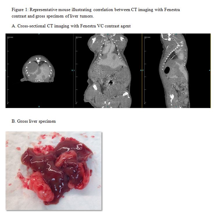
TT, a human MTC cell line, was genetically engineered to overexpress Notch3, a tumor suppressor in MTC, in the presence of doxycycline. Prior to injection, the cells were plated, and treated with doxycycline every 48 hours for 12 days to ensure adequacy of the inducible construct in vitro. 1×10⁷ TT-Notch3 cells were injected intra-splenically into male nu/nu mice. The cells were allowed 2 minutes to enter circulation, and a splenectomy was performed. The mice were then recovered, given a standard chow diet, and the tumors were allowed to propagate. Physical examination of the mice at 4, 6, and 8 weeks revealed no external signs of intra-abdominal tumor burden such as ascites or palpable masses. Each mouse was imaged via computerized tomography (CT) scan with Fenestra VC contrast agent (MediLumine Inc, Montreal, Quebec) to evaluate for presence of tumors at 12 weeks. The mice were then sacrificed to determined correlation with CT findings and gross specimens. Lastly, the liver tumors were quantified with the use of Inveon Research Workplace (IRW, Siemens Healthcare).
Results:
Western blotting analysis for the Notch3 protein revealed maintenance of induction in the TT-Notch3 cells over a period of 12 days in vitro. There was 100% survival of these mice at 12 weeks. When imaged, 100% of mice demonstrated the presence of liver metastasis on CT scanning with Fenestra VC contrast. The imaging results correlated well in 80% of mice on necropsy with grossly visible tumors (Figure 1). The tumors on CT scan were then quantified by volume measurements with the use of IRW software.
Conclusion:
The intrasplenic injection model is a feasible method in which to form, image, and quantify liver metastasis for MTC. This in vivo model is an important step in further understanding metastatic MTC, and in the discovery and development of future therapeutics.