C. A. Cooper1, G. Jiang2,3, Y. Liu2,3, T. A. Zimmers1,4,5 4Indiana University School Of Medicine,IU Simon Cancer Center,Indianapolis, IN, USA 5Indiana University School Of Medicine,Anatomy & Cell Biology,Indianapolis, IN, USA 1Indiana University School Of Medicine,Department Of Surgery,Indianapolis, IN, USA 2Indiana University School Of Medicine,Department Of Medical And Molecular Genetics,Indianapolis, IN, USA 3Indiana University School Of Medicine,Center For Computational Biology And Bioinformatics,Indianapolis, IN, USA
Introduction:
Clear cell renal cell carcinoma (ccRCC) is frequently associated with cachexia, a wasting syndrome characterized by the loss of skeletal muscle (SKM) and adipose tissue (AT) mass. In ccRCC patients, cachexia is correlated with decreased survival, while obesity is associated with increased survival. Cachexia results in part from host response to tumor-produced cytokines. Here we assessed the associations between body composition, tumor cytokine expression and overall survival (OS) in ccRCC.
Methods:
The Cancer Imaging Archive and the Cancer Genome Atlas were queried for ccRCC patients with available abdominal computed tomography (CT) imaging studies, survival information and tumor RNAseq data. All three datasets were available for 215 patients. SKM, intramuscular AT tissue (IMAT), visceral AT (VAT), and subcutaneous AT (SAT) area were measured at the L3 vertebral level in CT scans using Sliceomatic. Data were stratified by gender and tissue area quartiles. Kaplan-Meier survival analysis and log rank test (p<0.05) were used for OS comparisons between highest and lowest quartiles for each tissue area. Associations between tumor expression of 21 cachexia-associated cytokines and SKM were analyzed with Spearman’s rank correlation (rho>0.1, p<0.05) with False Discovery Rate for multiple comparisons. Identified genes were probed for OS differences between groups stratified below and above median gene expression.
Results:
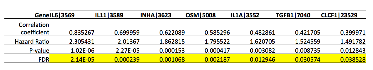
OS was lower in the lowest versus the highest quartile for SKM in both males and females (p=0.002 and p=0.010, respectively). OS was lower for low SAT and total AT areas in males only (p=0.003 and P=0.015, respectively). A difference in OS for VAT area approached significance in males only (p=0.069). No differences in OS for IMAT were noted in either sex. Correlation coefficients for gene expression and any tissue measurement were small (<0.2). For SKM, the tissue presumably most functionally important for survival, correlated genes were INHBB, CTF1, and TGFB2 (p=0.029, p=0.010 and 0.028). Gene expression of 7 other cytokines were robustly associated with reduced OS (Table).
Conclusions:
Body composition analysis showed sex-specific differences in body composition. Low SKM area was robustly associated with decreased OS in both males and females independent of stage in ccRCC, consistent with abundant data linking muscle loss and mortality. Differences in the other associations of body composition and OS indicated a possible gender-specific role of AT in ccRCCC. Tumor-derived mediators of cachexia are highly associated with survival in ccRCC, although not with body composition, suggesting they might act indirectly.