N. Bhutiani1, C. A. Doughtie1, R. C. Martin1 1University Of Louisville,Surgical Oncology,Louisville, KY, USA
Introduction:
Currently, the prediction of irreversible electroporation (IRE) ablation dimensions are modeled using algorithms derived from mathematical and ex-vivo models. These have not been validated using in-vivo studies. The aim of this study is to assess the correlation between the mathematical prediction model to and ultrasound and histopathology findings for in vivo ablations in a porcine model.
Methods:
IRE ablations were performed on porcine liver and spleen with probe spacings ranging from 0.6 to 2.6 cm. Pre and 2 hour post-ablation ultrasound images were recorded and validated with histopathology confirmation. Three dimensions of the ablation regions were recorded and ablation volumes were calculated and correlated with theoretical mathematical models for each given probe spacing.
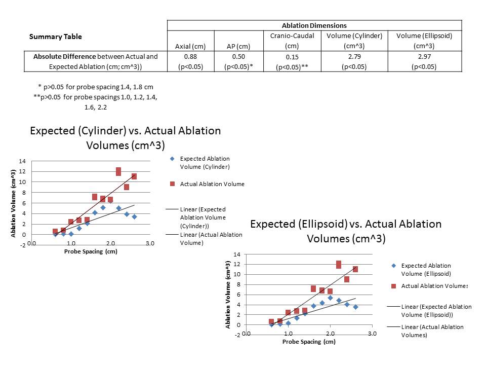
Results:
In-vivo axial and anterior-posterior (AP) ablation distances were significantly greater than predicted for nearly all probe spacings (p<0.05). Ablation volumes were significantly less than predicted for the all probe spacings when modeled using both a cylinder and an ellipsoid. Geometrically, mathematically derived ablation regions demonstrated more central tapering (‘necking’) and diminished volumes compared to their in-vivo counterparts. The relationships between probe spacing and AP ablation dimensions were less linear (r2=0.57) than the relationships observed via ultrasound.
Conclusion:
The current mathematical models poorly predict ablation regions observed in vivo. They underestimate ablation dimensions and, by extension, ablation volumes. Further work should be done to improve models for ablative planning, and physicians should recognize the limitations of existing models when planning ablative treatments.