C. J. Park1, M. P. Shaughnessy1, R. A. Cowles1 1Yale University School Of Medicine,Pediatric Surgery,New Haven, CT, USA
Introduction: Animal models allow researchers to study the effects of pharmacological and surgical interventions on the gastrointestinal (GI) mucosa. The benefits of using mice in the laboratory include low cost, ease of maintenance, and the applicability of a wide range of molecular tools. While the typical laboratory mouse reaches sexual maturity at 6-8 weeks, maturation ensues and mice are considered mature adults at 12-24 weeks. Meanwhile, the ages and genders of mice used for experiments reported in the literature vary drastically. We hypothesized that there is age-, but no gender-related variation in intestinal morphometric parameters, necessitating thoughtful experimental design.
Methods: With IACUC approval, C56Bl/6J mice of varying ages (6, 12, 18, 24 weeks; n=4/group) were euthanized and the small intestine was isolated from the ligament of Treitz to the ileocecal valve. 2 cm segments of proximal, middle and distal small intestine were harvested and morphometric parameters assessed with microscopy. The distal segments were also stained for Ki67 to determine the crypt proliferation index (CPI). Secondary analysis comparing males and females (n=4/group) of matched ages was performed. Means were compared with Student’s t-test and variance of proportions was assessed with the Chi-squared test to a significance of p<0.05.
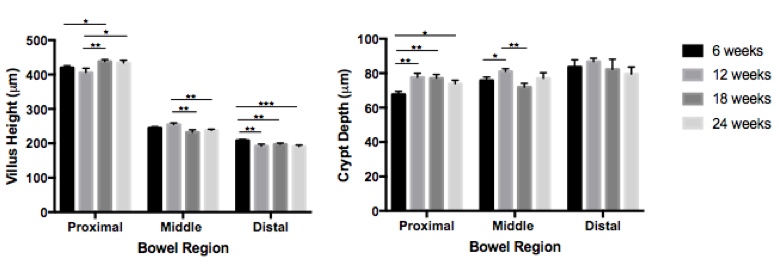
Results: There was variation in the measured morphometric parameters (Figure). In the proximal small intestine, 18-week old mice had taller villi compared to 6- and 12-week old mice and 24-week old mice had taller villi compared to 12-week old mice. In the middle segments, 12-week old mice had taller villi compared to 18- and 24-week old mice. In the distal segments, 6-week old mice had taller villi compared to all other age groups. When comparing crypt depth, 6-week old mice had shallower crypts compared to all other groups in the proximal region and 12-week old mice had deeper crypts compared to 6- and 18-week old mice in the middle segments. CPI was statistically lower only in 12-week old mice compared to all other age groups. When comparing age-matched males and females, there was no difference in villus height or crypt depth except for the middle small intestine where villus height was greater in females compared to males. There was no difference in CPI between genders.
Conclusion: Small, but statistically significant, differences in villus height, crypt depth and crypt proliferation are present in mice of different ages, while fewer differences exist between male and female mice of the same age. Investigators studying the GI mucosa should be aware of these differences and aim for consistent age-matching of experimental animals in order to avoid errors and allow direct comparison between studies.