J. Leckenby1,3, M. Chacon1, A. Grobbelaar3, J. Lichtman2 1University Of Rochester,Plastic Surgery,Rochester, NY, USA 2Harvard School Of Medicine,Molecular And Cell Biology,Cambridge, MA, USA 3Royal Free Hospital NHS Foundation Trust,Plastic Surgery,London, LONDON, United Kingdom
Introduction:
Peripheral nerve assessment has traditionally been studied through histological and immunological staining techniques in a limited cross-sectional modality. The introduction of transgenic species, such as YFP-H mice, has greatly increased our ability to observe axonal regeneration and subjectively comment on behavior. However, detailed analysis is still difficult to assess with either of these methods and understanding cellular interactions is almost impossible. A new application of serial section electron microscopy (SSEM) is presented to overcome these limitations and open the application of this technique to other areas of pathology.
Methods:
Direct nerve repairs (DNR) were performed on the posterior auricular nerve of transgenic YFP-H mice. Six weeks post-operatively the nerves were imaged using confocal fluorescent microscopy then excised and embedded in resin. Resin blocks were sequentially sectioned at 100nm and sections were serially imaged with an electron microscope (Magellan 400L, FEI) utilizing the following parameters: 100nm pixel size, 200ns Dwell Time, 8k x 8k pixel field of view with 7.0kV and 26nA. Images were aligned and auto-segmented to allow for 3D rendering using 3D Studio Max (Autodesk).
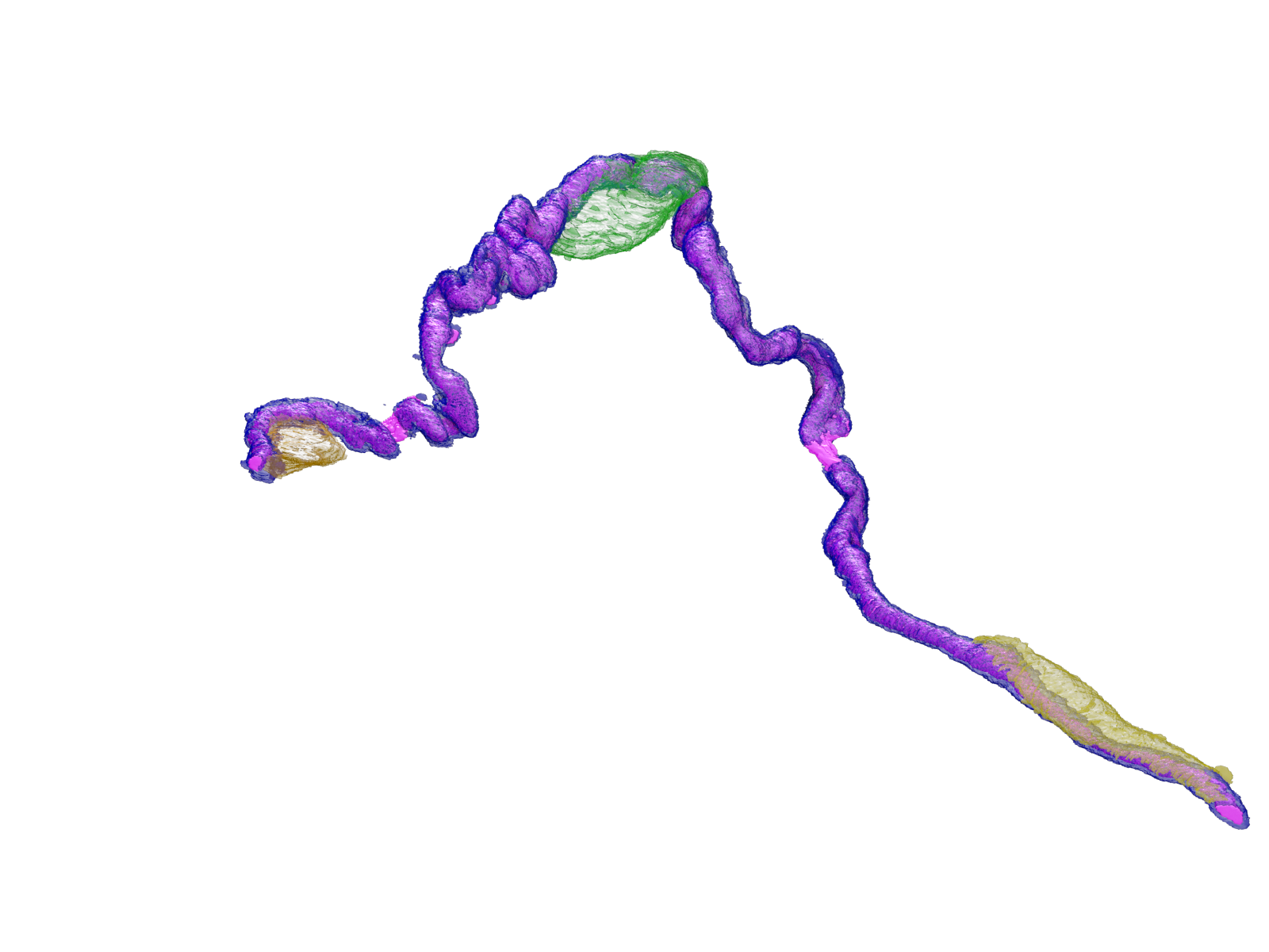
Results:
Basic morphometry and axonal counts were fully automated. Using full 3D reconstructions, the relationships between the axons, the Nodes of Ranvier, and Schwann cells could be fully appreciated (See Fig. 1). The quality of regeneration could be examined throughout the entire dataset providing a comprehensive analysis. The interactions of individual axons with their surrounding environment could be visualized and explored in a virtual three dimensional space.
Conclusion:
SSEM allows the detailed pathway of the regenerating axon to be visualized in a 3D virtual space in comparison to isolated individual traditional histological techniques. Fully automated histo-morphometry can now give accurate axonal counts, provide information regarding the quality of nerve regeneration and reveal the cell-to-cell interaction at a super-resolution scale. It is possible to fully visualize and ‘fly-through’ the nerve to help understand the behavior of a regenerating axon within its environment. Having established this technique it provides future opportunities to evaluate the affect different treatment modalities have on the neuro-regenerative potential and help us understand the impact different surgical techniques have when treating nerve injuries. Further investigations are being carried out to determine whether SSEM can be used to analyze cellular interactions in disease processes such as infection or tumor formation.