A. Al-Gahmi1, C. P. Jara1, M. A. Carlson1 1University Of Nebraska College Of Medicine, Surgery, Omaha, NE, USA
Introduction: Despite decades of research, survival from pancreatic cancer (PC) has been minimally impacted. Swine have anatomic, genetic, physiologic, metabolic, and immunologic similarities to humans, making them suitable for PC modeling and for testing novel therapeutics or devices. Our objective was to develop an orthotopic porcine PC model using KRASG12D-transformed ductal cells that also expressed PIDO (fusion protein of PD-L1 and indoleamine dioxygenase) and/or PD-L2. These proteins have been shown to induce immune evasion of allogeneic islet cell implants in immunocompetent mice. We hypothesized that the expression of PIDO/PDL2 in tumorigenic porcine pancreatic ductal cells would produce allograft survival through immune evasion and yield an orthotopic PC model in immunocompetent swine.
Methods: Anesthetized domestic swine (N = 4; ~42 kg, age 3-4 months) underwent upper midline laparotomy and pancreatic duodenal lobe exposure followed by four pancreatic injections per swine (two anterior and two posterior) of KRASG12D-transformed pancreatic ductal cells (107 in 0.2-1 mL of 5 mg/mL Matrigel) expressing (i) control (mutant KRAS only), (ii) PIDO, (iii) PD-L2, and (iv) PIDO + PD-L2. All swine were euthanized at four weeks and tissue was collected for histopathologic analysis.
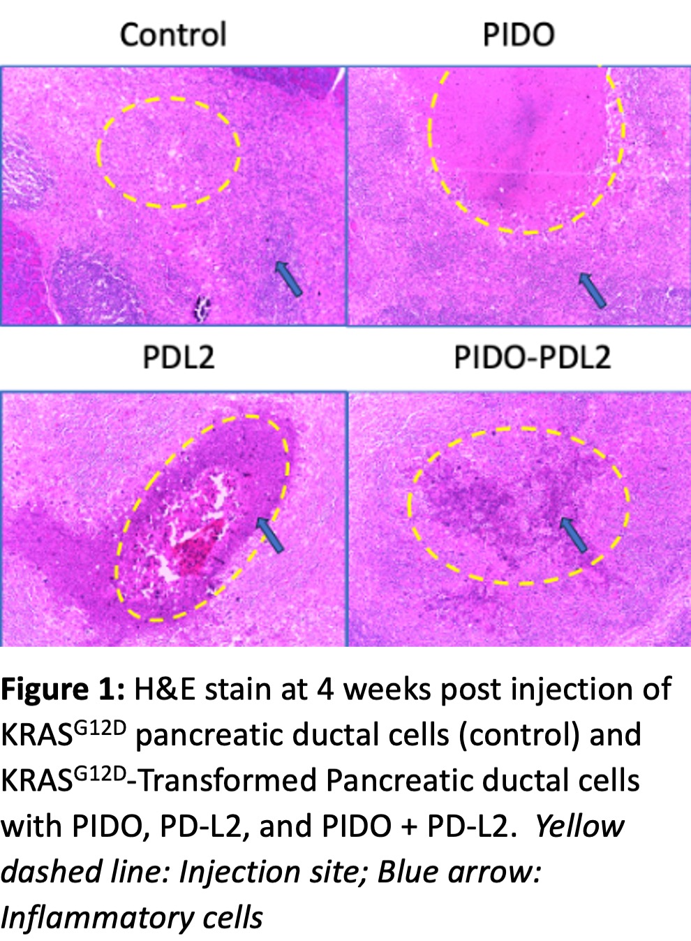
Results: Expression of mutant KRAS and PD-1 ligand proteins was confirmed in vitro prior to the in vivo implantations (data not shown). All four swine survived the four-week observation period without negative signs. Pre-implantation vs. necropsy serum studies (including CA 19-9, CAE, AFP) were not different. At necropsy, there was gross evidence of mild fibrosis/inflammation at the injection sites without palpable mass. Peripancreatic lymphadenopathy was present. All four injectate types (control, PIDO, PD-L2, and PIDO + PD-L2) had no microscopic evidence of transformed tumor cells (Fig. 1) at the injection or regional nodes, neither by H&E nor immunohistochemistry. However, each site did have prominent inflammatory infiltrate consisting of neutrophils, macrophages, and T-cells.
Conclusion: In this preliminary study, orthotopic implantation of allogenic KRASG12D-transformed porcine pancreatic ductal cells with various PD-1 ligand protein expression elicited an immune response but did not produce any observable tumor implants. The failure of various PD-1 ligands to promote immune evasion in this allogeneic orthotopic porcine tumor implantation model (in contrast to the previous non-tumor murine studies) may be secondary to inherent differences between swine and mice, inappropriate/inadequate implantation conditions, redundant immune response mechanisms specific to tumors, or other issues. Additional studies are underway to explore these possibilities.