25.02 PI3K/mTOR Inhibition Suppresses Pancreatic Cancer by Reprogramming Cancer-Associated Fibroblasts

J. L. Williams2, I. A. Elliott1, A. H. Nguyen1, C. Matsumura1, R. Ghukasyan1, P. A. Toste1, J. R. Capri3, S. G. Patel1, L. Li1, N. Wu1, C. G. Radu3, T. R. Donahue1,3  1David Geffen School Of Medicine, University Of California At Los Angeles,Department Of Surgery,Los Angeles, CA, USA 2Harbor-UCLA Medical Center,Department Of Surgery,Torrance, CA, USA 3David Geffen School Of Medicine, University Of California At Los Angeles,Department Of Molecular And Medical Pharmacology,Los Angeles, CA, USA

Introduction:  Pancreatic ductal adenocarcinoma (PDAC) is a highly lethal disease, with an overall survival of less than one year.  Contributing to this poor prognosis is PDAC’s characteristically dense stroma, which is comprised predominantly of cancer-associated fibroblasts (CAFs). CAFs promote tumor growth, metastasis, and treatment resistance because when activated by environmental factors such as cancer cell (CC) exposure, hypoxia, and cytotoxic chemotherapy, they secrete protumorigenic cytokines, growth factors, and extracellular matrix components.  However, elimination of CAFs also increases tumor growth and invasion.  Therefore, rather than ablation, reprogramming of CAFs to an inactive or quiescent state is a potential PDAC treatment strategy.  In this study, we aimed to determine the effect of PI3K/mTOR inhibition, a pathway known to be involved in fibroblast activation, on PDAC tumorigenicity.

Methods:  Immortalized CAFs were treated with NVP-BEZ235 (BEZ), a dual PI3K/mTOR inhibitor, and exposed to gemicitabine (GEM), hypoxia (1% O2), or PDAC CC conditioned media (CM) for 48 hours.  After treatment, markers of CAF activation were determined via reverse transcription polymerase chain reaction (RT-PCR), Western blot, and mass spectrometry proteomics analysis of CAF CM.  Additionally, PDAC CCs (Mia PaCa-2 and PANC-1) exposed to CM from treated CAFs were tested with in vitro viability, migration, and invasion assays. PDAC CCs were also co-injected with treated CAFs in vivo, and tumor size was assessed.  A 3D co-culture model was used to assess the effect of BEZ treatment on CCs grown together with CAFs.

Results: After treatment with BEZ, gene expression of known markers of CAF activation, including alpha-smooth muscle actin (αSMA) and type I collagen, were decreased, even after exposure to activating stimuli.  Similarly, αSMA protein levels were reduced in BEZ-treated CAFs. CM from BEZ-treated CAFs contained lower levels of extracellular matrix components, proinflammatory cytokines, and growth factors. CCs exposed to CM from BEZ-treated CAFs were less viable, migratory, and invasive in vitro. Pre-treatment of PDAC CAFs with BEZ restricted PDAC growth in our in vivo co-implantation model (Figure 1).  In CC-CAF 3D co-culture, combination treatment of BEZ with GEM was more effective at suppressing CC growth than GEM treatment alone.

Conclusion: Inhibition of PI3K/mTOR decreases CAF activation, resulting in reduced PDAC CC tumorigenesis in vitro and in vivo. Additionally, combination treatment with BEZ, a dual PI3K/mTOR inhibitor, and GEM synergistically inhibits PDAC CC growth in the presence of CAFs. These data suggest that the addition of BEZ may improve the effectiveness of cytotoxic chemotherapy in PDAC.
 

24.07 Nucleotide Depletion by Autophagy Inhibition Sensitizes Kras-driven PDAC to Replication Stress

I. A. Elliott1, J. L. Williams2, R. Ghukasyan1, C. C. Matsumura1, N. Wu1, L. Li1, W. Kim3, S. Poddar3, E. R. Abt3, A. M. Dann1, H. DeRubertis1, D. Braas4, T. M. Le3, C. G. Radu3, T. R. Donahue1,3  1University Of California – Los Angeles,Department Of Surgery,Los Angeles, CA, USA 2Harbor-UCLA Medical Center,Department of Surgery,Torrance, CA, USA 3University Of California – Los Angeles,Department Of Molecular And Medical Pharmacology,Los Angeles, CA, USA 4University Of California – Los Angeles,Metabolomics Center,Los Angeles, CA, USA

Introduction:
Autophagy is a critical source of nucleotides, which are rate-limiting for Ras-driven cancer cell proliferation. Genetically disabling autophagy impairs energy and redox homeostasis by depletion of nucleotide pools, and sensitizes cancer cells to radiation. We hypothesized that the autophagy inhibitor chloroquine (CQ) would deplete deoxyribonucleotide triphosphate (dNTP) availability for incorporation into DNA, thus sensitizing cancer cells to replication stress and revealing a targetable liability in Ras-driven pancreatic ductal adenocarcinoma. 

Methods:
Human (MiaPaCa-2) and murine (KPC) PDAC cells were treated with CQ +/- the replication stress response (RSR) inhibitor VE822. S-phase cells were labeled by pulsing with 5’-ethynyl-2’-deoxyuridine (EdU) and cell cycle progression measured by flow cytometry. Newly synthesized dNTP incorporation into DNA was measured via [13C6]glucose labeling and our novel liquid chromatography mass spectrometry (LC-MS) platform. Global metabolomics analyses were performed by detection of relative amounts of metabolites using LC-MS. Western blots (WB) were done on cell lysates. Cell viability was measured by Cell-Titer-Glo assay. MiaPaCa-2 or KPC cells were injected s.q. in the flanks of NSG or C57BL/6 mice. Mice were treated 5x/week with CQ+/-VE822 (60mg/kg p.o.). Immunohistochemistry (IHC) was performed on explanted KPC tumors after 3 days of CQ+VE822.

Results:
We found that CQ caused S-phase arrest in MiaPaCa-2, and impaired incorporation of newly synthesized dNTPs into replicating DNA(Fig.1a), indicating nucleotide pool insufficiency. Global metabolomic profiling of MiaPaCa-2 revealed that this was due to depletion of purine and pyrimidine substrates for dN synthesis under CQ, and this was exacerbated by addition of the RSR inhibitor VE822(Fig.1b). We then tested the impact of this nucleotide depletion on cell fate; CQ+VE822 treatment led to synergistic induction of DNA damage (reflected by pH2A.X WB), and decreased viability of MiaPaCa-2 and KPC in vitro (Fig.1c). We also observed increased IHC staining for pH2A.X in KPC tumors and impaired growth of MiaPaCa-2 tumors after CQ+VE822 treatment in vivo. Finally, we confirmed the ability of CQ to induce oxidative stress as indicated by HO-1 levels on WB. Management of redox balance and DNA damage are critical to recovery from radiation; accordingly, we found that CQ+VE822 profoundly impaired survival after radiation of MiaPaCa-2 and KPC cells.

Conclusion:
Pharmacologic inhibition of autophagy by CQ impairs incorporation of dNTPs into DNA by depleting substrates for nucleotide biosynthesis. When combined with an RSR inhibitor, this leads to induction of DNA damage and synthetic lethality in PDAC cells in vitro and in vivo.

02.03 The Tumor Microenvironment Stroma Promotes Cancer By Using Cell-Type Specific Stress Kinase Pathways.

S. G. Patel1, L. Li1, A. Maas1, R. Ghukasyan1, J. Williams1, P. Toste1, A. Nguyen1, I. Elliott1, N. Wu1, T. Donahue1  1University Of California Los Angeles,Surgery,Los Angeles, CA, USA

Introduction:
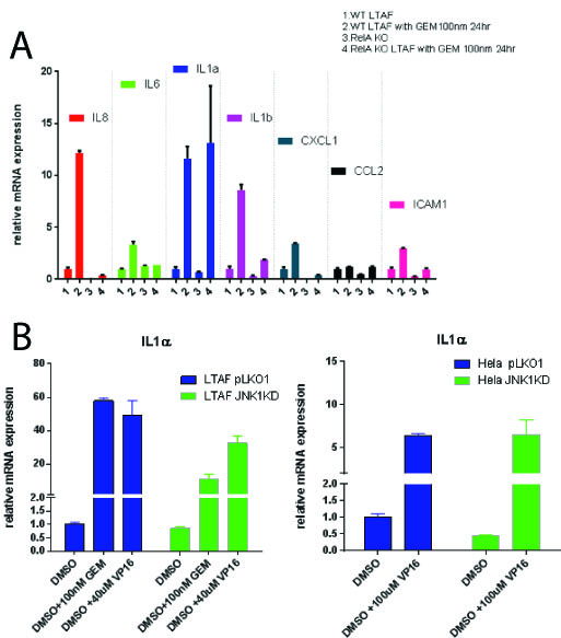
We have demonstrated that pancreatic cancer (PDAC) tumor associated fibroblasts (TAFs) support pancreatic cell growth and invasion from a pro-inflammatory, DNA damage (DDR) mediated pathway.  This pathway referred to as the senescence associated secretory phenotype (SASP) requires both stress kinases and Nfkb signaling for maximum signal production after stimulus.  Within the tumor microenvironment, genotoxic chemotherapies can stimulate the stromal cells to produce factors that promote tumor cell fitness.  Contrary to the prevailing model, we have identified that JNK isoforms are cell-type specific and that both JNK1 and JNK3 can transduce this inflammatory pathway in the tumor microenvironment.  We additionally have found that not all of the SASP is regulated by Nfkb, but rather a key mediator, IL-1 alpha, can be expressed in its absence.

Methods:
Immortalized pancreatic cancer tumor associated fibroblasts (Logsden Lab) as well as Hela cells were used to create both gene knockouts as well as stable knockdowns for JNK1, JNK3 and p65 (Nfkb signaling).  SASP induction was verified after a DNA damage response using RTPCR.  Small molecules specific to JNK isoforms were used to verify genetically manipulated cell lines in WT cells.

Results:

Gemcitabine treatment (100 nm) of PDAC TAFs induced a pro-inflammatory gene expression program including statistically significant (p < 0.05) upregulation of a number of cytokines: including IL-6, IL-8, IL-1 alpha and beta, CXCL1, CCL2 and ICAM1.   Homozygous knockout TAFs for p65 (TAF p65 -/-) stops the induction of these all genes tested (p<0.05) except IL-1 alpha (p = 1) [Figure 1A].   Nfkb (p65) is required for upregulation of IL-1 alpha in the presence of recombinant (10 ug/mL) TNF-alpha or IL-1 alpha (p <0.05).  We found that TAFs do not express JNK3, but rather JNK1 and 2.  We identified using a combination of shRNAs to JNK1 and JNK3 as well as small molecule inhibitors to JNK1/2 that TAFs require JNK1 for upregulation of IL-1 alpha after a DDR (p<0.05) [Fig 1B].   This implies that in the tumor microenvironment, the SASP is propagated by specific stress kinases expressed.

Conclusion:
Previous studies have attributed the SASP to be critically dependent on Nfkb signaling as well as amplified in culture with JNK3.  We identify that some elements of the SASP are not dependent on Nfkb signaling.   Furthermore, we find that JNK3 is not a ubiquitous stress kinase in this response, but rather JNK isoforms are cell-type restricted in expression.  These findings highlight the complexity of the pro-inflammatory signaling within the tumor microenvironment and suggest a more careful analysis of cell and tissue specific gene expression is needed for developing optimal treatment strategies.