28.05 Rapamycin Improves Adaptive Venous Remodeling and Decreases Arteriovenous Fistula Wall Thickening

A. Fereydooni1,2, X. Guo2,3, H. Hu2, T. Isaji2, N. Nassiri4, L. Zhang3, A. Dardik2,4  1Howard Hughes Medical Institute,Chevy Chase, MD, USA 2Vascular Biology And Therapeutics Program,Yale School Of Medicine,NEW HAVEN, CT, USA 3Renji Hospital, Shanghai Jiaotong University,Department Of Vascular Surgery,Shanghai, SHANGHAI, China 4Yale University School Of Medicine,Department Of Surgery, Section Of Vascular And Endovascular Surgery,New Haven, CT, USA

Introduction: Arteriovenous fistulae (AVF) continue to be the most common access created for hemodialysis, but up to 50% of AVFs fail to mature, suggesting a need to improve AVF maturation. In a mouse model, Akt1 expression increases during AVF maturation and reduced Akt1 expression in vivo reduces fistula wall thickness and diameter and improves long-term patency.  Mammalian target of rapamycin (mTOR) is a key regulatory protein that integrates signals from the Akt pathway to coordinate cell growth and proliferation. We hypothesized that inhibition of the Akt1-mTORC1 axis reduces pathologic venous remodeling that is associated with failure of AVF maturation.

Methods:  A C57BL6/J mouse aortocaval fistula model was used (male, 9–12 weeks). Mice were injected with 0 or 100 μg of rapamycin (intraperitoneal) daily.  The AVF (venous limb) of control- and rapamycin-injected mice were harvested at days 0, 3, 7 and 21 and for comparison analysis.  Post-operative vessel remodeling was assessed using serial ultrasound measurements of the AVF diameter and computer morphometry to measure vessel wall thickness.  AVF were compared for leukocyte, M1 and M2 macrophage surface markers and expression level of Akt1 signaling proteins using Western blot and immunofluorescence (IF) intensity.

Results: Rapamycin reduced AVF wall thickness (day 3, 4.4 μm vs 7.6 μm; day 7, 4.7 μm vs 17.8 μm; day 21, 6.2 μm vs 42.2 μm; p<0.01; n=4), without any change in AVF diameter (1-11% reduction in relative diameter; p>0.5 for day 21; n=6).  Rapamycin decreased PCNA expression (day 3 and 7, p< 0.05; n=3), but did not increase cleaved caspase-3 expression (day 3, 7, and 21 p>0.05; n=3) in AVF.  Deposition of collagen I, collagen III and fibronectin also decreased in AVF of rapamycin-treated mice, compared to control mice (41-63% reduction in IF intensity of all three markers at day 21, p< 0.05 for collagen I and III day 7 and 21; n=4; p< 0.01 for fibronectin day 3, 7 and 21; n=5).  Rapamycin treatment was associated with diminished phosphorylation of the mTORC1 pathway: Akt1, 4EBP1 and p70S6K (p<0.001; n=5-7), but not of the mTORC2 pathway: PKC-α  and SGK1 (p>0.4; n=4).  Both leukocyte CD45+ and macrophage CD68+ protein expression increased in AVF compared to sham-operated vein (days 3, 7 and 21; p<0.05).  Macrophage depletion with clodronate liposomes reduced AVF wall thickness compared to control veins (p< 0.01, day 21; n=3). Rapamycin also reduced macrophage CD68+ protein expression as well as both M1 and M2 macrophage activity in AVF (iNOS, TNF-α, IL-10 and CD206, day 7, p<0.04; n=4).

Conclusion: Rapamycin reduces inflammation and wall thickening during AVF maturation through the Akt1-mTORC1 signaling pathway.  Rapamycin may be a translational strategy to improve AVF patency.


28.04 The Critical Role of Autophagy and Mitochondrial Remodeling in Vascular Tissue Engineering

K. E. Hekman1, C. He2, J. A. Wertheim1  1Feinberg School Of Medicine – Northwestern University,Department Of Surgery,Chicago, IL, USA 2Feinberg School Of Medicine – Northwestern University,Department Of Cell And Molecular Biology,Chicago, IL, USA

Introduction:
Vascular tissue derived from patient-specific induced pluripotent stem cells (iPSCs) suffers from premature replicative senescence, creating a significant barrier to the advancement of organ and tissue bio-engineering. Viral overexpression of longevity gene sirtuin1 as well as nutrient starvation each attenuate this premature senescence. Both are known mediators of autophagy, which is the process of cellular self-digestion that recycles intracellular components. Selective autophagy of the mitochondria, i.e., mitophagy, regulates accumulation of dysfunctional mitochondria which contribute to endothelial cell (EC) senescence. The mechanisms of premature senescence in reprogrammed iPSC-derived ECs are currently unknown.

Methods:
The iPSC lines ACS1028 and Y6 were subjected to directed differentiation and acquired endothelial lineage markers over 6 days. Cells were then purified by positive selection for VE-cadherin. Autophagy activity was monitored by quantifying the key autophagy protein, microtubule-associated proteins 1A/1B light chain 3B (LC3), through Western blotting. Mitochondrial mass, morphology and membrane potential were evaluated with MitoTracker staining. Endothelial cell function was assessed through light microscopy and quantification of nitric oxide production using the 4-amino-5-methylamino-2',7'-difluorofluorescein diacetate substrate.

Results:
During directed differentiation from iPSCs to ECs, mitochondrial morphology evolved from globular to filamentous, and the membrane potential per mass decreased >50% (p=0.09 ACS1028, p<0.05 Y6). LC3 expression decreased by >40% (p<0.05 Y6), which correlated with increased mitochondrial mass on day 3. Mature iPSC-derived ECs had minimal autophagy activity within 5 days after purification, over which time nitric oxide production also declined by 60% (p<0.05). These iPSC-derived ECs senesced by day 13 after purification.

Conclusion:
The evolution of mitochondrial morphology and membrane potential represent remodeling during EC differentiation from iPSCs. The inverse correlation between mitochondrial mass and autophagy activity suggests this process is mediated by mitophagy. Mature iPSC-derived ECs demonstrate a significant decline in autophagy activity that correlates with the loss of mature EC function, evidenced by decreased nitric oxide synthesis, which precedes the onset of premature replicative senescence. This supports the role of mitophagy in mediating cellular senescence of iPSC-derived ECs, and renders autophagy induction a target for attenuating senescence. Overcoming this premature senescence would enable a more durable supply of patient-specific ECs for diagnostic and therapeutic applications in a wide range of disease states.
 

28.03 Targeted Spatiotemporal Delivery of Peptide Amphiphile to the Vasculature

H. A. Kassam1, C. Gillis1, N. Tsihlis1, S. Stupp2, M. Kibbe1  1University Of North Carolina At Chapel Hill,Surgery,Chapel Hill, NC, USA 2Northwestern University,Chicago, IL, USA

Introduction:   The aim of this study is to develop and evaluate a novel, systemically delivered, targeted therapy that will prevent restenosis following all cardiovascular interventions.  Increased fractalkine (CX3CL1) levels have been seen in atherosclerotic arteries; however, limited studies have demonstrated presence in injured vessels.  Recently, our lab discovered that fractalkine levels are increased after arterial injury.  Thus, for this study we identified a unique peptide sequence that binds to fractalkine with the goal of synthesizing a self-assembled peptide amphiphile (PA) nanofiber targeted to fractalkine after arterial injury.  In addition, our goal was to determine the optimal dose of the fractalkine-targeted nanofiber to bind to the site of injury, along with binding duration of the targeted nanofiber to the site of arterial injury.

Methods:   The fractalkine-targeted PA (C16-VVAASFPELDLENFEYDDSAEA) nanofiber containing a fluorescent TAMRA tag was synthesized using solid-phase peptide synthesis and purified by reversed-phase high-pressure liquid chromatography (HPLC).  Nanofiber formation was assessed via transmission electron microscopy (TEM).  Controls included a PA without the binding sequence (backbone PA, C16-VVAAK), a lower charged fractalkine PA (C16-VVAASFPELDLQNFQYNNSAEA), and injury alone.  Male Sprague Dawley rats (250-300g) underwent the carotid artery balloon injury model followed by intravenous injection of the PA (0.5-5.0 mg) 24 hours after injury.  Arteries were harvested after 5 hours of circulation time (n=3/group).  To assess binding duration, balloon-injured rats received intravenous injections of fractalkine PA (5 mg) 24 hours after injury and arteries were harvested after 5, 24, 48, and 72 hours of circulation time (n=3/group).  Harvested carotid arteries were frozen, cross-sectioned at 5 microns thick, and imaged for fluorescence.

Results:  HPLC confirmed PAs were >95% pure, and TEM showed PAs formed nanofibers.  Animals whose carotid arteries were injured and underwent injection of the fractalkine-targeted PA showed fluorescence at the site of injury in the area of the arterial media.  In contrast, no binding was observed with the backbone PA or lower charged fractalkine PA.  No significant fluorescent signal was detected in the uninjured arteries.  We observed binding of fractalkine-targeted PA at doses as low as 0.5 mg.  Furthermore, PA binding was seen at the injured site for up to 48 hours after injection. 

Conclusion:  We have demonstrated specific binding of our fractalkine-targeted PA nanofiber to the site of arterial injury compared to control PA nanofibers, with binding duration observed up to 48 hours after injection and at doses as low as 0.5 mg.  This research serves as the foundation upon which a targeted, drug-eluting therapy to prevent restenosis, and possibly prevent atherosclerosis, will be evaluated.


28.01 Endothelial CEPT1 Promotes Aortic Atherogenesis

C. Yang1,2, L. Belaygorod1, C. Feng3, C. F. Semenkovich3, M. Zayed1  1Washington University,Section Of Vascular Surgery,Department Of Surgery, Washington University School Of Medicine,St. Louis, MO, USA 2Huazhong University Of Science And Technology,Department Of Vascular Surgery,Union Hospital, Tongji Medical College,Wuhan, Hubei, China 3Washington University,Division Of Endocrinology, Metabolism, And Lipid Research, Department Of Medicine,,St. Louis, MO, USA

Introduction: Intracellular lipid metabolism is essential for endothelial cell activation and function. However, it is unknown to what extent this affects atheroprogression. We recently observed that choline ethanolamine phosphotransferase (CEPT1), a central enzyme in phospholipogenesis, is upregulated in peripheral arterial plaque of patients with clinical risk factors such as diabetes. In these patients, plaque phospholipid profiles were also reflective of increased CEPT1 activity. We hypothesize that CEPT1 may be an important EC regulator of plaque progression. To test this, we evaluated aortic atheroprogression in an adult apoe-/- mice following selective conditional endothelial knockdown of cept1.

Methods: Tamoxifen-induced VE-Cadherin driven cre expression and recombination of Loxp (Lp) flanked exon3 of the cept1 was used to induce selective gene knockdown in the endothelium of adult apoe-/- mice (cept1Lp/Lp apoe-/-). Six week old apoe-/- and cept1Lp/Lp apoe-/- mice were maintained on a western diet (42% fat content) for 12 weeks. Aortic atheroprogression was then evaluated and quantified following Oil red O staining. 

Results:Whole mount aortic staining revealed that cept1Lp/Lp apoe-/- mice had markedly reduced aortic plaque volumes, particularly in the areas of high sheer stress such as the aortic arch (p < 0.05; Fig1A) and infrarenal aorta (p < 0.05; Fig1B). Histologic analysis of the aortic root intima also revealed significantly reduced Oil red O staining in cept1Lp/Lp apoe-/- mice (p < 0.05; Fig1C).

Conclusion:Conditional knockdown of cept1 in the endothelium of adult apoe-/- mice is atheroprotective. This novel observation demonstrates the critical role of CEPT1 in plaque progression, and invites further investigation regarding its potential therapeutic targeting to reduce atheroprogression.
 

23.10 Inhibition of ADAMTS-4 Prevents the Development of Aortic Aneurysms and Dissections in Mouse

P. Ren1,2, M. Hughes1,2, L. Zhang1,2, Y. Zheng1,2, S. Krishnamoorthy1,2, J. Coselli1,2,3, Y. Shen1,2,3, S. LeMaire1,2,3  1Baylor College of Medicine,Cardiothoracic Surgery,Houston, TEXAS, USA 2Texas Heart Institute,Cardiovascular Surgery,Houston, TEXAS, USA 3Baylor College of Medicine,Cardiovascular Research Institute,Houston, TEXAS, USA

Introduction: We have previously shown that ADAMTS-4, a disintegrin and metalloproteinase with thrombospondin motifs 4, was significantly elevated in aortic tissue from patients with aortic aneurysms and dissections (AAD), and that ADAMTS-4 contributed to aortic destruction and sporadic AAD development in a mouse model. The current study aimed to determine whether pharmacologic inhibition of ADAMTS-4 can prevent AAD development in mice.

Methods: Four-week old male wild type C57BL/6J mice (n=83) were challenged with high-fat diet for 8 weeks and received angiotensin II infusion during the last 4 weeks. The challenged mice were randomly assigned to receive water (control group, n=50) or 75mg/kg/day of ADAMTS inhibitor pentosan polysulfate sodium (NaPPS) (treatment group, n=33). Incidence of AAD was evaluated by two independent observers. Aortic inflammatory cell infiltrate, ECM destruction, SMC apoptosis, and ADAMTS-4 levels were measured.

Results: NaPPS treatment reduced the development of AAD. The overall incidence of AAD decreased from 78% in the control group to 58% in the NaPPS treatment group (P=0.05). AAD reduction was particularly significant in the descending aorta (36% in control group vs 15% in NaPPS treatment group [P=0.05]) and in the suprarenal aorta (60% in control group vs 33% in NaPPS treatment group [P=0.03]). Furthermore, aortas from the NaPPS treatment group showed greater reductions in ADAMTS-4 expression, macrophage infiltration, elastic fiber destruction and SMC apoptosis as compared to controls. 

Conclusion: ADAMTS inhibitor NaPPS partially reduced the challenge-induced aortic destruction, inflammation, and the development of AAD in mice. Thus, pharmacologically targeting ADAMTS-4 may be a promising therapeutic strategy for preventing AAD formation and progression.
 

23.09 Endothelial CEPT1 Promotes Aortic Atherogenesis

C. Yang1,2, L. Belaygorod1, C. Feng3, C. F. Semenkovich3, M. Zayed1  1Washington University,Section Of Vascular Surgery,Department Of Surgery, Washington University School Of Medicine,St. Louis, MO, USA 2Huazhong University Of Science And Technology,Department Of Vascular Surgery,Union Hospital, Tongji Medical College,Wuhan, Hubei, China 3Washington University,Division Of Endocrinology, Metabolism, And Lipid Research, Department Of Medicine,,St. Louis, MO, USA

Introduction: Intracellular lipid metabolism is essential for endothelial cell activation and function. However, it is unknown to what extent this affects atheroprogression. We recently observed that choline ethanolamine phosphotransferase (CEPT1), a central enzyme in phospholipogenesis, is upregulated in peripheral arterial plaque of patients with clinical risk factors such as diabetes. In these patients, plaque phospholipid profiles were also reflective of increased CEPT1 activity. We hypothesize that CEPT1 may be an important EC regulator of plaque progression. To test this, we evaluated aortic atheroprogression in an adult apoe-/- mice following selective conditional endothelial knockdown of cept1.

Methods: Tamoxifen-induced VE-Cadherin driven cre expression and recombination of Loxp (Lp) flanked exon3 of the cept1 was used to induce selective gene knockdown in the endothelium of adult apoe-/- mice (cept1Lp/Lp apoe-/-). Six week old apoe-/- and cept1Lp/Lp apoe-/- mice were maintained on a western diet (42% fat content) for 12 weeks. Aortic atheroprogression was then evaluated and quantified following Oil red O staining. 

Results:Whole mount aortic staining revealed that cept1Lp/Lp apoe-/- mice had markedly reduced aortic plaque volumes, particularly in the areas of high sheer stress such as the aortic arch (p < 0.05; Fig1A) and infrarenal aorta (p < 0.05; Fig1B). Histologic analysis of the aortic root intima also revealed significantly reduced Oil red O staining in cept1Lp/Lp apoe-/- mice (p < 0.05; Fig1C).

Conclusion:Conditional knockdown of cept1 in the endothelium of adult apoe-/- mice is atheroprotective. This novel observation demonstrates the critical role of CEPT1 in plaque progression, and invites further investigation regarding its potential therapeutic targeting to reduce atheroprogression.
 

22.10 Rapamycin Improves Adaptive Venous Remodeling and Decreases Arteriovenous Fistula Wall Thickening

A. Fereydooni1,2, X. Guo2,3, H. Hu2, T. Isaji2, N. Nassiri4, L. Zhang3, A. Dardik2,4  1Howard Hughes Medical Institute,Chevy Chase, MD, USA 2Vascular Biology And Therapeutics Program,Yale School Of Medicine,NEW HAVEN, CT, USA 3Renji Hospital, Shanghai Jiaotong University,Department Of Vascular Surgery,Shanghai, SHANGHAI, China 4Yale University School Of Medicine,Department Of Surgery, Section Of Vascular And Endovascular Surgery,New Haven, CT, USA

Introduction: Arteriovenous fistulae (AVF) continue to be the most common access created for hemodialysis, but up to 50% of AVFs fail to mature, suggesting a need to improve AVF maturation. In a mouse model, Akt1 expression increases during AVF maturation and reduced Akt1 expression in vivo reduces fistula wall thickness and diameter and improves long-term patency.  Mammalian target of rapamycin (mTOR) is a key regulatory protein that integrates signals from the Akt pathway to coordinate cell growth and proliferation. We hypothesized that inhibition of the Akt1-mTORC1 axis reduces pathologic venous remodeling that is associated with failure of AVF maturation.

Methods:  A C57BL6/J mouse aortocaval fistula model was used (male, 9–12 weeks). Mice were injected with 0 or 100 μg of rapamycin (intraperitoneal) daily.  The AVF (venous limb) of control- and rapamycin-injected mice were harvested at days 0, 3, 7 and 21 and for comparison analysis.  Post-operative vessel remodeling was assessed using serial ultrasound measurements of the AVF diameter and computer morphometry to measure vessel wall thickness.  AVF were compared for leukocyte, M1 and M2 macrophage surface markers and expression level of Akt1 signaling proteins using Western blot and immunofluorescence (IF) intensity.

Results: Rapamycin reduced AVF wall thickness (day 3, 4.4 μm vs 7.6 μm; day 7, 4.7 μm vs 17.8 μm; day 21, 6.2 μm vs 42.2 μm; p<0.01; n=4), without any change in AVF diameter (1-11% reduction in relative diameter; p>0.5 for day 21; n=6).  Rapamycin decreased PCNA expression (day 3 and 7, p< 0.05; n=3), but did not increase cleaved caspase-3 expression (day 3, 7, and 21 p>0.05; n=3) in AVF.  Deposition of collagen I, collagen III and fibronectin also decreased in AVF of rapamycin-treated mice, compared to control mice (41-63% reduction in IF intensity of all three markers at day 21, p< 0.05 for collagen I and III day 7 and 21; n=4; p< 0.01 for fibronectin day 3, 7 and 21; n=5).  Rapamycin treatment was associated with diminished phosphorylation of the mTORC1 pathway: Akt1, 4EBP1 and p70S6K (p<0.001; n=5-7), but not of the mTORC2 pathway: PKC-α  and SGK1 (p>0.4; n=4).  Both leukocyte CD45+ and macrophage CD68+ protein expression increased in AVF compared to sham-operated vein (days 3, 7 and 21; p<0.05).  Macrophage depletion with clodronate liposomes reduced AVF wall thickness compared to control veins (p< 0.01, day 21; n=3). Rapamycin also reduced macrophage CD68+ protein expression as well as both M1 and M2 macrophage activity in AVF (iNOS, TNF-α, IL-10 and CD206, day 7, p<0.04; n=4).

Conclusion: Rapamycin reduces inflammation and wall thickening during AVF maturation through the Akt1-mTORC1 signaling pathway.  Rapamycin may be a translational strategy to improve AVF patency.


21.04 Inhibition of ADAMTS-4 Prevents the Development of Aortic Aneurysms and Dissections in Mouse

P. Ren1,2, M. Hughes1,2, L. Zhang1,2, Y. Zheng1,2, S. Krishnamoorthy1,2, J. Coselli1,2,3, Y. Shen1,2,3, S. LeMaire1,2,3  1Baylor College of Medicine,Cardiothoracic Surgery,Houston, TEXAS, USA 2Texas Heart Institute,Cardiovascular Surgery,Houston, TEXAS, USA 3Baylor College of Medicine,Cardiovascular Research Institute,Houston, TEXAS, USA

Introduction: We have previously shown that ADAMTS-4, a disintegrin and metalloproteinase with thrombospondin motifs 4, was significantly elevated in aortic tissue from patients with aortic aneurysms and dissections (AAD), and that ADAMTS-4 contributed to aortic destruction and sporadic AAD development in a mouse model. The current study aimed to determine whether pharmacologic inhibition of ADAMTS-4 can prevent AAD development in mice.

Methods: Four-week old male wild type C57BL/6J mice (n=83) were challenged with high-fat diet for 8 weeks and received angiotensin II infusion during the last 4 weeks. The challenged mice were randomly assigned to receive water (control group, n=50) or 75mg/kg/day of ADAMTS inhibitor pentosan polysulfate sodium (NaPPS) (treatment group, n=33). Incidence of AAD was evaluated by two independent observers. Aortic inflammatory cell infiltrate, ECM destruction, SMC apoptosis, and ADAMTS-4 levels were measured.

Results: NaPPS treatment reduced the development of AAD. The overall incidence of AAD decreased from 78% in the control group to 58% in the NaPPS treatment group (P=0.05). AAD reduction was particularly significant in the descending aorta (36% in control group vs 15% in NaPPS treatment group [P=0.05]) and in the suprarenal aorta (60% in control group vs 33% in NaPPS treatment group [P=0.03]). Furthermore, aortas from the NaPPS treatment group showed greater reductions in ADAMTS-4 expression, macrophage infiltration, elastic fiber destruction and SMC apoptosis as compared to controls. 

Conclusion: ADAMTS inhibitor NaPPS partially reduced the challenge-induced aortic destruction, inflammation, and the development of AAD in mice. Thus, pharmacologically targeting ADAMTS-4 may be a promising therapeutic strategy for preventing AAD formation and progression.
 

103.18 Use of Thrombolysis in Acute Lower Extremity Ischemia with Known Distal Target Vessel for Revascularization

G. AlAwwa1, L. Flores1, P. Haser1, A. Tortolani1, F. J. Veith1, M. Khalil1, N. J. Gargiulo1  1The Brookdale Hospital and Medical Center,Vascular,New York, NY, USA

OBJECTIVES: Thrombolysis is an important therapeutic tool in localizing a distal arterial target vessel in patients presenting with acute lower extremity ischemia. In some patients, however, thrombolysis fails to identify a suitable target vessel for lower extremity bypass often resulting in amputation. Pre-existing knowledge of the distal target vessel may obviate the need for thrombolytic-mediated distal arterial recanalization. We report our five year experience in managing patients with acute lower extremity ischemia with a known distal arterial target vessel without thrombolytic therapy.

Methods: Over a five year period, 27 patients presented with varying degrees of lower extremity ischemia SVS I (7), SVS II (18), SVS III (2). All patients with emboli were excluded from this experience. Twenty three of 27 patients had acute graft occlusions and the remaining four patients had native arterial occlusions. In 14 of 27 patients, the distal peripheral arterial status was unknown and required thrombolytic therapy. In the other 13 patients, a known distal arterial target vessel was used for successful revascularization without the aid of thrombolysis.

Results: The 14 patients who failed to demonstrate a distal target vessel after thrombolysis ultimately required below-knee or above-knee amputation despite surgical intervention exploring distal arterial target vessels for revascularization. The other 13 patients with a known distal arterial target did not require thrombolysis and underwent successful revascularization. Despite this, 3 of these 13 patients ultimately required amputation between 3 and 6 months.
 

Conclusions: Pre-existing knowledge of the distal arterial target vessel obviates the need of thrombolytic therapy in those patients presenting with acute lower extremity ischemia. Additionally, amputation may be avoided in those patients who fail to manifest a distal arterial target after thrombolytic therapy in which the distal arterial target vessel is already known.


103.17 Bolton Aortic Endograft Demonstrates Significant Aneurysmal Sac Shrinkage.

M. D. Balceniuk1, P. Zhao1, L. Cybulski1, M. C. Stoner1  1University Of Rochester,Division Of Vascular Surgery,Rochester, NY, USA

Introduction:
Aneurysmal sac shrinkage is associated with successful aneurysm exclusion following endovascular aortic repair (EVR). There are a variety of approved aortic endografts, with recent data demonstrating device-specific sac shrinkage. The Bolton Treo endograft is a modular two-docking limb EVR device which has completed Phase II trial in the United States and is freely available in other markets. The objective of this study is to evaluate sac shrinkage of the Bolton Medical endograft.

Methods:
This is a retrospective analysis of EVRs at a single institution over a one-year period in which the Bolton graft was used. The change in sac size and rate of sac shrinkage (mm/month) was evaluated between Bolton and non-Bolton grafts. All Bolton grafts were included in the analysis. Non-Bolton grafts were matched a priori for Bolton IFU anatomic specifications. Only elective, intact aneurysm cases were selected. The primary outcome was sac shrinkage and secondary outcomes were composite complication profile within 30 days of operation.

Results:
Six Bolton grafts and 19 non-Bolton grafts were included for analysis (Table). The groups were similar in age, gender and race. The groups were also similar in aortic anatomy prior to EVR. The aneurysm sac shrinkage rate (mm/month) is significantly greater in the Bolton group compared to the non-Bolton group (0.504 ±0.109 vs 0.033 ±0.100, p=0.016). The total average size of sac shrinkage was also greater for the Bolton group (-0.571 ±2.422 vs 12.33 ±2.71, p<0.001). The composite complication profile of stroke, myocardial infarction, death and respiratory complications was not different between groups.

Conclusion:
The Bolton Treo is a pre-market modular endograft with several unique engineering properties. These data demonstrate a potential advantage the Bolton graft has with increased sac shrinkage while maintaining minimal complication rates, compared to other grafts. This study adds to the growing body of literature supporting Bolton graft use for EVR.
 

103.16 Efficacy and Safety of Interventional Approach for Pulmonary Embolism

A. A. Siddiq1, W. Qu1, H. Maldonado1, A. K. Baothman1, M. Osman1, F. Brunicardi1, M. Nazzal1  1University Of Toledo Medical Center,Department Of Surgery,Toledo, OH, USA

Introduction:

Pulmonary embolism (PE) is the third most common cause of cardiovascular-related death in the United States. Endovascular techniques have been suggested as an option to treat submissive and massive PE with lower hemorrhagic complications than systemic thrombolysis. We aim to evaluate the outcomes of different endovascular techniques of in the management of submissive and massive PE.

Methods:

This is a single center retrospective review. All adult (age≥18 years) patients with a diagnosis of acute massive or sub-massive PE during Jan 2012 and Jan 2017 were identified. Medical records were reviewed for demographics, medical history, and treatment modality of PE. The outcomes of PE including ICU stay, mortality, and postoperative complications were analyzed. Statistical analyses were performed using IBM SPSS Statistics, Version 24 (IBM Corp., Armonk, NY) as appropriate.  

Results:
 

There were 36 PE patients enrolled in our study. One third of patients (n=12 were had massive PE, the rest (n=24) were sub massive PE. In 32 (88.9%) patients, PE  involved both sides of the pulmonary artery.

The most common symptoms presented were dyspnea (58.3%, n=21), chest pain (25%, n=9), and syncope (16.7%, n=6). The average age was 54.8±12.6 years old. There were 28 Females (58.3%) and 15 males (41.7%). The majority of the patients were Caucasian (88.9%, n=32), 3 (8.3%) of the patients were African Americans. There were no significant differences in demographics between patients with massive PE and those with sub massive PE.  Four groups were identified on the basis of the therapy received: Catheter Directed Thrombolysis (CDT) only (n=9), CDT + EkoSonic Endovascular System (EKOS) (n=15), other( which includes; CDT with mechanical thrombectomy or maceration suction) (n=10). One patient had systemic thrombolysis while another one had open embolectomy. No significant differences were identified in outcomes (mortality, complications, and echocardiographic results) between those groups. Patients received EKOS have 30 days mortality rate 0% (P-value = 0.027) compared to those did not receive EKOS intervention (30%). Post-hoc pairwise comparison with Fisher exact test of 30 days mortality showed no significant difference between any pair of the endovascular intervention groups. There is no significantly difference in ICU or hospital length of stay between the different intervention (median 3 and 6 days respectively).

Conclusion:
 

Massive and sub massive PE can be safely treated with a variety of endovascular techniques. All endovascular techniques had comparable outcomes, although EKOS thrombolysis may have lower mortality.

103.15 The Effect of Social Determinants on In-patient Complications and Management of Diabetic Foot Ulcer.

A. R. ALDHAHERI1, M. F. Osman1, J. Ortiz1, F. C. Brunicardi1, W. Qu1, K. Bauer1, M. Shanidze1, M. Nazzal1  1University Of Toledo Medical Center,Vascular Surgery/ Department Of Surgery/ College Of Medicine,Toledo, OH, USA

Introduction:

Complications of diabetes (DM), such as diabetic foot ulcers (DFU), are common in hospital settings and are expected to increase in the future. Our goal is to analyze the prevalence, demographics, revascularization, and amputation associated with DFU.

Methods:

All diagnoses and procedures were identified with ICD-9-CM code from the National Inpatient Sample (NIS) database (2008-2014). All statistical analyses were done with IBM SPSS statistical software ver.24. Type I error level was set at 0.05.

Results:

The total number of DM and DFU in this study was (9567169 & 309496) respectively. Prevalence of DM was higher in the patients 65yr or older than younger patients, males (28.7% vs. 22.5%), Native Americans (NAA) and African Americans (AA) compared to Caucasians (CA, 31.1% and 30.6% vs. 23.3%), and patients in the 0-25thpercentile of the income scale (IT1) than those at 76th-100thpercentile (IT4, 28% vs 20.9%,all p<.001). DFU was most prevalent in those 45-64yr (IT3, 4.3%), males (4.4% vs. 2.2%), in NAA (4.1%) than AA (3.4%) and CA (3.2%) and higher in IT1 compared to the IT4 (3.4% vs 3.1%,all p<.001). Annual rates of minor amputation (MIA) increased from 13.7% to 16.4% over the study period (coeff=0.55%, P=.002) while major amputations (MAA) was relatively unchanged. The MAA rate in age group 18-24yr was 1.5% compared to 6.3% in age group >=65yr (p<.001). MIA was more common in age groups of 25-44yr (17.4%) and 45-64yr (17.3%). Both MIA and MAA were higher in males (6.1% and 16.9% vs 5.3% and 11.5%,both p<.001). MAA was higher in AA than CA (7.8% vs 5.2%, p<.001). Medicare patients had a higher rate of MAA than private insurance patients (6.3% vs 4.7%, p<.001). MAA and MIA were higher in IT1 group than the IT4 group (6.6% and 15.2% vs 4.8% and 14.2%, both p<.001).

Annual rate of revascularization (RV) decreased over time (11.2 to 10.5%, coeff = -0.16%, p=.011). RV for the DFU was higher in patients >=65yr (14.7%), females (11.3% vs 10.4%), Medicare patients than private insurance (12.8% vs 8.8%) and in IT4 than IT1 (11.5% vs 10.3%, all p<.001). Both open RV and endovascular interventions were higher in the IT4 group (8.3% and 3.7% respectively) compared to the IT1 group (7.8% and 3.1%, respectively, all p <.001).

Post-hospital discharge to nursing home was higher in females (33.4% vs 28.1%), higher in IT4 than IT1 (32.5% vs 28.5%, both p<.001), with no differences seen between AA and CA (31.5% vs 31.1%, P>.05).

Conclusion:

The prevalence of DM is higher in patients >=65yr, males, NAA, AA and low-income patients. DFU has a higher incidence in the IT3 group 45-64yr, males, NAA, AA and the low-income group. Amputation rate was high in the older age group, males, AA, and the low-income group. This study demonstrates variations in DM and DFU as well as amputations based on (ethnicity, income, age and gender), and also suggests that there are disparities of health in the prevalence and management of DFU.
 

103.14 Patients’ Experiences During Treatment for Critical Limb Threatening Ischemia: A Qualitative Study

O. Choy1, S. Monaro3,4, S. Aitken1,2  1The University of Sydney,Concord Clinical School,Sydney, NSW, Australia 2Concord Repatriation General Hospital,Institute Of Academic Surgery,Sydney, NSW, Australia 3Concord Repatriation General Hospital,Department Of Vascular Surgery,Sydney, NSW, Australia 4The University of Sydney,Susan Wakil School Of Nursing,Sydney, NSW, Australia

Introduction:

Critical Limb Threatening Ischemia (CLTI) is a debilitating vascular condition, often with patients requiring multiple operations associated with a high risk of adverse events and poor outcomes. Few studies examine patient experiences of treatment for CLTI, with most published outcome measures determined by researching clinicians. This study aims to describe patients’ concerns and values related to CLTI treatment, informing patient-centered care for CLTI patients.

Methods:
An exploratory qualitative study design was piloted to describe the experiences of CLTI patients undergoing elective vascular surgery. In-depth, semi-structured patient interviews were recorded preoperatively and three months post discharge. Transcribed interviews were analyzed by two independent researchers, using content analysis, to derive patient-centered themes. Findings were mapped to the patient-centered care framework described by Doyle et al (2013) with themes of relational care: supportive, participative, educative and trustworthy, and functional care: effective, holistic, situated, and integrated. The Consolidated Criteria for Reporting Qualitative research checklist (2007) informed qualitative rigor.

Results:
Six patients were recruited, comprising 12 interviews. Procedures included angioplasty, minor amputation and open surgical bypass. For patients with CLTI, the negative experience of multiple procedures (5, 83%), impaired mobility (5, 83%), pain (4, 67%) and ulceration (4, 67%) were frequently expressed concerns. Mobility and ulceration improved after surgery but pain and sleep disturbance varied. Only two (33%) patients had concerns about amputation, with current symptoms of greater concern for patients than possible future outcomes. Patient-centered values closely reflected the analysis framework, with patients valuing strong relationships with their surgeon, and holistic, effective care. Failure to integrate care or address other health issues was seen as a negative outcome. Contrary to other surgical patients, CLTI patients did not strongly desire shared decision-making.

Conclusion:

If confirmed in larger studies, these preliminary findings suggest for patients with CLTI, strong relationships with their treating surgeon, involving trust and support are of equal importance as functional symptom relief. CLTI patients want to be informed; however, often feel without a choice in treatment options, limiting their participation in decision making. Immediate functional and symptom related outcomes were prioritized over hypothetical future outcomes like amputation. This exploratory pilot study is feasible to assess patient-centered values.


Doyle C, Lennox L, Bell D. A systematic review of evidence on the links between patient experience and clinical safety and effectiveness. BMJ Open 2013;3:e001570.


Tong A, Sainsbury P, Craig J. Consolidated criteria for reporting qualitative research (COREQ). International Journal for Quality in Health Care; 2007; 19(6): 349

103.13 Superior Cardiovascular Morbidity and Mortality in Patients Undergoing PTFE Tibial/Peroneal Bypass

M. Khalil2, F. J. Veith2, G. J. AlAwwa2, L. Flores2, P. Haser2, A. J. Tortolani2, N. J. Gargiulo2  2The Brookdale University Hospital and Medical Center,Surgery,New York, NY, USA

Background:  Polytetrafluoroethylene (PTFE) tibial and peroneal bypasses without vein cuffs, patches or arteriovenous fistulas have been advocated for critical limb ischemia.  We hypothesize superior short (30 day) and long-term (1/5 years) cardiovascular morbidity and mortality in those patients undergoing PTFE grafting as compared to those undergoing primary amputation.  This reviews our 40-year experience.

Methods: A retrospective analysis was performed on a cohort of 23,391 patients with critical limb ischemia requiring revascularization between July 1977 and January 2017.  In this cohort, 443 (1.89%) of the 23,391 patients underwent 443 PTFE bypasses to a tibial or peroneal artery without any adjunctive procedure.  In this same cohort, 103 (0.44%) patients had no distal target vessel for revascularization despite thrombolytic and/or anticoagulation/anti-platelet therapy and required primary amputation.  Cumulative life table primary and secondary patency and limb salvage rates were calculated for all PTFE tibial and peroneal artery bypass procedures.  Short (30 day) and long-term (1 year/5 year) cardiovascular morbidity and mortality were compared between the PTFE and primary amputation group.  In addition, ethnic background, TASC distribution, hemoglobin A1C levels, and inflammatory mediators (CRP, IL-6, and IL-10) were compared.  Multivariate logistic regression was used to calculate intergroup differences with significance determined as P<0.02.

Results: Five- and 10-year primary patency and five- and 10-year limb salvage for the PTFE cohort was 34.0%, 33%, 73.3%, and 33% respectively.   Short term (30 day) cardiovascular outcome in the PTFE (0.3%, 0.5%, 0%) cohort far exceeded that in the primary amputation (3%, 7%, 21%) group with regards to stroke, MI and death.    These short term cardiovascular benefits were also observed long term both at 1 and 5 years in the PTFE (0.7%, 1.1%, 9%) cohort as compared to the primary amputation (5%, 13%, 17%) cohort. 

Conclusions: PTFE bypasses without adjunctive procedures to infrapopliteal arteries is an acceptable alternative option for those patients without autologous vein facing imminent amputation.  Several important perioperative strategies may help improve PTFE graft patency and overall limb salvage.  We have observed a superior short (30 day) and long-term (1/5 years) cardiovascular morbidity and mortality in those patients undergoing PTFE grafting as compared to those undergoing primary amputation. 

103.12 Catheter Directed Lysis & Thrombectomy Equally Effective for Extensive Deep Vein Thrombosis

L. Loss1, S. Stefanopoulos1, A. Siddiq1, S. Markowiak1, W. Qu1, M. F. Osman1, J. Ortiz1, F. C. Brunicardi1, M. Nazzal1  1University Of Toledo Medical Center,Department Of Surgery,Toledo, OH, USA

Introduction:

The optimum treatment of deep vein thrombosis (DVT) remains elusive and practice guidelines continue to evolve.  For acute, extensive, proximal DVT, there is currently little consensus on the role of thrombectomy compared to catheter directed lysis (CDL).  We sought to determine whether any differences in outcomes exist between thrombectomy and CDL in terms of postoperative venous patency, recurrence rate, pulmonary embolism (PE), and bleeding/hematoma.

Methods:

An IRB approved, retrospective cohort study was performed at a single academic medical center for patients with radiographically confirmed lower extremity DVT during the period of 2012-2015.  The patients were divided into thrombectomy and CDL cohorts. The thrombectomy cohort included patients treated with Angiojet™, Pulse Spray Angiojet™, or Trellis Infusion Catheter™.  Demographic information, comorbidities and laboratory data, postoperative patency, postoperative bleeding, postoperative pulmonary embolism, popliteal hematoma, and recurrence of DVT were collected. X2 tests were used with p value set at 0.05.

Results:

35 patients were identified, 54.2% (n=19) received CDL (31.6% female (n=6), average age 58.6 years, median age 64 years) and 46% (n=16) underwent thrombectomy (37.5% female (n=6), average age 46 years).  Patient comorbidities and hypercoagulable states were not significantly different among the groups (Table 1). The two groups had the same indications for treatment as they did not differ significantly in terms of their presenting symptoms (Table 1). 

The two techniques did not have significantly different postoperative patency (p=0.871), bleeding (p=0.863), PE (p=0.930), popliteal hematoma (0.367), or recurrence of DVT (p=0.849, Table 1). 5.3% (n=1) of CDL patients had postoperative compartment syndrome while no thrombectomy patients had this complication. 6.7% (n=1) of thrombectomy patients had postoperative hematuria while no CDL patients had this complication. 63.2% (n=12) of CDL patients had no postoperative complications and 60% (n=9) of thrombectomy patients had no postoperative complications (p=0.927, Table 1).  This study was powered to detect a 33% difference in outcome based on a p-value of 0.05 and power of 0.80.

Conclusions:

In selecting optimum treatment for acute, extensive, proximal DVT our retrospective cohort study found no significant differences among treatment groups in safety, efficacy, recurrence, and progression to PE.  We conclude that modality of treatment should be decided upon based on hospital resources, surgeon experience and comfort with each technique, and the patient’s physiologic status.

103.11 Brachial Vessel Injuries: An Analysis of the National Trauma Data Bank

D. R. Rigg1, A. Person1, M. Dale1, G. Mendoza1, S. Brown1, D. Keleny1, J. Dabestani1, D. Agrawal1, J. Asensio1  1Creighton University Medical Center,Department Of Trauma Surgery And Surgical Critical Care,Omaha, NE, USA

Introduction:  Brachial vessel injuries remain uncommon, even in busy urban trauma centers, and therefore many trauma surgeons have limited experience with these types of injuries. Despite being a relatively uncommon injury, the brachial artery is the most frequently damaged artery in the upper extremity due to its vulnerability and close proximity to upper extremity bony structures. Therefore, trauma surgeons must be familiar with this injury and its associated outcomes. The objective of this study are: 1) review of the nationally reported experience with these injuries; 2) Identify predictors of outcome; 3) Compare predictors of outcome; 4) Report total charges incurred in the management of these injuries.

Methods:  The National Trauma Data Bank (NTDB) was queried for pre-hospital admission data for brachial vessel injuries. Data extracted included demographics, vital signs on admission, and mechanism of injury. Univariate and stepwise logistics regression statistical analysis was used

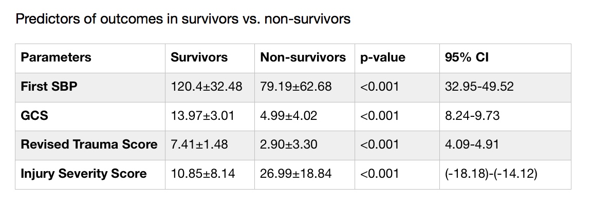
Results: There were 1,799 patients sustaining 1,900 brachial vessel injuries out of 1,466,887 patients registered in the NTDB from 2001 to 2005. Incidence was calculated to be 0.12%. Mean age at injury was 30 ± 14, mean RTS 7.23 ± 1.8, mean Glasgow Coma Scale 13.6 ± 3.5, mean ISS 11.5 ± 9.5. Mechanism of Injury: penetrating n=1,114 (61.9%), blunt n=699 (37.1%), and non-specific n=16 (1%). Mean initial SBP 119 ± 32.5; overall and adjusted survival rates: survivors 94.7%; non-survivors 5.3%. Patients underwent surgical repair n=671. Stratified as primary arteriorrhaphy 369 (55%), RSVG 165 (24.5%), resection of upper limb vessel with replacement 116 (17.3%), PTFE 21 (3.2%). Analysis revealed an increased ISS in the blunt injury group vs penetrating (10.65±8.82 vs 13.09±10.43; p<0.001). In survivors vs non-survivors, initial systolic blood pressure, glasgow coma scale, revised trauma score, and injury severity score were all statistically significant between the two groups (see table).

Conclusion: Brachial vessel injuries remain a low incidence injury as is shown in the NTDB. The injury severity score has a higher correlation with morbidity based on mechanism of injury. The overall cost of all brachial injuries was $22,954,998 in patient with extractable information (n=727).


103.10 Malnutrition in Vascular Surgery Patients: Looking Beyond Serologic Markers

J. Miranda1, H. Younes1, L. Le1, L. Probstfeld2, J. Braun1, N. Barshes1, P. Kougias1, H. S. Tran Cao1  1Baylor College Of Medicine,Surgery,Houston, TX, USA 2Michael E. DeBakey Veterans Affairs Medical Center,Nutrition,Houston, TX, USA

Introduction:   Malnutrition is a known risk factor for poor wound healing and surgical wound infections, especially important clinical outcomes for vascular surgery patients.  In 2012, leading dietetic organizations issued a consensus definition of clinical malnutrition based on 6 clinical parameters (AND/ASPEN criteria).  We sought to assess the incidence of malnutrition for vascular surgery patients using this definition compared to serologic markers commonly used as indicators.

Methods:   This is a retrospective cohort study of patients undergoing elective vascular surgical procedures at a single institution (2015-2017) who received malnutritional screening via a comprehensive nutritional program that included AND/ASPEN criteria.  These criteria include weight loss, decreased oral intake, muscle mass loss, loss of subcutaneous fat, fluid accumulation, and decreased hand grip strength.  Per AND/ASPEN guidelines, at least two deficits must be met to establish a diagnosis of malnutrition.  Correlation with serologic markers was measured.

Results:  Among 65 patients admitted for elective vascular procedures who received a comprehensive nutritional screening by a trained dietitian, 16 (24.6%) did not meet criteria for malnutrition, 21 (32.3%) were moderately malnourished, 11 (16.9%) were severely malnourished, and 17 (26.2%) were not fully assessed due to missing anthropometric data or key elements of the history and physical examination.  Although serum albumin was higher among non-malnourished patients than moderately and severely malnourished patients (3.12 ± 0.64 vs. 3.04 ± 0.51 vs. 2.71 ± 0.68, respectively, p=0.202), this difference failed to reach significance.  Moreover, 10/21 (47.6%) of moderately and 3/11 (27.3%) of severely malnourished patients had serum albumin > 3.0 g/dL, a cut-off often used to represent malnutrition.  Serum prealbumin was not routinely obtained, but was likewise non-discriminatory in detecting clinical malnutrition (e.g. exceeding 18 mg/dL in malnourished patients by AND/ASPEN criteria).  In turn, no surgical site infection was encountered in the clinically non-malnourished cohort, compared to 3 detected in clinically malnourished patients, including one without hypoalbuminemia.

Conclusion:  The prevalence of clinical malnutrition is significant among vascular surgery patients, and may not be fully appreciated with serologic markers alone.  As malnutrition may be a modifiable preoperative risk factor, efforts to comprehensively screen for this condition relying on a combination of clinical and serologic markers may be beneficial.
 

103.09 Examination of Race and Conduit Use for Lower Extremity Bypass in the SVS Vascular Quality Initiative

L. Stewart1, A. Beck1, E. Spangler1  1University Of Alabama at Birmingham,Department Of Surgery, Division Of Vascular Surgery And Endovascular Therapy,Birmingham, Alabama, USA

Introduction: Vein conduit for infrainguinal bypass (IB) has better patency than prosthetic conduits.  We explore if racial disparities exist in use of vein conduit for IB, and examine associations of patient factors and systems of care factors with racial disparities in conduit use. 

Methods: We analyzed a retrospective cohort of 23,959 IBs originating from the common femoral artery performed for occlusive disease with non-missing race/ethnicity in the SVS Vascular Quality Initiative (VQI) from 2003-17.  Demographics of patients receiving vein vs other conduit were compared by t test and X2 testing, while univariate and multivariate logistic regression analyses were performed to evaluate for predictors of vein conduit use, with the pseudo-R2 used as an assessment of the model. 

Results:Lower proportions were found in vein bypass patients of women, minorities, emergent cases, patients with ASA class>3, prior CABG, prior IB or ipsilateral IB, dialysis, or COPD.  A higher proportion of vein bypass patients had vein mapping, distal target below the knee, or diabetes. 

Unadjusted regression of vein use by race showed black patients were 88% as likely (p<.001), Hispanic patients 90% as likely (p=.08), and non-white/non-black/non-Hispanic patients 93% as likely (p=.37) to have a vein IB compared to white patients;  however race alone explains only 0.04% of variation in vein conduit use.  Adjusted models after backward stepwise regression demonstrated black patients were 76% as likely (p<.001), Hispanic patients 79% as likely (p=.003), and non-white/non-black/non-Hispanic patients 83% as likely (p=.09) to have a vein IB compared to white patients; however other factors had greater weight within the model.  Factors most correlating with vein use included vein mapping and more distal target.  Factors most strongly against vein use included higher age, ASA class 4, bedridden mobility status pre-op, any prior CABG, prior ipsilateral IB, or bypass performed before 2012; however the entire adjusted model still explained only 15% of variation in vein conduit use.  Due to the importance of vein mapping, we examined the racial breakdown of vein mapping by target level (Figure) and saw that while black patients were less likely to receive vein IB, they were vein mapped at similar or higher rates than other groups.

Conclusion: Racial disparities exist in conduit use for IB from the common femoral artery for occlusive disease, with blacks less likely to receive vein bypasses, however the contribution of race to conduit selection is small in adjusted and unadjusted models.  Overall, pre-operative variables captured in the VQI poorly predicted vein conduit use for IB.


103.08 Factors Contributing to Chronic Venous Disease and Venous Leg Ulcers

F. Alanazi1, A. Baothman1, W. Qu1, M. Osman1, K. Bauer1, J. Ortiz1, F. C. Brunicardi1, M. Nazzal1  1University Of Toledo Medical Center,Department Of Surgery,Toledo, OH, USA

Introduction:

Chronic venous disease (CVD) and venous leg ulcers (VLU) are common medical problems that cause loss of work hours and require lengthy medical treatment. In this study we analyzed the social determinants of health associated with factors that contribute to both CVD and VLU.

Methods:
National Inpatient Sample (NIS) database (2008-2014) of the Healthcare Cost and Utilization Project (HCUP) was used to select adult (≥18 years) CVD and VLU patients in the study. All diagnoses and procedures were identified with ICD-9-CM code. Statistical analyses were done with IBM SPSS statistical software ver. 24. Type I error level was set at 0.05.

Results:

Total number of patients with CVD and VLU in the study was 2,418,709 and 610,895, respectively. While the decrease in general hospital admission rate in the period from 2008 to 2014, the prevalence of CVD increased by 51.1% (from 0.87% to 1.32%, coeff=0.07, P<.001), and the prevalence of VLU increased by 57% (from 0.22% to 0.34%, coeff=0.02, P<.001). The prevalence of VLU in CVD patients increased by 3.9% in the same period (from 24.8% to 25.7%, coeff=0.135, p=.012).

Prevalence of CVD increased with age (from 0.3% in 18-24 years group to 1.7% in ≥65 years group, Cochran–Armitage test p<.001). Other risks for CVD included: male gender (1.37% vs 0.94%, P<.001 ), Caucasian (CA) vs. African American (AA) and Hispanics (HIS, 1.29% vs. 0.85% and 0.65%, both P<.001), patients in the 26th-50thpercentile (IT2) and 51st-75thpercentile of the income scale (IT3, 1.15 and 1.13%, respectively) compared to those in 0-25thpercentile (IT1, 1.08%) or76th-100thpercentile of the income scale (IT4, 1.11%)(all P<.001), and patients with BMI ≥ 40 compared to those with BMI<40 (5.5% vs. 0.90%,P<.001).

The prevalence of VLU in CVD patients was highest in patients of age 45-64 years (28.5%), gradually decreased to 25.8% (age 25-44 years), 23.8% (age ≥65 years), and 19.2% (age 18-24years, all P<.01). Other risk factors for VLU were: male gender (28.1% vs 22.7%, P<.001), AA and HIS compared to CA (36.2% and 27.4% vs. 23.7%, both P<.001), IT1 compared to IT2 -IT4 (27.6% vs. 24.8%, 24.6%, and 23.6%, all P<.001); patients with BMI ≥ 40 compared to those with BMI<40 (26.9% vs. 24.9%,P<.001).

Conclusion:

This paper reflects demographic and social status variations in patients with CVD and VLU. The prevalence of both CVD and VLU increased over the years of the study. Prevalence of CVD is higher in elders, in CA compared to HIS and AA, in patients in intermediate household incomes compared to higher and lower household income groups, and morbidly obese patients. The prevalence of VLU is higher in the middle age group, in men, in AA and HIS, in low income patients and morbidly obese patients. Analysis of the NIS data indicates an increasing burden of chronic venous disease and venous leg ulcers, particularly among populations at risk for poor social determinants of health.

103.07 Chronic Renal Failure is Not a Contraindication for Femoral Endarterectomy in Claudicants

L. A. Huntress1, J. Kalenik2, V. Dombrovskiy1, S. G. Huang1, R. Shafritz1, S. Rahimi1  1Rutgers RWJMS,Division Of Vascular Surgery,New Brunswick, NJ, USA 2University of Georgia,Athens, GA, USA

Introduction:  Femoral endarterectomy has proven to be a durable vascular reconstructive procedure for patients with critical limb ischemia (CLI). Our objective was to evaluate its applicability in patients with chronic renal failure that have severe claudication.

Methods:  Patients 18 years or older with severe claudication who underwent femoral endarterectomy were selected from the 2012-2015 National Inpatient Sample using the appropriate ICD-9 and ICD-10 diagnosis and procedure codes. Postoperative outcomes in those with and without renal failure were compared using the Chi square test, multivariable logistic regression analysis, and Wilcoxon rank sum test. Patients with acute renal failure were excluded from the analysis

Results: Among the 30,805 patients in the study population, 2,705 (8.8%) had chronic renal failure. The likelihood of this comorbid disease was greater in older patients (70 years or older) compared to younger counterparts (OR [odds ratio]=1.74; 95%CI [confidence interval] 1.61-1.88), greater in females as compared to males (OR=1.22; 95%CI 1.12-1.33), and greater in Blacks as compared to Whites (OR=1.96; 95%CI 1.73-2.22). In the multivariable logistic regression analysis with control for age, gender, race, and comorbidities, chronic renal failure did not affect the rates of cardiac and respiratory complications, postoperative stroke, sepsis, or embolism/thrombosis of lower extremity arteries. No patient with chronic renal failure who underwent femoral endarterectomy required a major amputation as a complication of the procedure. However, patients with renal failure were more likely to develop bleeding (OR=1.35; 95%CI 1.20-1.51), or require blood transfusions (OR=1.89; 95%CI 1.69-2.13). Multivariable analysis showed equivalence in all cause hospital mortality between two groups. However, patients with chronic renal failure had longer hospital length of stay (median= 3 days, IQR [interquartile range] 2-4 days vs median= 2 days, IQR 1-3 days; P<0.0001) and greater total hospital cost (median= $14,235; IQR $9,621-20.878 vs median= $12,810; IQR $8,824-18,953; P<0.0001) compared to patients with normal renal function.

Conclusion: Femoral endarterectomy is a safe procedure in severe claudicants with chronic renal failure, but is associated with greater hospital resource utilization. Because of greater hospital LOS and increased bleeding complications, femoral endarterectomy in patients with chronic renal failure and claudication should be offered with caution.