02.14 Xanthohumol Increases DR5 Expression and Enhances Growth Suppression with TRAIL in Neuroblastoma

S. Engelsgjerd1, S. Kunnimalaiyaan1, T. Gamblin1, M. Kunnimalaiyaan1  1Medical College Of Wisconsin,Surgical Oncology/Surgery,Milwaukee, WI, USA

Introduction:  High-risk neuroblastoma (NB) is a lethal childhood cancer. Published data have reported the anti-proliferative effect of Xanthohumol (XN), a prenylated chalcone, in various cancer types suggesting that XN could be a useful small molecule compound against cancer.  The effect and mechanism of XN on NB cell proliferation is unknown. This study hypothesizes that XN will inhibit NB growth, and the effects of XN on cellular proliferation as well as the mechanism of action in NB cell lines were examined. The TNF-Related Apoptosis Inducing Ligand (TRAIL) is an endogenous ligand that is expressed in monocytes, macrophages, dendritic cells, natural killer (NK) cells, and activated T cells. TRAIL mediates apoptosis through binding of transmembrane receptors, death receptor 4 (DR4) and/or death receptor 5 (DR5). Cancer cells are frequently resistant to TRAIL-mediated apoptosis, and the cause of this may be decreased expression of death receptors. This study hypothesizes that XN increases DR5 expression in NB cells thus sensitizing them to TRAIL.

Methods:  The effect of XN in human NB cell lines NGP, SH-SY-5Y, and SK-N-AS was determined via MTT assay. Cell confluency assay by cell live imaging was also carried out for SK-N-AS and NGP cell lines after XN treatment. Cell lysates were analyzed through Western blotting for pro- and anti-apoptotic markers as well as death receptor (DR5). Synergistic analysis of XN and TRAIL in SK-N-AS cells was performed via MTT assay.

Results: XN treatment causes a statistically significant decrease in the viability of NB cells with IC50 values of approximately 12 µM for all three cell lines. Inhibition of cell proliferation via apoptosis was evidenced by an increase in pro-apoptotic markers, including cleaved PARP, cleaved caspase-3, and Bax, and a reduction in anti-apoptotic markers BcL-2 and survivin. Importantly, XN treatment increased expression of DR5. Furthermore, statistically significant synergistic reduction was observed following pretreatment with XN (41%) compared to either TRAIL or XN alone (10%) in SK-N-AS cells.

Conclusion: XN treatment reduces NB cell growth via apoptosis in a dose-dependent manner. Treatment is associated with an increase in DR5 expression. Most importantly, enhanced growth reduction was observed in combination with TRAIL. This is the first study to demonstrate that XN alters the expression of DR5 as well as the synergistic effect of XN on TRAIL in NB. This study provides a strong rationale for further preclinical in vitro and in vivo analysis of XN in combination with TRAIL. 


02.16 Tumor Derived Microparticles and Exosomes Induce Invasion in Pancreatic Cancer

I. A. Naqvi1, R. Gunaratne1, J. Yeh4, D. Pisetsky5, B. Sullenger2, R. White3  1Duke University Medical Center,School Of Medicine,Durham, NC, USA 2Duke University Medical Center,Department Of Surgery,Durham, NC, USA 3University Of California – San Diego,Department Of Surgery,San Diego, CA, USA 4University Of North Carolina At Chapel Hill,School Of Medicine,Chapel Hill, NC, USA 5Duke University Medical Center,Department Of Medicine,Durham, NC, USA

Introduction:  Pancreatic cancer (PC) has the worst prognosis of the major cancers. Metastatic progression is one of the main reasons PC is so deadly. Microparticles (MPs) and exosomes (EXOs) are cell-derived lipid particles derived from the plasma membrane and endoplasmic reticulum, respectively. Recent work has shown that intercellular communication via MPs and EXOs may play a role in tumor progression. We investigated the functional effects of tumor-derived MPs and EXOs on a pancreatic cancer cell line derived from a genetically engineered mouse model (KPC).

Methods:  KPC cells were cultured in EXO-free media at 100% confluence for 72 hours after which cell supernatant was collected and centrifuged at 20,000g to isolate MPs and further at 120,000g to isolate EXO. Effects of MPs and EXO on cellular invasion were quantified by Transwell Matrigel-Invasion assay. Briefly, cells were plated in the upper chamber of a Transwell chamber with either MPs, EXO, or vehicle control and allowed to invade for 24 hours. The Transwell membrane was then fixed and stained with crystal violet. Invaded cells were quantified using Image-J software. The effects of MPs and EXO on intracellular signaling were investigated via western blot.

Results: We observed that treating KPC cells with tumor cell-derived MPs and EXOs significantly increased their invasiveness in the Transwell Matrigel-Invasion Assay Figure 1A and 1B). We also observed that treating with MPs and EXOs caused increased phosphorylation of both p65 NFkB and p-38 MAPK, both known to be important mediators of tumor progression. 

Conclusion: Our results are consistent with recent work showing that MPs and EXOs may be playing a critical role in cancer progression and metastasis. Ongoing work is focused on inhibiting the effects of MPs and EXOs on cancer cells and understanding the molecular underpinnings of the effects induced by MP and EXO treatment. 


02.17 Examining the Role of Telomere Length and Inflammation in Glioma Oncogenesis with an Agent-Based Model

D. Kokosi2, G. An1  1University Of Chicago,Surgery,Chicago, IL, USA 2National And Kapodistrian University Of Athens,School Of Medicine,Athens, ATHENS, Greece

Introduction:  Gliomagenesis is a phenomenon of complex molecular etiology, with recent genomic screening supporting the importance of telomere biology. There is a suggestion of a the correlation between long telomere length predisposition and increased risk of glioma. The hypothesis that longer telomere length enhances the proliferative capacity of each cell and therefore increases the possibility of malignant transformation is backed by various studies related to different types of carcinogenesis; however, a systemic approach to understanding the dynamics of this correlation is difficult to accomplish in vivo due to the complexity of the constituting factors. Agent-based modeling, a computational modeling method, can be applied to examine hypotheses related to specific elements of gliomagenesis in a dynamic and systemic fashion, and evaluate macroscopic evidence through cell-level systems components and functions. Consequently, we have developed an abstract agent-based model (ABM) that encompasses the fundamental aspects of neural tissue function in healthy and injured states, and incorporated a possible role of telomere length and inflammation in glioma oncogenesis. 

Methods:  The ABM that was developed includes agents representing neurons, astrocytes, microglia and stem cells. The inter-agent interaction via signaling molecules in both healthy and injured states is based on published mechanisms. The model was calibrated to simulate the generation and accumulation of mutations in astrocytes in relation to their degree of activation during injury response. Important parameters that were incorporated and examined during experimental runs include: activation of glial cells, astrocyte proliferation, lifespan of cells, telomere length and mutation levels of astrocytes, neuron damage and degeneration, inflammation and chemotaxis. Stem cell differentiation was triggered via a counter or due to local injury. 

Results: The ABM identified that the ability of astrocytes to acquire, accumulate, and pass on mutations was enhanced during an activated state due to elevated levels of stress and proliferation caused by inflammation-induced injury. The proliferative capacity of astrocytes was increased by longer telomere length as a larger number of cell divisions were permitted before cell senescence. Simulations of 80-year trials suggest that this mechanism is plausible in explaining the role of longer telomere length in gliomagenesis.

Conclusion: Gliomagenesis is comprised of an intricate network of pathways that are the subject of ongoing research. The ABM effectively represents fundamental responses of neural tissue to inflammation-induced injury and indicates a plausible explanation for the role of longer telomere length in glioma oncogenesis. ABMs provide a means of defining a basic biological framework into which detailed elements can be dynamically incorporated in order to visualize, evaluate, and reshape hypotheses.


02.18 HDAC Inhibitors Modulate Notch3 Expression though a Unique Transcriptional Motif in NE Cancer Cells

S. Jang1, H. Jin1, R.Jaskula-Sztul1, H. Chen1  1University Of Alabama At Birmingham,Surgery,Birmingham,, AL, USA

Introduction: It is known that Notch signaling is minimally active in neuroendocrine (NE) cancer cells and the induction of Notch isoforms alter the malignant neuroendocrine phenotype. The induction of Notch3 by Histone Deacetylase (HDAC) Inhibitors appears to be the result of increased mRNA expression at the transcriptional level, which leads to suppression of cancer cell proliferation and to apoptosis. The aim of our study is to investigate the molecular mechanism of HDAC inhibitor activation on the Notch3 pathway.

Methods: We functionally characterized the Notch3 promoter using deletion mapping. The mapping started with the truncated genomic DNA fragment fused with a luciferase reporter in plasmid vector, transfected into BON cell, a carcinoid cell line, screened for luciferase activity. Protein-DNA binding was then performed by electrophoretic mobility shift assay (EMSA).

Results: One HDAC inhibotor, Thailandepsin-A, was shown to induce luciferase activity controlled by a small distal region of Notch3 promoter, from -120 to +1 of the start codon ATG.  Further, we identified a functional DNA motif that is responsible for HDAC induction located at -120 to -100 of the Notch3 promoter region. Thus, an in vitro assay, EMSA revealed the transcription factor-DNA complex formed in the flanking sequence.

Conclusion: We have identified the DNA motif located in the Notch3 promoter region that is responsive to HDAC inhibitor. This understanding of how HDAC inhibitor act on the Notch3 promoter may lead to the development of novel therapies for neuroendocrine cancers.


02.19 Sphingosine-1-phosphate in the Lymphatic Fluid Determined by Novel Methods

M. Nagahashi1, A. Yamada2, T. Aoyagi2, J. Allegood3, T. Wakai1, S. Spiegel3, K. Takabe4  1Niigata University Graduate School Of Medical And Dental Sciences,Division Of Digestive And General Surgery,Niigata, NIIGATA, Japan 2Virginia Commonwealth University School Of Medicine,Division Of Surgical Oncology And The Massey Cancer Center,Richmond, VA, USA 3Virginia Commonwealth University School Of Medicine,Department Of Biochemistry And Molecular Biology And The Massey Cancer Center,Richmond, VA, USA 4Roswell Park Cancer Institute,Breast Surgery, Department Of Surgical Oncology,Buffalo, NY, USA

Introduction:

Sphingosine-1-phosphate (S1P) is a pleiotropic bioactive lipid mediator that regulates many physiological and pathological processes. S1P produced by sphingosine kinases (SphK1 and SphK2) is secreted from cells and signals in autocrine and/or paracrine manners by binding to its specific cell surface receptors. We have recently reported that S1P is strongly associated with lymphatic network development (FASEB J 2013) and lymphatic metastasis in cancer patients (Cancer Research 2012, JSR 2016). Further, we have shown that S1P links inflammation and cancer in colitis-associated colon cancer (Cancer Cell 2013). It has been suggested that S1P gradient with high concentrations in the blood and lymphatic fluid and low concentrations in the peripheral tissue plays important roles in immune cell trafficking and potentially cancer progression. Although S1P levels in blood have been published to be associated with lymphatic metastasis by our group and others, only a few reports have assessed its levels in lymphatic fluid due to lack of established method in experimental setting. Here, we report simple technique for collection of lymphatic fluid to measure sphingolipids in murine models.

Methods:

The lymphatic fluid was collected directly with a catheter needle (classical method) or was absorbed onto filter paper after incision of cisterna chyli (new method). Whole blood, serum, lymphatic fluid and mesenteric lymph nodes were corrected from WT and SphK2 knockout mice to determine levels of sphingolipids including S1P by mass spectrometry.

Results:

S1P levels were measured in the lymphatic fluid collected either by classical and new methods. The volume of the lymphatic fluid collected by the new method was at least three times greater than those collected by the old one. S1P levels in lymphatic fluid corrected by both classical and new methods showed consistent results with minimal variation. In 8 weeks old mice, S1P concentrations in lymphatic fluid were in the range of 100 to 400 nM, compared to more than 1500 nM in whole blood and more than 600 nM in serum, and less than 50 nM in the mesenteric lymph node tissue. SphK2 knockout mice showed higher levels of S1P in whole blood and serum than WT mice, partially due to overexpression of SphK1 in the blood endothelial cells. Interestingly, S1P levels in lymphatic fluid from SphK2 knockout mice were also significantly higher than those in the WT mice, suggesting an important role of SphK2 and/or SphK1 in the lymphatic endothelial cells to provide S1P into the lymphatic fluid.

Conclusion:

We determined the levels of S1P in lymphatic fluid, which is lower than blood and higher than lymph nodes. In agreement with the previous theory, our results confirm the “S1P gradient” among blood, lymphatic fluid and the peripheral lymphatic tissues. Convenient methods for collection and measurement of sphingolipids in lymphatic fluid are expected to provide new insights on functions of bioactive sphingolipids.


02.20 A Pre-Metastatic Niche in the Omentum of Human Pancreatic Cancer Patients

J. C. King1, X. Jung1, M. Xu1, A. Schmidt1, C. Chou1, O. J. Hines1, G. Eibl1  1University Of California – Los Angeles,Surgery,Los Angeles, CA, USA

Introduction: Pancreatic cancer (PC) is almost universally metastatic to the liver and / or peritoneum and current methods for detection and prognostication like serum tumor markers or peritoneal cytology are poor predictors of outcome following curative resection. Recent animal research demonstrates extracellular matrix protein deposition and cellular changes such as stellate cell activation in organs at risk for metastatic spread may predict metastasis-termed the pre-metastatic niche. We sought to identify features of the pre-metastatic niche in human PC patients.

Methods: Histologically verified non-tumor tissue samples from 9 human PC patients were stratified by the distribution of metastases: no metastases (n=3), liver only metastases (n=3), liver and peritoneal metastases (n=3). We identified pre-metastatic niche changes by staining uninvolved omentum and liver by immunofluorescence for fibronectin (FN) and smooth muscle actin (SMA). Samples were scored by fluorescence signal intensity in three independent high power fields and normalized to total cell count by DAPI staining.

Results:FN expression was increased in the uninvolved omentum of patients with metastatic disease (33.2±16.7 vs 71.7±23.6; p=0.04). Omentum SMA expression was similar between local only and metastatic patients (56.8±27.1 vs 60.4±36.0; p=0.81). There was a trend toward increased expression of FN (11.3±4.1 vs 67.6±46.3; p=0.10) and SMA (20.0±20.4 vs 50.9±43.4; p=0.10) in uninvolved liver of patients with metastatic disease compared to those without metastases.

Conclusion:We found the presence of metastatic disease was associated with increased FN expression in uninvolved tissues of the omentum. There appeared to be a trend towards increased FN and SMA expression in the livers of patients with metastatic disease with no association between metastatic disease and SMA expression in the omentum. These findings typify the pre-metastatic niche found in animal models. FN expression in the omentum may be an early indicator metastagenesis in patients with pancreatic cancer.


02.04 Roles of Sphingosine-1-Phosphate Produced by Sphingosine Kinases in Pancreatic Cancer Progression

M. Nakajima1, Y. Miura1, T. Ando1, K. Yuza1, J. Tsuchida1, Y. Tajima1, M. Abe2, K. Sakimura2, T. Wakai1, M. Nagahashi1  1Niigata University Graduate School Of Medical And Dental Sciences,Division Of Digestive And General Surgery,Niigata City, , Japan 2Brain Research Institute, Niigata University,Department Of Cellular Neurobiology,Niigata City, , Japan

Introduction: Pancreatic cancer is one of the most lethal diseases known, and it is important to develop new therapeutic agents. Sphingosine-1-phosphate (S1P) is a pleiotropic lipid mediator that regulates cell survival, migration, immmune cell recruitment, angiogenesis and lymphangiogenesis, which are all factors involved in cancer progression. S1P, which functions intra- and extracellularly, is generated inside the cell by two sphingosine kinases (SphK1 and SphK2). We have reported that SphK1 plays an important role in S1P secretion (J Biol Chem 2010) and cancer progression (Cancer Res 2012, J Surg Res 2016), and that SphK2 has a unique role in regulating cellular functions in the liver (Hepatology 2015). Little is known, however, about the role of SphK1 and SphK2 in pancreatic cancer progression. The aim of this study is to investigate the role of SphK1 and SphK2 in pancreatic cancer progression using SphK-knockout (KO) cells generated by CRISPR/Cas9 technology.

Methods: We generated Pan02 murine pancreatic cancer cell lines with a CRISPR/Cas9 mediated targeted deletion of the SphK1 or SphK2 gene. Western blotting and RT-qPCR determined the expression levels of SphKs in these cell lines. To investigate the role of SphK1 or SphK2 in cellular proliferation, we assessed cell growth by a spectrophotometric technique using the water-soluble tetrazolium salt, WST-8. Cell migration was measured by an in vitro scratch assay.

Results: We confirmed the knockout of SphK1 or SphK2 in Pan02 pancreatic cancer cells by western blotting and RT-qPCR. SphK2 KO cells were significantly less proliferative than wild type (WT) cells. Unexpectedly, SphK1 KO cells were significantly more proliferative than WT cells. The in vitro scratch assay indicated that SphK2 KO cells were less migratory than WT cells, and that SphK1 KO cells had greater migratory ability than WT cells. These results indicate that S1P produced by SphK2, rather than by SphK1, may have important roles in proliferation and migratory behavior in pancreatic cancer cell lines.

Conclusion: Our findings indicate that S1P produced by SphK2, rather than by SphK1, promotes pancreatic cancer cell proliferation and migration. Targeting SphK2 may be a potential strategy for pancreatic cancer treatment. Further studies that include in vivo models are needed to explore this therapeutic possibility.
 

02.05 COMP Gene Is Over-expressed in Early-Onset Colon Cancer and Associated with Poor Survival.

V. N. Nfonsam1, J. Koblinski1, J. Jandova1  1University Of Arizona,Surgery,Tucson, AZ, USA

Introduction: Although the overall incidence of colon cancer (CC) has steadily declined over the last three decades, the incidence has increased in patients younger than 50. The etiology of early-onset (EO) CC is not fully understood. COMP gene has been shown to confer tumor aggressiveness in pancreatic cancer. The aim of this study was to elucidate gene expression patterns in EOCC and show its uniqueness compared to late-onset (LO) disease.

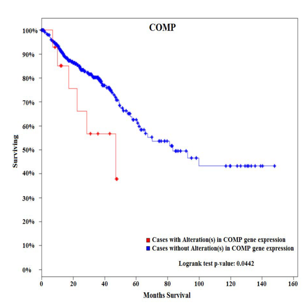
Methods: Two cohorts of patients with sporadic CC were identified. Tumors and matching noninvolved tissues from six EOCC patients (<50) and six late-onset colon cancers (LOCC) patients (>65) were obtained from pathology archives. De-paraffinized tissues were macro-dissected from FFPE sections, RNA isolated, and used for expression profiling of 770 cancer-related genes representing 13 canonical pathways. Survival analysis was performed using the cBioPortal for cancer genomics using 382 CRC patients from the TCGA Provisional database.

Results:Among 770 genes assayed, changes in expression levels of 93 genes were statistically significant between EOCC and matching noninvolved tissues. There were also significant differences in expression levels of 118 genes between LOCC and matching noninvolved tissues. Detailed comparative gene expression analysis between EOCC and LOCC normalized to their matching noninvolved tissues revealed that changes in expression of 88 genes were unique to EOCC using the cutoff criteria of expression levels difference >2 fold and P value <0.01. From these differentially expressed genes specific to EOCC, 28 genes were upregulated and 60 genes downregulated. At the pathway level, PI3K-AKT signaling was the most deregulated pathway in EOCC and cell cycle in LOCC. COMP glycoprotein was one of the genes that were uniquely over-expressed in EOCC. Survival analysis of 382 patients with CRC tumors using the cBioPortal for cancer genomics showed that CRC patients with alterations (all of them with up-regulated COMP) in COMP expression present with poorer overall survival compared to patients without alterations in COMP expression.

Conclusion:
These results suggest that sporadic EOCC is characterized by distinct molecular events compared to LOCC. In addition, COMP glycoprotein is associated with poor overall survival. COMP gene can potentially serve as a novel biomarker associated with EOCC as COMP glycoproteins could be detected in serum and urine.  
 

02.06 Obesity-induced Sphingosine-1-phosphate in Breast Cancer Interstitial Fluid Promotes Angiogenesis

J. Tsuchida1, M. Nakajima1, K. Moro1, A. Ohtani1, M. Endo1, T. Niwano1, K. Tatsuda1, C. Toshikawa1, M. Hasegawa1, M. Ikarashi1, Y. Koyama1, J. Sakata1, T. Kobayashi1, H. Kameyama1, T. Wakai1, K. Takabe2, M. Nagahashi1  1Niigata University Graduate School Of Medical And Dental Sciences,Division Of Digestive And General Surgery,Niigata, NIIGATA, Japan 2Roswell Park Cancer Institute,Breast Surgery, Department Of Surgical Oncology,Baffalo, NY, USA

Introduction: It is well established that obesity evokes chronic inflammation, which aggravates cancer progression by facilitating interactions between cancer cells and stromal cells including blood endothelial cells. Sphingosine-1-phosphate (S1P) is a bioactive lipid mediator produced by sphingosine kinases (SphKs) that plays critical roles in inflammation and cancer progression. We have recently discovered that the SphK1/S1P/S1PR1 axis play critical roles in inflammation-associated cancer progression, and that the S1PR1 functional antagonist, FTY720, suppresses cancer progression by inhibition of the S1P axis during chronic inflammation. We have recently developed a method to measure S1P levels in tumor interstitial fluid (IF), which is a component of the tumor microenvironment that bathes cancer cells in the tumor. Our findings suggested the possibility that S1P secreted from tumor cells to IF may be important for metastasis. We hypothesized that obesity and its related inflammation up-regulate SphK1, which produces more S1P, and the elevated levels of S1P in both tumor and tumor IF stimulate angiogenesis and progression of breast cancer. In this study, we test this hypothesis utilizing animal models with obesity.

Methods: Orthotopically-implanted E0771 syngeneic breast cancer mouse models were used. Mice were fed with normal or high-fat diet (HFD). FTY720 was administered orally (1 mg/kg/day). Angiogenesis in the primary tumor was determined by measurement of microvessel density. S1P levels in tumor and tumor IF were measured by mass spectrometry.

Results: HFD induced obesity significantly worsened the cancer progression. Obesity elevated inflammatory cytokines such as IL-6 and TNF-α in both circulation and in the tumor. Mass spectrometry analysis revealed that while S1P levels in the normal breast mammary fat pad were increased with HFD feeding, S1P levels were even higher in breast tumors. Consistent with increased SphK1 and S1P in tumors, S1P was also significantly increased in the tumor IF. Further, microvessel density analysis revealed that significantly more angiogenesis in peri-tumor area in mice fed with HFD than in mice fed with normal diet. Importantly, FTY720 dramatically reduced the levels of cytokines as well as SphK1, S1PR1 and S1P in primary tumor. Further, FTY720 suppressed not only primary tumor growth, but also angiogenesis in the primary tumor with suppression of aggregation of tumor-associated macrophages.

Conclusion: Our results indicate an important role of S1P in tumor microenvironment, which is affected by obesity, for obesity-related angiogenesis and breast cancer progression. S1P will be one of the promising targets for breast cancer patients with obesity.


02.07 Peri-tumor Sphingosine-1-phosphate Levels Are Affected by the Tumor

J. Tsuchida1, K. Moro1, A. Ohtani1, M. Endo1, T. Niwano1, K. Tatsuda1, C. Toshikawa1, M. Hasegawa1, M. Ikarashi1, M. Nakajima1, Y. Koyama1, J. Sakata1, T. Kobayashi1, H. Kameyama1, K. Takabe2, T. Wakai1, M. Nagahashi1  1Niigata University Graduate School Of Medical And Dental Sciences,Division Of Digestive And General Surgery,Niigata, NIIGATA, Japan 2Roswell Park Cancer Institute,Breast Surgery, Department Of Surgical Oncology,Buffalo, NEW YORK, USA

Introduction: Sphingosine-1-phosphate (S1P), a pleiotropic bioactive lipid mediator, has been implicated as a key regulatory molecule in cancer through its ability to promote cell proliferation, migration, angiogenesis and lymphangiogenesis. Although many researchers including us have reported important roles of S1P in breast cancer progression utilizing in vitro and in vivo models, there have been very few data on human patients due to the difficulty in accurate measurements of S1P levels in the clinical samples. Recently, we have reported that high S1P levels in the tumor determined by mass spectrometry are associated with lymph node metastasis in breast cancer patients. In this study, to reveal further roles of sphingolipids in the patients, we measured the levels of sphingolipids including S1P in plasma and breast tissue including peri-tumor tissue.

Methods: Forty-nine serum prior to operation and 20 tumors from invasive cancer larger than 1.5cm were collected from breast cancer patients who underwent operation from November 2015 to March 2016 in Niigata University Medical and Dental Hospital. The levels of sphingolipids were determined by mass spectrometry.

Results: The levels of sphingolipids including sphingosine, dihydro-sphingosine, S1P, dihydro-S1P were detected successfully in the plasma from 49 breast cancer patients and in the tissue samples from 20 patients. Despite our expectation, no significant differences in plasma S1P levels were identified by hormone receptors (ER, PgR) and HER2 status, Ki-67 index, nuclear grade, lymphatic and vascular invasion of the tumor, pT, pN, and pStage. The levels of sphingosine in patients with lymph node metastasis (pN1,2) were significantly higher than those in patients without metastasis (pN0) (P = 0.023). Interestingly, dihydro-S1P levels in patients with high nuclear grade (NG2,3) were significantly lower than those in patients with low nuclear grade (NG1) (P = 0.025). Moreover, there were significant differences among S1P levels in tumor, peri-tumor, and normal breast tissue (P = 0.0004). Significant differences were also seen among sphingosine (P = 0.0007) and dihydro-sphingosine levels (P = 0.0236) in the three different tissue types.

Conclusion: This is the first report to measure the S1P levels in peri-tumor tissue compared to the tumor and normal breast tissue. This is in agreement with our notion that S1P that is generated in cancer is secreted out to peri-tumoral microenvironment where it aggravates cancer progression, such as lymphangiogenesis. Further study will be needed to reveal the role of S1P in breast cancer patients.

02.08 Paradoxical Association of Postoperative Sphingosine-1 Phosphate and Breast Cancer Aggressiveness

R. Ramanathan1, A. Raza1, J. Young2, J. Sturgill1, D. Lyon1, K. Takabe2  1Virginia Commonwealth University,Richmond, VA, USA 2Roswell Park Cancer Institute,Breast Surgery,Buffalo, NY, USA

Introduction: Sphingosine-1 phosphate (S1P) is bioactive lipid mediator that has been shown to serve an important regulatory function in breast cancer progression. The dynamics of the circulating S1P levels during the postoperative period, however, have yet to be delineated. In this study, we analyze plasma S1P levels in breast cancer patients undergoing adjuvant therapy as compared to control healthy volunteers.

Methods: With institutional review board approval, plasma S1P was measured in 20 healthy women without breast cancer (control) as well as 158 patients with breast cancer who underwent adjuvant chemotherapy with or without irradiation after surgical resection. Among patients with breast cancer, postoperative plasma S1P was measured two weeks prior to adjuvant therapy (baseline), prior to the 4th cycle of chemotherapy (midpoint) and two weeks after completion of adjuvant therapy (completion). Correlations with demographics, treatments and inflammatory markers were analyzed with the use of chi-square tests, paired and non-paired t-tests and ANOVA analyses.

Results: 452 plasma S1P samples among 158 breast cancer patients were analyzed, along with 20 control healthy volunteers. Mean S1P levels did not differ between cancer patients and controls (1221.7 vs. 1139 pmol/mL, p=NS). S1P levels similarly did not associate with age or race in our cohort. Smoking was associated with higher S1P levels (1445.4 vs. 1163.3 pmol/mL, p<0.05). Midpoint S1P levels during adjuvant therapy were lower than baseline (1088.8 vs. 1221.8, p<0.05), with near return to baseline after completion (1121.5 vs. 1221.8, p=0.06), indicating a relationship between chemotherapy and circulating S1P. Furthermore, while stage of disease did not correlate with plasma S1P levels, they were lower among patients with Her2 enriched and triple negative breast cancer as compared to luminal type cancer (1119.2 and 1167.1 vs. 1280.8, p<0.05). Additionally, patients with intermediate and high grade tumors similarly had lower S1P than those with low grade tumors (1230.0 and 1176.5 vs. 1570.8 pmol/mL, p<0.05), further suggesting an inverse relationship between plasma S1P levels and tumor aggressiveness. There were no significant differences based on chemotherapy regimen and radiation.

Conclusions: We found that plasma S1P levels are paradoxically suppressed in aggressive breast cancer and during adjuvant chemotherapy, which raises the possibility that plasma S1P levels do not reflect S1P secretion from cancer cells.

02.09 cFLIPS is a Critical Regulator of EMT-Associated Resistance to Apoptosis

C. Padmanabhan1, E. J. Rellinger1, H. An1, A. G. Waterson2, C. W. Lindsley2, A. Means1, R. D. Beauchamp1  1Vanderbilt University Medical Center,Surgery,Nashville, TN, USA 2Vanderbilt University Medical Center,Chemistry,Nashville, TN, USA

Introduction:  

Epithelial cancers comprise the top four causes of cancer related deaths in the United States. Epithelial-to-mesenchymal transition (EMT) is a major cellular reprogramming of epithelial cancers associated with loss of E-cadherin expression and resistance to apoptosis. The mechanism that drives this phenotype, however, is unclear and this knowledge gap has hindered the development of apoptosis inducing therapeutics. It has previously been reported that E-cadherin expression is necessary for apoptosis induction by the tumor necrosis factor related apoptosis inducing ligand (TRAIL). Using a small molecule, ML327, that we previously showed to partially reverse EMT, we have identified the short variant of the cellular FLICE-like inhibitory protein (cFLIP) as a critical regulator of EMT-associated resistance to TRAIL that is independent of E-cadherin re-expression. 

Methods:

Trail Sensitization: Solid tumor cancer cell lines were treated with either ML327 (10 μ M) or vehicle control for 24 hours. TRAIL was added at 24 hours (50 – 500 ng/mL) for 4 hours. Cells were lysed and analyzed via western blot for cFLIP and markers of apoptosis. Cells were also fixed and stained with propidium iodide (PI) and analyzed via FACS cell cycle analysis.

cFLIP Knockdown: si-RNA knockdown of cFLIP was performed and cells were subsequently treated with TRAIL as above. Cells were lysed and analyzed via western blot for cFLIP and markers of apoptosis.

cFLIP Overexpression: cFLIPS was exogenously overexpressed using a CMV promoter based plasmid. Transfected cells were treated with ML327 and TRAIL as above. Cells were lysed and analyzed via western blot for cFLIP and markers of apoptosis.

E-Cadherin Independence: si-RNA knockdown of E-cadherin was performed in cancer cells and cells were subsequently treated with ML327 or vehicle and TRAIL as above. Cells were lysed and analyzed via western blot for e-cadherin, cFLIP, and markers of apoptosis. 

Results:

ML327 treatment caused a significant reduction of cFLIPS protein at 24 hours. This reduction in cFLIPS was associated with TRAIL sensitization as measured by cleaved PARP and sub G0 population on cell cycle analysis. si-RNA knockdown of cFLIP mimicked ML327 treatment and sensitized cancer cells to TRAIL-induced apoptosis. cFLIPS overexpression blunted apoptosis induction by ML327 and TRAIL. E-cadherin re-expression was not required for ML327-induced TRAIL sensitization.

Conclusion:

EMT-associated resistance to apoptosis in epithelial cancers is well described but the mechanism that drives this phenotype is unclear. Here, we demonstrate that cFLIPS is a critical regulator of this phenotype and that E-cadherin expression may not be necessary for apoptosis induction. Further study is required to determine the exact mechanism of cFLIP loss with EMT reversal and whether this finding can be exploited for therapeutic development.

02.11 Inhibitor of NF-κB Enhances the Antitumor Effect of Radiation Therapy in Colorectal Cancer.

H. Sugano1,2, Y. Shirai1, N. Saito1,2, T. Horiuchi1,2, H. Shiba1, K. Eto1, T. Uwagawa3, T. Ohashi2, K. Yanaga1  1The Jikei University School Of Medicine,Department Of Surgery,Minato-ku, TOKYO, Japan 2The Jikei University School Of Medicine,Department Of Gene Therapy, Research Center For Medical Science,Minato-ku, TOKYO, Japan 3The Jikei University School Of Medicine,Department Of Hematology And Medical Oncology,Minato-ku, TOKYO, Japan

Introduction:

Colorectal cancer is the third most commonly diagnosed malignancy and the fourth leading cause of cancer death in the world. Most patients with CRC are diagnosed at advanced stages, and the prognosis of such patients remains very poor. Neoadjuvant chemoradiothrapy (CRT) followed by radical surgery is currently the standard of care for patients with locally advanced low rectal cancer. CRT has the potential to downsize tumors before surgery and to decrease locoregional recurrence. However, previous findings indicate that irradiation promotes tumor migration, distant metastasis, and the invasive potential of cancer cells. This radiation-induced invasiveness is associated with an increased expression of matrix metalloproteinase (MMP) through nuclear factor kappa B (NF-κB) pathways. Irradiation also activates NF-κB that plays an important role in the regulation of cell apoptosis, inflammation, and oncogenesis including invasion and angiogenesis. We previously reported that nafamostat mesilate, a synthetic serine protease inhibitor, inhibited NF-κB activation and induced antitumor effects for pancreatic cancer. We hypothesized that nafamostat mesilate may inhibit radiation-induced NF-κB activation and tumor invasiveness, and improve therapeutic outcome of colorectal cancer. 

Methods:

We assessed NF-κB activity, cell viability, induction of caspase cascade,  quantification of apoptosis, and cell invasiveness of human colorectal cancer cell line (SW620) in the following four groups: 1) radiation alone, 2) nafamostat mesilate alone, 3) combination (radiation and nafamostat mesilate), or 4) vehicle as control. In combination therapy, we incubated the cells with nafamostat mesilate at 3 hour before radiation therapy.

Results:

NF-κB activity in radiation group was higher than that in vehicle group (p<0.001). NF-kB activity was significantly surpressed in nafamostat mesilate group and combination group as compared with radiation group (p<0.001). Cell viability in combination group was lower than that in radiation group (p<0.001). Cleaved caspase-3, 8, and 9 level in combination group was the greatest in the 4 groups. Apoptosis in combination group was significantly higher than that in radiation group. In the assessment of cell invasiveness, invasive ability in radiation group was higher than that in vehicle group (p<0.01). Invasive ability was significantly suppressed in nafamostat mesilate group and combination group as compared with radiation group (p<0.001). MMP expression in nafamostat mesilate and combination groups was lower than that in radiation group.

Conclusion:

Nafamostat mesilate enhanced the antitumor effect of radiation therapy,  degraded radiation-induced tumor invasiveness, and inhibited MMP through  downregulation of NF-κB in colorectal cancer cell.


02.12 Sphingosine Kinase 1 Contributes to Breast Cancer Cell Metabolism and Survival

M. Nagahashi1, M. Nakajima1, T. Saito2, M. Komatsu2, T. Soga3, R. Zhao4, H. Zhou4, S. Okuda5, K. Takabe6, T. Wakai1  1Niigata University Graduate School Of Medical And Dental Sciences,Division Of Digestive And General Surgery,Niigata, NIIGATA, Japan 2Niigata University Graduate School Of Medical And Dental Sciences,Department Of Biochemistry,Niigata, NIIGATA, Japan 3Keio University,Institute For Advanced Biosciences,Tsuruoka, YAMAGATA, Japan 4Virginia Commonwealth University School Of Medicine,Department Of Microbiology And Immunology, Medical College Of Virginia Campus,Richmond, VA, USA 5Niigata University Graduate School Of Medical And Dental Sciences,Bioinformatics,Niigata, NIIGATA, Japan 6Roswell Park Cancer Institute,Breast Surgery, Department Of Surgical Oncology,Buffalo, NY, USA

Introduction: Sphingosine-1-phosphate (S1P) is a pleiotropic bioactive lipid mediator that regulates many physiological and pathological processes. S1P exerts its function either intracellularly or extracellularly after produced by sphingosine kinases (SphK1 and SphK2) inside the cells. Previous studies suggested that S1P and SphKs play important roles in cancer cell survival. Indeed, expression of SphK1 has been reported to be associated with worse prognosis of breast cancer patients. We hypothesized that SphK1 affects cancer cell-specific metabolism, such as “Warburg effect” in which cancer cells predominantly produce energy by a high rate of glycolysis, and plays a role in cancer cell survival. The aim of this study is to test the hypothesis that SphK1 regulates breast cancer cell metabolism by metabolome analysis.

Methods: SphK1 was downregulated by SphK1-shRNA in 4T1 murine mammary adenocarcinoma cell line. Proliferation and migration assays were performed to examine the cell biology. Metabolic changes in 4T1 cells were analyzed using capillary electrophoresis time-of-flight mass spectrometry (CE-TOFMS) between sh-control (sh-Ct) or sh-SphK1 treated cells.

Results: Proliferation assays revealed significantly less cell proliferation of 4T1 sh-SphK1 cells compared to sh-Ct cells under serum free condition. Cell migration was also suppressed with down-regulation of SphK1. CE-TOFMS analysis revealed the metabolomics profiles of 4T1 sh-SphK1 cells, which were dramatically changed in the glycolysis pathway, amino acid synthesis, and tricarboxylic acid (TCA) cycle compared to the sh-Ct cells. The levels of most nucleotides such as ATP, ADP, GTP, and GDP in 4T1 sh-SphK1 cells were significantly lower than those in sh-Ct cells. Moreover, 4T1 sh-SphK1 cells contained lower amount of glutathione (GSH) than sh-Ct cells, which indicate an important role of SphK1 in not only cell survival, but also oxidative stress and drug resistance.

Conclusion: Our results indicate that SphK1 plays pivotal roles in cancer specific metabolism, which strengthen resistance to oxidative stress and cancer cell survival. SphK1 will be a promising target for patients with breast cancer.


02.13 Clinical Relevance of Annexin A1 in Triple-negative Breast Cancer Patients

M. Okano1,2, H. Okayama1, T. Ohtake1, T. Kawaguchi2, L. Yan3, Q. Qi3, S. Liu3, K. Takabe2  1Fukushima Medical University School Of Medicine,Department Of Organ Regulatory Surgery,Fukushima, FUKUSHIMA, Japan 2Roswell Park Cancer Institute,Department Of Surgical Oncology,Buffalo, NY, USA 3Roswell Park Cancer Institute,Department Of Biostatistics & Bioinformatics,Buffalo, NY, USA

Introduction: Annexin A1 (ANXA1) is a calcium-dependent phospholipid-linked protein, involved in anti-inflammatory effects, regulation of cellular differentiation, proliferation and apoptosis. Recently, we reported that ANXA1 is associated with triple-negative breast cancer (TNBC) and its poor prognosis. Inhibition of programmed death-1 (PD-1) receptor/PD-1 ligand (PD-L1) was reported to suppress progression of TNBC. I In the present study, we evaluated the association between ANXA1 and PD-L1 expression in TNBC.

Methods: The ANXA1 and PD-L1 protein expression in the breast cancer (BrCa) tissues was assessed by IHC staining using an anti-ANXA1 and PD-L1 antibody in 48 TNBC patients and compared the levels with clinicopathological factors. We also validated the result using The Cancer Genome Atlas (TCGA) including RNA-seq data and clinical data.

Results:ANXA1 was positively expressed in 17 of the 48 cases (35.4%). Overall survival was significantly shorter in patients with ANXA1 positive tumors compared with ANXA1 negative tumor (p = 0.0082). No other clinicopathological factor was associated with ANXA1 expression. Utilizing TCGA dataset (n = 1060) for a validation study, high expression of ANXA1 associated with worse overall survival in TNBC patients (n = 111, p = 0.0459), while high expression of ANXA1 demonstrated conversely better overall survival in non-TNBC patients (n = 785, p = 0.0287). There was no survival difference with PD1 expression in our cohort, while high expression of PD-L1 associated with better prognosis in all BrCa patients in TCGA cohort (p = 0.0246). There was no association between ANXA1 and PD-L1 expression in the primary cohort, while high expression levels of ANXA1 demonstrated significant association with high PD-L1 expression in TCGA cohort (p < 0.0001).

Conclusion:These results suggested that ANXA1 is associated with poor prognosis of TNBC .


95.17 Evaluating Cancer Knowledge and Practice of Physicians and Medical Students at a Hospital in Nigeria

P. N. Eneh1, D. Hogan3, S. Theiler1, J. O’Donnell1, C. Nwogu2,4  1Dartmouth Medical School,Lebanon, NH, USA 2Roswell Park Cancer Institute, University At Buffalo SUNY,Departments Of Thoracic Surgery And Cancer Prevention,Buffalo, NY, USA 3Havana Specialist Hospital,Lagos, LAGOS, Nigeria 4Lakeshore Cancer Center,Lagos, LAGOS, Nigeria

Introduction: Cancer incidence rates are still on the rise around the world with developing countries experiencing the most deaths from preventable cancers. Nigeria is one of such nations currently experiencing an increase in cancer mortality rates. A contributing factor to these high rates may be the lack of knowledge about cancer screening and care among the caregivers. Cancer control efforts in developing countries must include education of healthcare providers who are in a position to positively influence patient screening behavior.

Methods: A needs assessment was completed at the Lagos State University Teaching Hospital in Lagos Nigeria to investigate the level of cancer knowledge, and evaluate attitudes of physicians and medical students. 51 medical students and 48 physicians completed the surveys.

Results: Out of the 23 cervical and breast cancer knowledge questions, the mean percent correct responses were 76% for the physicians and 71% for the students. While many physicians recognized the need for early screening and treatment, only 38% said they perform routine breast and cervical cancer screening in their practice. The surveys also showed that the majority of responders are interested in cancer education programs.

Conclusion: This gap between knowledge and practice needs further investigation. As cancer education programs in Nigeria continue, it is important to assess the level of knowledge among healthcare providers in order for these efforts to be designed to meet local needs.
 

02.01 MicroRNA 451 and 203 Regulation of Radiation Sensitivity in Rectal Carcinomas

K. A. Kelley1, R. Ruhl3, S. Rana2, C. R. Thomas2, S. Anand2,3, V. L. Tsikitis1  1Oregon Health And Sciences University,Department Of Surgery,Portland, OREGON, USA 2Oregon Health And Sciences University,Department Of Radiation Medicine,Portland, OREGON, USA 3Oregon Health And Sciences University,Department Of Cell, Developmental, Cancer Biology,Portland, OREGON, USA

Introduction:
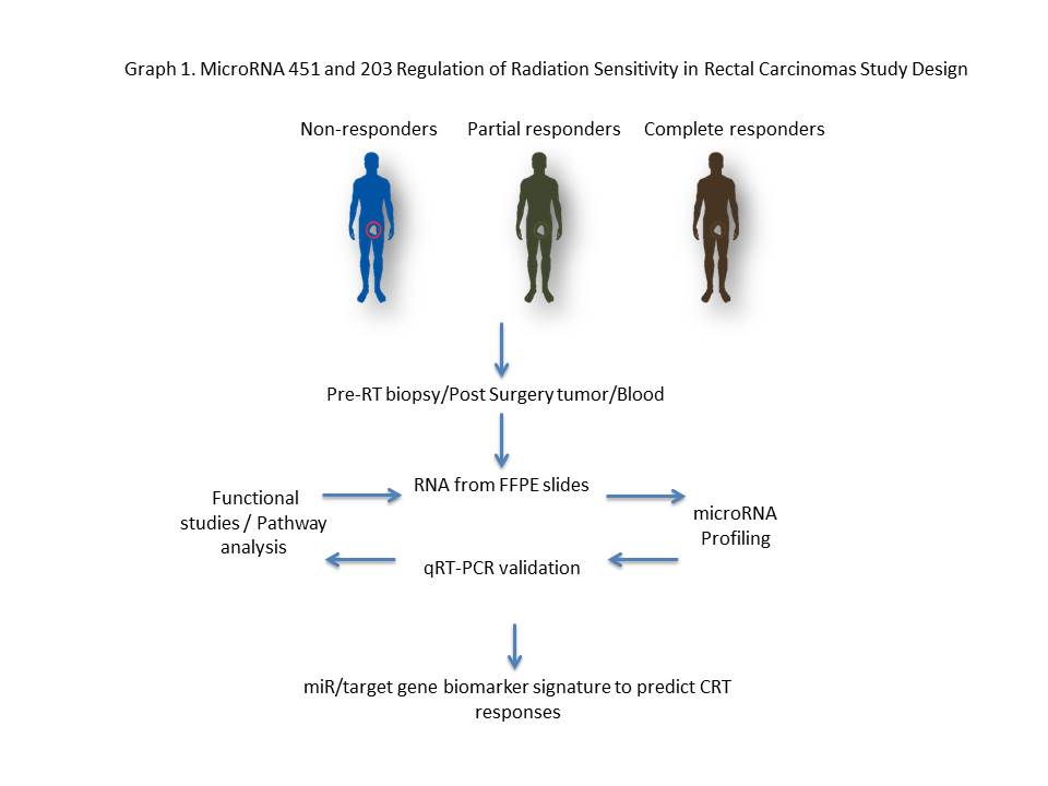
Chemoradiotherapy (CRT) response is an independent predictor of overall survival in locally advanced rectal cancer, highlighting the importance of improving CRT response rates. It is known that several tumor intrinsic factors govern responses to radiation therapy.  Emerging evidence suggests that microRNAs (miRs) modulate gene expression profiles in response to radiation.

Methods:
microRNA profiles were examined and analyzed in approximately forty rectal cancer patients that had either a pathological partial response (PR) or no response (NR).  Using Nanostring technology, a miR profiling platform, miRNAs significantly down and up regulated were identified in FFPE biopsy specimens.   mRNA differentially expressed was followed by in vitro radiation models and tumor sphere assays.  Graph 1.

Results:
miR-451a was among the most upregulated miRs in the PR group whereas miR-203 was among the most downregulated miR.  Inhibition of miR-451a in HCT-116 significantly increased survival in response to a 2 Gy dose of radiation; conversely, transfection of a miR-451 mimic significantly decreased cell survival, with more than a 12-fold decrease when combined with a 5 Gy dose fraction.  Consistent with these results, miR-451a inhibition increased proliferation in 2D as well as in a tumor sphere assay, whereas transfections of a miR-451a mimic robustly decreased proliferation.  In contrast to miR-451a, miR-203 was downregulated in patients with a partial response to CRT.  Inhibition of miR-203 increased apoptosis in combination with a 5 Gy dose fraction.

Conclusion:
A gain of miR-451a and an inhibition of miR-203 in tumor cell lines enhanced cell death and decreased survival in combination with radiation. In this context, we hypothesize that differential expression of miRs regulates rectal cancer radiation sensitivity and therefore can be used as a biomarker to predict therapeutic responses to radiation therapy.
 

02.02 LY2090314 suppresses cholangiocarcinoma cell growth via apoptosis and reduction in GSK-3β

K. M. Sokolowski1, S. Kunnimalaiyaan1, T. C. Gamblin1, M. Kunnimalaiyaan1  1Medical College Of Wisconsin,Surgical Oncology/Surgery,Milwaukee, WI, USA

Introduction:

Cholangiocarcinoma (CCA) is characterized by poor prognosis and therapeutic inefficacy. The current standard of practice for advanced CCA is systemic chemotherapy with gemcitabine and cisplatin. However, a limited survival benefit along with increasing resistance has ushered in a renaissance in alternative approaches. Increased understanding and application of new technologies has led to the identification of more pathway-focused treatment strategies. The glycogen synthase kinase-3 (GSK-3) pathway is a potential therapeutic target as it is overexpressed in various cancer types including CCA. However, the role of targeting GSK-3 in CCA progression remains elusive. We hypothesize that GSK-3 stabilizes cellular growth thereby promoting proliferation and with subsequent inhibition, leading to a reduction in cellular growth in vitro. For this, we have used a clinically well-characterized GSK-3 inhibitor, LY2090314. 

Methods:

The effects of LY2090314 on CCA cell lines (CCLP-1 and SG-231) were assessed by MTT and colonogenic assay. Cell lysates were analyzed via Western blotting for pro-apoptotic, anti-apoptotic and cell cycle proteins. Apoptotic induction was further evaluated through caspase-glo 3/7 assay. Mechanism of LY2090314 administration on CCA cellular proliferation was also examined. 

Results:

Increasing LY2090314 treatment (0μM – 20μM) had a significant dose and time-dependent growth reduction (p<0.01) compared to control. The concentration at which 50%growth inhibition (IC50) was observed in vitro following 96hr treatment for CCLP-1 and SG-231 is 13.7µM and 9µM respectively. Similar concentrations inhibited colony formation in both CCA cell lines.  Western analysis indicated that growth suppression was due to apoptosis as well as  cell cycle arrest as evidenced by increased expression of pro-apoptotic (cleaved PARP and cleaved caspase-3), reduced anti-apoptotic (survivin) proteins and mitigation of cell cycle arrest proteins (p21 and cyclin D1). Functionally, this was confirmed by an increase in caspase activity. Importantly, LY2090314 treatment reduced the expression levels of only GSK-3β in CCA cell lines. 

Conclusion:

LY2090314, a GSK-3 inhibitor, effectively reduces CCA growth in vitro and appears through reduction of GSK-3β. To our knowledge, this is the first study on the effect of LY2090314 in cholangiocarcinoma cell lines in vitro and provides rationale for further preclinical analysis.

02.03 The Tumor Microenvironment Stroma Promotes Cancer By Using Cell-Type Specific Stress Kinase Pathways.

S. G. Patel1, L. Li1, A. Maas1, R. Ghukasyan1, J. Williams1, P. Toste1, A. Nguyen1, I. Elliott1, N. Wu1, T. Donahue1  1University Of California Los Angeles,Surgery,Los Angeles, CA, USA

Introduction:
We have demonstrated that pancreatic cancer (PDAC) tumor associated fibroblasts (TAFs) support pancreatic cell growth and invasion from a pro-inflammatory, DNA damage (DDR) mediated pathway.  This pathway referred to as the senescence associated secretory phenotype (SASP) requires both stress kinases and Nfkb signaling for maximum signal production after stimulus.  Within the tumor microenvironment, genotoxic chemotherapies can stimulate the stromal cells to produce factors that promote tumor cell fitness.  Contrary to the prevailing model, we have identified that JNK isoforms are cell-type specific and that both JNK1 and JNK3 can transduce this inflammatory pathway in the tumor microenvironment.  We additionally have found that not all of the SASP is regulated by Nfkb, but rather a key mediator, IL-1 alpha, can be expressed in its absence.

Methods:
Immortalized pancreatic cancer tumor associated fibroblasts (Logsden Lab) as well as Hela cells were used to create both gene knockouts as well as stable knockdowns for JNK1, JNK3 and p65 (Nfkb signaling).  SASP induction was verified after a DNA damage response using RTPCR.  Small molecules specific to JNK isoforms were used to verify genetically manipulated cell lines in WT cells.

Results:

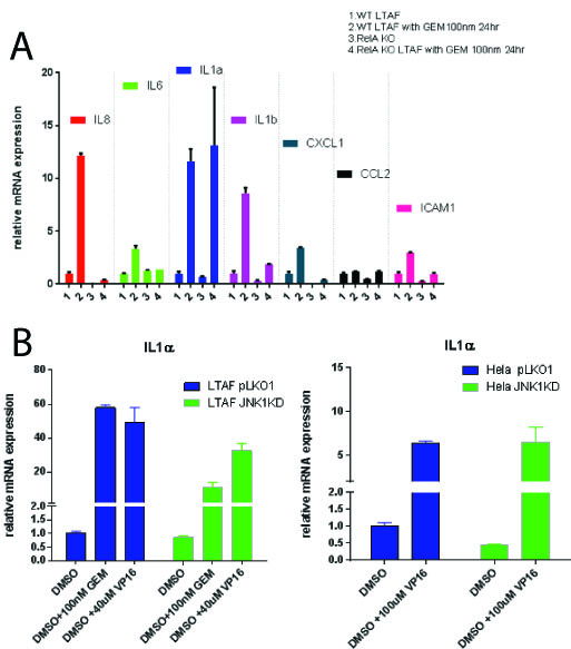
Gemcitabine treatment (100 nm) of PDAC TAFs induced a pro-inflammatory gene expression program including statistically significant (p < 0.05) upregulation of a number of cytokines: including IL-6, IL-8, IL-1 alpha and beta, CXCL1, CCL2 and ICAM1.   Homozygous knockout TAFs for p65 (TAF p65 -/-) stops the induction of these all genes tested (p<0.05) except IL-1 alpha (p = 1) [Figure 1A].   Nfkb (p65) is required for upregulation of IL-1 alpha in the presence of recombinant (10 ug/mL) TNF-alpha or IL-1 alpha (p <0.05).  We found that TAFs do not express JNK3, but rather JNK1 and 2.  We identified using a combination of shRNAs to JNK1 and JNK3 as well as small molecule inhibitors to JNK1/2 that TAFs require JNK1 for upregulation of IL-1 alpha after a DDR (p<0.05) [Fig 1B].   This implies that in the tumor microenvironment, the SASP is propagated by specific stress kinases expressed.

Conclusion:
Previous studies have attributed the SASP to be critically dependent on Nfkb signaling as well as amplified in culture with JNK3.  We identify that some elements of the SASP are not dependent on Nfkb signaling.   Furthermore, we find that JNK3 is not a ubiquitous stress kinase in this response, but rather JNK isoforms are cell-type restricted in expression.  These findings highlight the complexity of the pro-inflammatory signaling within the tumor microenvironment and suggest a more careful analysis of cell and tissue specific gene expression is needed for developing optimal treatment strategies.