81.08 Laparoscopic Surgery for Rectal Prolapse: Short-Term Outcomes Should Not Dictate the Approach

P. L. Rosen1, D. J. Gross1, H. Talus5, V. Roudnitsky2, M. Muthusamy3, G. Sugiyama4, P. J. Chung3  1State University Of New York Downstate Medical Center,Department Of Surgery,Brooklyn, NY, USA 2Kings County Hospital Center,Division Of Trauma And Acute Care Surgery,Brooklyn, NY, USA 3Coney Island Hospital,Department Of Surgery,Brooklyn, NY, USA 4Hofstra Northwell School Of Medicine,Department Of Surgery,Hempstead, NY, USA 5Kings County Hospital Center,Department Of Surgery,Brooklyn, NY, USA

Introduction:
Full thickness rectal prolapse is a debilitating condition for which multiple surgical approaches have been described. Laparoscopic transabdominal approaches are frequently employed, but there is a paucity of data comparing outcomes between laparoscopic transabdominal rectopexy (LR) and laparoscopic transabdominal rectopexy with sigmoidectomy (LRS). Using the American College of Surgeons National Surgical Quality Improvement Program (ACS NSQIP) database, we compared outcomes between these two commonly employed modalities. 

Methods:
Using ACS NSQIP 2010-2015, we identified cases in which LR (CPT 45400) or LRS (CPT 45402) were performed for a postoperative diagnosis of rectal prolapse (ICD 9 569.1). We excluded cases with missing sex, race, BMI, functional status, and ASA classification data. Outcomes of interest included length of stay (LOS), postoperative major morbidity (wound infections, pulmonary complications, cardiovascular complications, renal complications, sepsis/septic shock, bleeding, return to OR) and mortality.  LR and LRS cases were matched using propensity scores. Matching diagnostics were performed and outcomes were evaluated using conditional logistic regression or the Wilcoxon rank-sum test.

Results:
We identified 1,397 patients of which 841 (60.2%) underwent LR and 556 (39.8%) underwent LRS. Patients undergoing LR tended to be older (mean 61.6 vs 55.8 years, p<0.0001), had lower rates of independent functional status (95.7% vs 98.6%, p=0.0072), had higher proportion of African American race (3.8% vs 2.5%, p<0.0001), diabetes treated with medication (7.3% vs 3.8%, p=0.022),  CHF (1.31% vs 0.0%, p=0.0043), bleeding disorders (2.3% vs 0.72%, p=0.031), and ASA class 3 (39.6% vs 30.4%, p=0.0045). Unadjusted comparison between LRS and LR showed increased LOS (median 4 vs 2 days, p<0.0001), increased rates of superficial surgical site infection (SSI) (2.7% vs 0.6%, p=0.0019), bleeding (3.1% vs 1.3%, p=0.036), and sepsis (1.8% vs 0.5%, p=0.025). Propensity scores were then used to match 509 LRS to 509 LR cases with diagnostics showing that the groups were well-balanced across covariates. Conditional logistic regression demonstrated that LRS compared to LR had no statistically significant increased risk of 30-day postoperative complications or mortality. However LRS was associated with increased LOS compared to LR (median 4 vs 2 days, p<0.0001). 

Conclusion:
In this large observational study utilizing a national clinical database we found no differences in 30-day postoperative outcomes between laparoscopic transabdominal rectopexy without sigmoidectomy versus laparoscopic transabdominal rectopexy with sigmoidectomy after propensity score matching. This suggests that long-term outcomes should dictate the choice between these two procedures.

81.06 Emergency Room Admits and Complicating Factors are Linked to Poor Outcomes in Colorectal Surgery

M. Kanneganti1, P. Friedmann1, R. Levine1, H. In1  1Albert Einstein College Of Medicine,Surgical Oncology,Bronx, NY, USA

Introduction:
The circumstance of a person’s presentation at time of colorectal cancer (CRC) surgery is likely to influence outcomes. We aim to examine whether an admission through the emergency department and the presence of complicating factors (gastrointestinal bleeding (GIB), obstruction, and perforation) influence outcomes of CRC surgery.

Methods:
Nationwide Inpatient Sample (NIS) database was used. Patients who underwent surgery for CRC between 2007 and 2011 were included. CRC surgery patients were examined according to their admission circumstance; ER admission with complicating factors (ER-Cx), ER admission without complicating factors (ER-nonCx), non-ER admission with complicating factors (nonER-Cx) and non-ER admission without complicating factors(nonER-nonCx). Logistic regression models were used to estimate odds ratios of in-hospital death for admission status after adjusting for patient and hospital characteristics. Analyses were performed using SAS 9.4 (Cary, NC).

Results:
Of 81,774 patients, 5.2% were ER-Cx, 14.6% were ER-nonCx, and 3.8% were nonER-Cx. Patients with any ER admission or any complicating factors were more likely to be older (>75: 43.1% vs. 23.5%), black (12.7% vs. 5.9%) or Hispanic (7.2% vs. 4.1%), and have more comorbidities (3 or more: 22.9% vs.8.2%) compared to nonER-nonCx. These patients were also more likely to have complications (16.5% vs. 9.1%) and ostomies (26.6% vs. 15.0%). They had higher length of stay (LOS) as compared to all other patients [13.3 days (ER-nonCx), 12.9 days (nonER-Cx), 14.3 days(ER-Cx) vs. 7.9 days (nonER-nonCx)) and were less likely to be to be discharged home (65.0% vs. 85.8%). Adjusted logistic model shows that compared to nonER-nonCx, ER-nonCx (aOR 1.50, CI 1.29-1.75), nonER-Cx (aOR 2.06, CI 1.71-2.49) and ER-Cx (aOR 2.07, CI 1.73-2.47) were more likely to have in-hospital death.

Conclusion:
The circumstances for admission for CRC surgery independently influence outcomes. In our analysis, surgery after an ER presentation or surgery in the setting of GIB, obstruction or perforation is associated with worst outcomes. Future investigation should explore modifiable factors of ER admissions and admission with complicating factors, such as the timeliness of surgery. A patient’s presenting circumstance should be considered when evaluating outcomes and developing treatment plans following CRC surgery.
 

81.07 Revolving Door: The Impact of Length of Stay on Readmissions After Colon and Rectal Operations.

D. Peterson1,3, F. Guzman1, L. Yu4, W. Cirocco1, A. Harzman1, A. Traugott1, M. Arnold1, S. Husain1  1Ohio State University,Colon And Rectal Surgery,Columbus, OH, USA 3Penn State Hershey Medical Center,Surgery,York, PA, USA 4Ohio State University,Biostatistics,Columbus, OH, USA

Introduction:
With recent emphasis on pay for performance model for surgery, length of stay and readmission rates have come under renewed scrutiny. An inverse relationship between length of stay and readmission rates has been suggested raising concerns that early discharges may in fact lead to higher readmission rates. We sought to evaluate the relationship between length of stay and readmission rates and the impact of surgical approach, patient demographics and postoperative complications.  

Methods:
Retrospective chart review was conducted of all colorectal surgeries from September 1, 2011-August 31, 2016 at a tertiary medical center. Patient demographics, comorbidities, postoperative complications, length of stay and readmission rates were evaluated. Logistic regression used to evaluate continuous predictors and Fisher exact test used to evaluate categorical predictors.

Results:
A total of 1319 patients were included. The average length of stay was 10.3 days (median: 7) and 260 (19.7%). At least one complication was noted in 226 (17.36%) patients. Univariate analysis revealed that longer hospital stays correlated with higher readmission rates (R= 0.015, p= 0.00953). However, this association lost its statistical significance with multivariate analysis (p=0.858). Multivariate analysis also revealed that the both increased length of stay and readmissions were strongly associated with pre-existing patient comorbidities (age, diabetes, BMI, COPD, renal dysfunction) as well as postoperative complications (wound infection, abdominal abscess, SVT, PE, pneumonia, UTI). Furthermore, utilization of laparoscopic surgery had statistically significant association with shorter length of stay and lower readmission rates (p=0.004 and 0.02 respectively).

Conclusion:

While length of stay is associated with readmission rates in univariate analysis, this relationship is lost when factors like patient comorbidities, operative outcomes and surgical approach are taken into consideration. Our results also indicate that pre-existing comorbidities and postoperative complications result in prolonged hospitalization and increased readmission rates. Finally, our study not only confirms the well documented beneficial effect of laparoscopic approach on length of stay, it also indicates that minimally invasive approach results in lower readmission rates. While most pre-existing medical conditions leading to longer hospital stays and readmissions are not modifiable, a concerted effort is necessary to minimize postoperative complications and to promote utilization of minimally invasive platform.
 

81.05 Risk factors for 30-day readmission after colorectal surgery: does transfer status matter?

S. T. Lumpkin1, P. Strassle1,2, N. Chaumont1  1University Of North Carolina At Chapel Hill,Department Of Surgery,Chapel Hill, NC, USA 2University Of North Carolina At Chapel Hill,Department Of Epidemiology,Chapel Hill, NC, USA

Introduction: Rates of readmission after colorectal surgery (CRS) range from 9-25%, and cost the US $300 million annually. Our hypothesis is that transfer from an outside hospital prior to CRS, as one potential indicator of preoperative access to care, increases odds of 30-day readmission.

Methods: Using the Healthcare Cost and Utilization Project Nationwide Readmissions Database, a retrospective analysis of surviving adult patients who underwent inpatient CRS from 2010-2014 was performed. The primary outcomes were 30-day risks of cause-specific readmissions, listed in Figure 1. A composite ‘any surgical indication’ variable was created to assess if the primary indication for surgery was also a cause for readmission. Using multivariable logistic regression, we assessed the direct effect of potential risk factors for readmission, including demographics, hospital characteristics, comorbidities, indication for CRS, and initial transfer status to the index hospital where the CRS was performed.

Results: Total n=357,696 patients. The cause-specific rate of readmission was 7.1%, n=25269 (Figure 1). Primary indications for CRS were independent risk factors for readmission: relative to cancer, patients with IBD (OR 2.10, 95% CI 1.99, 2.22) and trauma (OR 1.24, 95% CI 1.08,1.43) were more likely to be readmitted, whereas patients with infectious indications (OR 0.74, 95% CI 0.72, 0.77) and non-infectious/vascular indications (OR 0.77, 95% CI 0.73, 0.81) were less likely to be readmitted. Patients treated at small hospitals were less likely to be readmitted (OR 0.94, 95% CI 0.90, 0.98) than patients treated at large hospitals. Treatment at a rural-nonteaching hospital compared to urban-teaching hospital, (OR 0.75, 95% CI 0.71, 0.79) decreased odds of readmission. Younger patient age, 18-34 years old compared to age 35-49 years old (OR1.13, 95% CI 1.05, 1.21), public primary insurance compared to private insurance (OR 1.27, 95% 1.22, 1.31), and multiple comorbidities were also significantly associated with increased odds of readmission. Two percent of patients were transferred from another hospital to the hospital where CRS was performed; this did not affect odds of 30-day readmission (OR 0.97, 95% CI 0.89, 1.06).

Conclusion: Preoperative considerations, such as primary indications for CRS, are important risk factors for readmission, but transfer status was not significant. At large, urban-teaching hospitals, where patients are at higher risk of readmission, targeting interventions towards patients between the ages 18-34, with public insurance, who have comorbidities, or whose primary indication for surgery is cancer, trauma, or IBD patients may reduce readmissions.

81.04 Treatment & Prognosis of Rectal Squamous Cell Carcinoma: Analysis of the National Cancer Database

K. E. Koch1, P. Goffredo1, A. Beck1, P. Kalakoti1, I. Hassan1  1University Of Iowa,Surgery,Iowa City, IA, USA

Introduction:  Rectal squamous cell carcinoma (RSCC) is a rare malignancy, accounting for approximately 0.01–0.025% of all colorectal cancers. As a result, literature regarding its treatment and prognosis is mainly based on small single institutional series. Our aim was to describe patient characteristics, stage specific management, and outcomes utilizing a national database.

Methods:  We identified 2915 patients with stage I, II, and III RSCCs from the National Cancer Database treated between 2004 and 2014. Management strategies were categorized as local excision or chemoradiation alone, chemoradiation with local excision, and chemoradiation with radical resection. These modalities accounted for approximately 80% of patients in the cohort. Stratified survival analyses were adjusted by gender, age, and race. Data were examined using simple summary statistics, chi-square, student’s-T tests, Kaplan-Meier analysis, and Cox proportional hazards regression.

Results: The majority of patients were females (69%) and Caucasian (86%) with a mean age at diagnosis of 61 years (SD=13). The Charlson/Deyo score was 0 in 81% of patients. Mean tumor size was 41 mm (SD=25).  The 5-year overall survival (OS) was 70% for stage I (1021 pts), 55.8% for stage II (711 pts), and 54.7% for stage III (809 pts, p<0.001). In univariate analysis for patients with stage II and III disease, no difference was observed in the 5-year OS among management strategies (p=0.90 and 0.07, respectively). However, for stage I disease, the combination of chemoradiation and local excision was associated with improved outcomes compared to chemoradiation or local excision alone (p <0.001) (Table 1). The results of the univariate analysis were confirmed in the multivariate model after adjustment for available demographic confounders.

Conclusion: Our data suggest that for stage I disease the combination of chemoradiation with local excision may be the optimal oncologic treatment. Conversely, the addition of local excision or radical surgery to a treatment strategy based on chemoradiation for stage II and III disease was not associated with a survival benefit. Therefore, a treatment approach based primarily on chemoradiation should be considered the optimal management strategy for squamous cell carcinomas of the rectum.

81.03 Incidence and Risk Factors of C. difficile Infection in Patients with Ileal Pouch-Anal Anastomosis

P. D. Strassle1,3, J. Samples1, E. E. Sickbert-Bennet2,3, D. J. Weber2,3, T. S. Sadiq1, N. Chaumont1  1University Of North Carolina At Chapel Hill, School Of Medicine,Department Of Surgery,Chapel Hill, NC, USA 2University Of North Carolina At Chapel Hill, School Of Medicine,Department Of Medicine,Chapel Hill, NC, USA 3University Of North Carolina At Chapel Hill, Gillings School Of Global Public Health,Department Of Epidemiology,Chapel Hill, NC, USA

Introduction:  In the last 10 years, recognition of Clostridium difficile infection (CDI) in patients with ileal pouch-anal anastomosis (IPAA) has been increasingly recognized. Despite the growing body of literature, conclusions about the incidence and risk factors of CDI in IPAA patients have been limited by single-institution studies, small sample sizes, and short follow-up. The goal of this study was to estimate the incidence and potential risk factors of CDI in patients with IPAA.

Methods: Patients diagnosed with ulcerative colitis, Crohn’s disease, or familial adenomatous polyposis, and undergoing an ileal pouch procedure between 2004 and 2013 in the Truven Health Analytics MarketScan® database were eligible for inclusion. Patients were required to have health insurance coverage for at least 6 months before and 30 days after surgery.  

Kaplan-Meier survival curves were used to estimate the 2-year risk of infection. CDI was identified using ICD-9 CM code 008.45, which has 78.0% sensitivity and 99.7% specificity. Multivariable Cox proportional hazard regression was used to assess the effect of potential risk factors. Risk factors included patient demographics, Charlson comorbidity score, pre-operative CDI (within 6 months of surgery), recent hospitalization (within 30 days of surgery), and use of corticosteroids, biologics, and immunomodulators (within 30 days of surgery). Inverse-probability of censor weights were used to account for differential follow-up. Age was modeled as a linear variable and centered at 40 years old. 

Results: 2,900 patients were included in the analysis. The median follow-up time was 628 days (IQR 287-730). The 2-year cumulative incidence of C. difficile was 3.3% (n=77). Twelve cases (15.6%) occurred during the surgical hospitalization. Patients with previous CDI (HR 7.33, 95% CI 3.85, 13.94) and patients taking corticosteroids (HR 2.19, 95% CI 1.30, 3.71) or biologics (HR 3.59, 95% CI 1.39, 9.24) prior to surgery were significantly more likely to have a CDI after IPAA. No significant differences in the risk of CDI across gender (p=0.51), age (p=0.70), Charlson score (p=0.99), history of recent hospitalization (p=0.50), or immunomodulator use (p=0.30) were seen.

Conclusion: The 2-year incidence of CDI after IPAA is at least 3%. Patients with a history of pre-operative CDI, and those taking corticosteroids or biologics before surgery are more likely to develop a CDI after surgery. 

81.02 The Association Between Sarcopenia And Myosteatosis And Post-operative Outcomes in patients with IBD.

S. J. O’Brien1,2, O. J. O’Connor3, E. J. Andrews2  1University Of Louisville,Surgery,Louisville, KY, USA 2University College Cork,Surgery,Cork, CORK, Ireland 3University College Cork,Radiology,Cork, CORK, Ireland

Introduction:
Sarcopenia has been defined by the European working group on Sarcopenia in Older People (EWGSOP) as a low muscle mass and either decreased muscle strength or low physical performance. Skeletal muscle index is use to measure muscle mass. Recent studies have demonstrated the association that sarcopenia has with adverse post-operative outcomes in patients with cancer. Few studies have examined the role of sarcopenia or myosteatosis, fatty infiltration of the muscle, in the setting of non-oncological surgery. The aim of this study was to assess the prognostic significance of sarcopenia and myosteatosis in patients with inflammatory bowel disease undergoing surgical resection with respect to post-operative complications.

Methods:
A retrospective analysis of a prospectively maintained surgical database was examined. All patients who underwent an elective or emergent colonic resection for IBD between 2011 and 2016 were included. Patient demographics, clinical indices and peri operative CT scans were collected. Skeletal muscle index was calculated by measuring the total muscle area (cm2),at the level of the L3 vertebra,  and normalising to the patients height squared (m2?) using the Osirix image analysis software (Figure 1). Myosteatosis is calculated by measuring the average Hounsfield unit at the same vertebral level. Regression analysis was used to identify predictors of outcomes.

Results:
39%(30/77) of patients were sarcopenic. Both sarcopenic and non- sarcopenic groups were equally matched with the exception of weight and BMI (p=0.014 and 0.009). There was a significant difference in hospital readmission between sarcopenic and non-sarcopenic patients and between myosteatotic and non-myosteatotic patients (p=0.03 and p=0.018). On univariate analysis, sarcopenia and myosteatosis were risk factors for hospital readmission (OR= 4.778, 95CI: 1.121-20.361 p=0.034 and OR= 6.24 95CI: 1.224-31.811, p=0.028). There was no difference in the incidence of major complications, anastomotic leaks and length of stay between the study groups.


Conclusion:
Sarcopaenia and myosteatosis were associated with hospital readmission in this study. As the cut-off values for a low skeletal muscle index are calculated from a cohort of oncology patients, this may accout for the lack of difference in the incidence of major complications, anastomotic leaks and length of stay. Further research is required to elucidate the role of myopenia and myosteatosis in patients undergoing surgery for non-malignant disease. 

81.01 Identifying Factors that Decrease Utilization of Adjuvant Chemotherapy in Stage III Colon Cancer

P. M. Schroder1, M. C. Turner1, B. Ezekian1, Z. Sun1, M. A. Adam1, C. R. Mantyh1, J. Migaly1  1Duke University Medical Center,Department Of Surgery,Durham, NC, USA

Introduction:  Standard of care for stage III colon cancer includes adjuvant chemotherapy, which increases survival by nearly 30%. Despite these results, many patients fail to receive adjuvant chemotherapy. We aim to describe the benefit of adjuvant chemotherapy for stage III colon cancer and to determine factors that influence the likelihood of receiving this treatment. 

Methods:  We queried the National Cancer Data Base 2006-2013 for patients with a single primary stage III colon adenocarcinoma and defined two groups: patients who did and did not receive adjuvant chemotherapy. Subgroup analyses were performed for healthy patients (Charlson-Deyo [CD] score = 0), comorbid patients (CD score ≥ 2), and those with post-operative complications (readmitted within 30 days of surgery). Kaplan-Meier (KM) curves were generated and Cox proportional hazard ratios (HR) were calculated to compare overall survival. Odds ratios (OR) for receiving chemotherapy were calculated to identify factors associated with failure to receive adjuvant chemotherapy. 

Results: Of the 74,588 patients included in this study, 54,235 received adjuvant chemotherapy and 20,353 did not. Overall survival was significantly better in the group that received adjuvant chemotherapy (HR of 0.477, p<0.001). Similar results were obtained in our subgroup analyses (see Figure). Adjuvant chemotherapy conferred a survival advantage for healthy patients (HR 0.485, p<0.001), comorbid patients (HR 0.492, p<0.001), and those with post-operative complications (HR 0.358, p<0.001). Several factors were associated with a reduced likelihood of receiving chemotherapy including older age (OR 0.9, p<0.001), black race (OR 0.728, p<0.001), comorbid patients with CD score ≥2 (OR 0.563, p<0.001), positive surgical margins (OR 0.83, p<0.001), and those with post-operative complications (OR 0.605, p<0.001). Patients with private insurance (OR 1.997, p<0.001) or Medicare (OR 2.184, p<0.001) were comparatively more likely to receive adjuvant chemotherapy.

Conclusion: We demonstrate a consistent survival benefit with adjuvant chemotherapy for patients with stage III colon cancer, even for comorbid patients or those with early post-operative complications. Factors such as older age, black race, more comorbidities, positive margins, post-operative complications, and lack of insurance were associated with a reduced likelihood of receiving adjuvant chemotherapy. These data suggest that adjuvant chemotherapy remains critically important for all patients with stage III colon cancer, but particular attention should be paid to utilizing this therapy in higher risk and underserved patients to avoid undertreating these vulnerable populations.

80.11 Impact of the Gut Microbiome-Host Interaction on Postoperative Complications: An Exploratory Review

V. M. Gershuni1,2, G. D. Wu2, R. R. Kelz1  1Hospital Of The University Of Pennsylvania,Department Of Surgery,Philadelphia, PA, USA 2Hospital Of The University Of Pennsylvania,Division Of Gastroenterology,Philadelphia, PA, USA

Introduction:
Composed of numerous bacteria, their genes, and bioactive metabolites, the gut microbiome is a significant contributor to overall health and disease. The gut microbiome plays a critical role in mucosal integrity and inflammation. The microbiome is an attractive target to modify surgical outcomes. The purpose of this review was to describe current knowledge on the relationship between the gut microbiome and GI surgical outcomes.

Methods:
Using defined search terms, an exploratory literature review was performed in PubMed and Google Scholar (January 1, 2012 to July 31, 2017) to identify articles on the gut microbiome and GI surgical outcomes, specifically including anastomotic leak (AL) and surgical site infection (SSI). After title and abstract review, articles were selected for data abstraction based on discussion amongst the co-authors. Data was not abstracted from review articles although primary sources were examined. The following study characteristics were recorded: article type, human or animal study, number of study subjects, scientific methods and outcomes.

Results:
Among 384 references identified, the majority (n=344) were not related to GI surgery and one article was a retrospective study of surgical outcomes. A total of 39 articles reported on aspects of the microbiome and GI surgical outcomes. Twenty-five articles specifically examined anastomotic leak and surgical site infection. Eighteen of the 39 were literature reviews (including one systematic and one meta-analysis), 13 studies reported primary data from animal models, and four reported primary data in human subjects. A summary of the findings from human studies can be seen in the Table. Animal data suggests that the microbiota may play an integral role in the host response to surgical injury. 

Conclusion:
Evidence on the association between the host-microbiome and GI surgical outcomes is limited in humans. Preliminary evidence suggests that the gut microbiota balance may be disturbed in patients who experience anastomotic leak and SSI. Further studies focused on the relationship between the microbiome and these outcomes may allow us to curtail the morbidity and mortality associated with GI surgery. Some of the interesting potential targets for perioperative intervention include non-absorbable oral antibiotic use, local delivery of phosphate-based therapy, and exploration into the mechanism of refaunation after microbiome disturbance.

80.09 The Impact of DNA Repair Genes and Microsatellite Instability on Survival in Colorectal Cancer

S. Narayanan1, T. Kawaguchi1, L. Yan1, X. Peng1, Q. Qi1, K. Takabe1  1Roswell Park Cancer Institute,Surgical Oncology,Buffalo, NY, USA

Introduction:  Microsatellite instability (MSI) occurs via inactivation of DNA mismatch repair genes resulting in impaired DNA repair function with subsequent accumulation of abnormal genes. Heterogeneity in MSI status is common in colon cancers and has been demonstrated to be an independent predictor of survival, with MSI-high tumors having a better overall prognosis. Previous studies have correlated upregulation of DNA repair genes with increased ability for tumors to metastasize and with chemotherapy and radiation resistance, thereby resulting in poorer survival. We, thus, hypothesized that low expression of DNA repair genes would result in improved survival secondary to increased MSI. 

Methods:  The Cancer Genome Atlas (TCGA) was used to evaluate a cohort of 283 patients with colorectal cancer. RNA sequence gene expression quantification data for colon cancer was retrieved from the Genomics Data Commons (GDC) data portal. Gene expression levels were derived using normalization methods provided in the DESeq2 package and designated as low or high. The expression of DNA repair genes was then compared between MSI-High and Microsatellite stable (MSS) cohorts. Overall survival for patients for each DNA repair gene was determined via Kaplan-Meier analysis. 

Results: We found that low expression of several DNA repair genes correlated with high levels of microsatellite instability. Some of these were the expected mismatch repair genes- MLH1 (p< 0.0001), PMS1 (p= 0.002), PMS2 (p< 0.0001) and MLH3 (p=0.036). However, others were double stranded break DNA repair genes such as ATM (p< 0.0001), PRKDC (p< 0.0001), ATR (p< 0.0001) and BRCA2 (p= 0.0081). Significantly improved overall survival was demonstrated in patients with low expression of ATM (p= 0.04), MLH1 (p= 0.028), PMS2 (p= 0.003) and MLH 3 (p= 0.0029). There was a trend towards improved survival in patients with low expression of BRCA2 and ATR without achieving statistical significance. 

Conclusion: In this analysis of patients with colorectal cancer we found that diminished expression of several DNA repair genes correlated with increased microsatellite instability even those that were not known mismatch repair (MMR) genes. However, improvement in survival primarily correlated with MMR-deficiency and with low expression of other DNA repair genes to a lesser extent. This phenomenon may relate to the increased immunogenicity of tumors with genomic instability and may have implications for further development of immunotherapy targets in the future. 


80.07 Survival Mouse Model of Intraperitoneal Perfusion Mimicking Hyperthermic Intraperitoneal Chemotherapy (HIPEC)

M. Peterson1, E. MCabe-Lankford1, B. McCarthy1, N. Levi-Polyachenko1  1Wake Forest University School Of Medicine,Winston-Salem, NC, USA

Introduction:
Peritoneal carcinomatosis (PC) is a late stage presentation of multiple gastrointestinal and gynecologic organ malignancies. In the past three decades, surgical debulking combined with locoregional administration of chemotherapy in a procedure termed hyperthermic intraperitoneal chemotherapy (HIPEC) has emerged as a promising treatment strategy, extending survival times and, in some cases, offering a cure for this previously uniformly terminal condition. Owing to its relative newness, HIPEC remains the subject of extensive research to optimize drug regimens and technique as well as to design chemotherapeutic agents specifically for intraperitoneal use. Mouse models for study of peritoneal carcinomatosis (PC) treatment have been validated as translatable and inexpensive, however, technical description of intraperitoneal perfusion procedures in mice is lacking in published literature. We present techniques for closed abdomen perfusion with a single or double outflow port and open abdomen (“coliseum”) perfusion mimicking hyperthermic intraperitoneal chemotherapy (HIPEC).

Methods:
Female Balb/C mice underwent 30-minute perfusion using Masterflex L/S Digital Pump, XX8000004 Head, 96400-15 tubing as pump system. For closed abdomen perfusion with both single and double outflow, small incisions were made in upper and lower abdomens and inflow and outflow tubes were inserted and secured with suture to perfuse at 50 mL/hr (Fig. 1 A, B). For open abdomen perfusion, vertical midline incision was made to expose abdominal cavity and edges of incision were secured to ring stand to create a “coliseum” to perfuse at 50-100 mL/hr (Fig. 1 C). 

Results:

Closed perfusion with single outflow: Of 12 mice, 11 survived to 24-hour post-operative end-point with 1 intra-op death. Intraoperative complications included suction of organs into outflow and incomplete circuit closure at outflow.

Closed perfusion with double outflow: Of 4 mice, 3 survived to 24-hour post-operative end-point. Incomplete circuit closure at outflow was the only prominent complication.

Open coliseum: Of 3 mice, 3 survived to 30-day post-operative end-point. This design presented excellent perfusate distribution, no organ suction, and allowed intraoperative organ manipulation.

Conclusion:
Survival mouse model closely mimicking HIPEC is achievable and can be customized to either open or closed perfusion techniques. 

80.06 High Expression of Sphingosine Kinase 1 in Colitis-Associated Cancer

K. Yuza1, M. Nagahashi1, Y. Shimada1, M. Nakano1, Y. Tajima1, H. Kameyama1, M. Nakajima1, H. Ichikawa1, J. Sakata1, T. Kobayashi1, K. Takabe2,3, T. Wakai1  1Niigata University Graduate School Of Medical And Dental Sciences,Division Of Digestive And General Surgery,Niigata City, NIIGATA, Japan 2Roswell Park Cancer Institute,Breast Surgery, Department Of Surgical Oncology,Buffalo, NY, USA 3University At Buffalo Jacobs School Of Medicine And Biomedical Sciences, The State University Of New York,Department Of Surgery,Buffalo, NY, USA

Introduction:
Colorectal cancer (CRC) that developed from inflammatory bowel disease (IBD), including ulcerative colitis and Crohn’s disease, is known as colitis-associated cancer (CAC). Significant advances have been made in understanding the link between chronic inflammation and cancer by advancements in molecular biology and animal models that recapitulate human diseases in recent years. It has been implicated that sphingosine-1-phosphate (S1P), a bioactive lipid mediator, is a key mediator of inflammation, tumorigenesis and cancer progression. We have recently shown that S1P produced by upregulation of sphingosine kinase 1 (SphK1) links chronic intestinal inflammation to CAC utilizing animal models (Cancer Cell 2013). However, the role of S1P in human CAC has never been investigated due to the difficulty to measure S1P levels directly in human samples. We have previously shown that high expression of phosphorylated-SphK1 (pSphK1) is associated with higher levels of S1P in human tissue, and detecting pSphK1 by immunohistochemistry could be an alternative method to examine the role of S1P in human patients (JSR 2016). The aim of this study is to determine the expression levels of pSphK1 in sporadic CRC and CAC, and to clarify the importance of S1P in CAC patients.

Methods:
Tissue samples of sporadic CRC (N = 10) and ulcerative colitis-associated cancer (N = 10), respectively, were randomly selected from patients who underwent curative resection between April 2012 and May 2017. The expression of pSphK1 in these samples was examined by immunohistochemistry staining. Semi-quantitative evaluation of the staining of pSphK1 in CRC cells was performed as follows; a staining score was calculated by multiplying two points of staining intensity (0-3) and the ratio (1-4). The staining score of 0 to 5 points was considered as pSphK1-negative, and 6 to 12 was considered as pSphK1-positive.

Results:
Immunohistochemistry analysis of pSphK1 revealed that sporadic CRC had a median staining score of 4 (range 0-12), while ulcerative colitis-associated cancer had a median staining score of 12 (range 4-12) (Mann Whitney test, P<0.05). Based on the staining scoring described in Methods, three out of 10 (30%) patients with sporadic CRC, and nine out of 10 (90%) patients with ulcerative colitis-associated cancer were considered as pSphK1-positive (Fisher’s exact test, P<0.05).

Conclusion:
To our knowledge, this is the first report that compared pSphK1 expression levels in CAC and those in sporadic CRC. The high levels of pSphK1 expression in CAC implicate an important role of S1P in the disease process of CAC.
 

80.05 DNA Repair Genes Differentiate Colitis from Neoplasia in Colitis-Associated Colon Cancer

*R. A. Malizia2, *S. P. Sharp2, T. Walrath1, D. Shanti1, C. J. Booth3, E. C. Lee2, S. C. Stain2, W. O’Connor1  1Albany Medical College,Department Of Immunology And Microbial Disease,Albany, NY, USA 2Albany Medical College,Department Of Surgery,Albany, NY, USA 3Yale University School Of Medicine,Department Of Comparative Medicine,New Haven, CT, USA

*Co-First Authors

Introduction:

Chronic intestinal inflammation predisposes patients with Inflammatory Bowel Disease (IBD) to Colitis-Associated Cancer (CAC).  Fewer than half of CAC cases are diagnosed early, leading to a high associated morbidity and mortality.  Despite continued efforts to improve early detection of high risk, pre-dysplastic regions in IBD patients, current macroscopic and genetic surveillance modalities remain limited.  DNA damage repair (DDR) genes and their role in the formation of sporadic colorectal cancer have been well characterized.  However, limited data exists regarding these genes in CAC.  DDR genes are of paramount importance as they govern microsatellite instability and responsiveness to chemotherapeutic agents.  We investigated the regulation of genes controlling DDR during the transition from chronic colitis to CAC in a preclinical model of disease.

Methods:

As recurrent colitic episodes increase the risk for CAC, we utilized the best characterized murine model for CAC to study colitis-associated tumorigenesis in C57BL/6 mice.  Mice were separated into four distinct cohorts; unmanipulated controls, Azoxymethane (AOM) only, Dextran Sodium Sulfate (DSS) only, or with the combination of AOM/DSS.  Colonic tumors were visualized and graded utilizing high-resolution murine colonoscopy. Upon sacrifice, histopathological analysis was conducted for each cohort. Distal colon segments were analyzed for the presence of dysplasia, adenoma, and adenocarcinoma. Gene expression was evaluated using semi-quantitative RT-PCR.

Results:

Tumors were observed in the AOM/DSS cohort by 5 weeks as expected.  No tumor development occurred in the control (0/5), AOM only (0/5), or DSS only (0/5) cohorts, as expected. Exposure to the AOM mutagen was sufficient to significantly induce mRNA expression of DNA repair genes. Colons exposed to DSS were grossly edematous and foreshortened as compared to control. Upon colonic resection, half of the AOM/DSS cohort displayed macroscopically visible tumor (MVT). The remaining population (7/14) was devoid of visible tumor. Importantly, histological evaluation displayed microscopic evidence of dysplasia, and adenocarcinoma.  Therefore, this group was denoted as the non-macroscopically visible tumor (NMVT) group.  Interestingly, both MVT and NMVT tumor-developing groups showed reduced mRNA expression of mlh1, anapc1, and ercc4 relative to DSS or mutagen alone. Moreover, colitis alone was able to reduce mRNA expression of ercc4.  

Conclusion:

In this study, for the first time, ercc4 has been shown to be down-regulated in chronic colitis and marks the early transition to dysplasia in murine colonic tissue. In addition, our results illustrate changes in DDR gene expression are specific and not universal in the progression to neoplasia.  This phenomenon has not been previously illustrated in CAC and validates the AOM/DSS model of CAC as a means to further investigate potential markers of early malignant transformation. 


80.04 Myofibroblasts Enhance Colorectal Cancer Growth In A Novel Mouse Model Utilizing Murine Colonoscopy

M. Papageorge1, T. Liu1, L. Chen1, J. Yoo1  1Tufts Medical Center,Surgery,Boston, MA, USA

Introduction: Communication between colorectal cancer cells and adjacent stromal cells alter the tumor microenvironment to regulate loco-regional disease and cancer progression.  However, existing mouse models are limited in their ability to study tumor-stromal cell interactions.  Additionally, they often use immunocompromised mice, or lead to tumors that do not grow in the colon.  Our goal was to develop a novel mouse model of colorectal cancer that is capable of studying tumor-stromal cell interactions in the native colon of immune-competent mice.

Methods: Primary mouse myofibroblasts (MFB) were isolated from the colon of C57BL/6 mice and grown in cell culture.  Genetically defined (Apc-null; Kras mutant;Trp53-null) primary mouse colon cancer cells (CRC) were suspended in serum-free media (20  μL) at varying concentrations (5×10³-2×10⁴ CRC cells) either alone or in combination with syngeneic MFB (2×10CRC cells + 2×10⁵ MFB).  Following isoflurane anesthesia, a colonoscopy was performed on immune-competent 8-10 week-old C57BL/6 mice with endoscopic microinjection of the cell suspension which also contained 20% Spot® to “tattoo” the bowel wall for subsequent evaluations.  Microinjections were performed using a Karl Storz Coloview miniendoscopic system, a manufactured 33-gauge needle (Hamilton®) and a 100 μL syringe (Hamilton®).  Proper injection into the colon wall was confirmed endoscopically by the development of a submucosal wheal.  A repeat colonoscopy was performed weekly for 3 weeks with photo and video documentation of the endoscopic findings.  Tumor size is presented as average diameter relative to colon circumference ± SD, n≥3. 

Results:A total of 20 mice were injected with a survival rate of 80% (16/20).  Endoscopic microinjection of CRC cells led to dose-dependent tumor growth in the distal mouse colon (Table 1) that could be assessed regularly without animal sacrifice.  Microinjection of 2×10⁴ CRC cells led to reliable (100%, 5/5 mice) tumor growth that was present at 1 week (0.05±0.06) with persistent growth at week 2 (0.11±0.01) and week 3 (0.11±0.01).  Microinjection of 2×10⁴ CRC cells with 2×10⁵ MFB resulted in much larger tumors that persisted over the 3 week time period (0.2±0.06 at week 1, 0.19±0.06 at week 2, 0.35±0.17 at week 3). 

Conclusion:Endoscopic microinjection of primary mouse CRC cells is feasible and leads to reliable and reproducible growth of colon tumors beginning 1 week post-injection.  Co-injection of primary mouse CRC cells with syngeneic MFB leads to enhanced tumor growth.  Co-implantation of CRC cells with syngeneic MFB provides a novel platform to study tumor-stromal interactions in the native colon of immune-competent mice.


80.03 Downregulation of Protein Tyrosine Phosphatase Receptor F Inhibit WNT and Cell Proliferation in CRC

T. Gan1, P. D. Stevens1, Y. Wen1, X. Xing1, S. Golshani1, B. M. Evers1, T. Gao1  1University Of Kentucky,Markey Cancer Center,Lexington, KY, USA

Introduction: Wnt signaling is a highly regulated and evolutionarily conserved pathway that plays an important role in normal intestinal stem cell renewal and differentiation. Hyperactivation of Wnt signaling is associated with 90% of colorectal cancers (CRCs). Many signaling molecules in the Wnt pathway are known to be regulated by protein kinase-mediated phosphorylation; however, it remains unclear whether the Wnt pathway in CRCs is regulated by protein phosphatases. In this study, we investigated the role of a transmembrane receptor tyrosine phosphatase, PTPRF, in promoting the activation of Wnt signaling in CRC. 

Methods:  The expression of endogenous PTPRF was silenced using lentivirus-mediated RNAi in CRC cell lines: SW480, HCT116, and Caco2. Stable PTPRF knockdown and control cell lines were generated by puromycin selection. Two different shRNA targeting sequences for PTPRF were used to control for specificity. The knockdown efficiency of each shRNA targeting sequence was confirmed using western blot and real-time PCR (RT-PCR) analyses. The expression of Wnt target genes was determined using Quantitative RT-PCR analysis.  Growth assays over 72 hours was performed assessing cell proliferation in control and PTPRF knockdown lines. Furthermore, we developed an orthotopic murine model of CRC by endoscopic colonic submucosal injection of 1 x 10­5 live human cancer cells. Endoscopic evaluation of tumor size was performed weekly. 

Results: Knockdown of PTPRF in CRC cells significantly decreased Wnt target gene expression (eg, LGR5, Axin2, TCF7, Cyclin D1 and CD44) as noted by RT-PCR, suggesting a positive role of PTPRF in regulating the canonical Wnt pathway. Moreover, growth assays consistently demonstrated a marked reduction in cell proliferation in PTPRF knockdown cells. The levels of phosphorylated LRP6, a co-receptor for Wnt, and activated β-catenin proteins were downregulated in PTPRF knockdown cells as noted by western blot, demonstrating a potential interaction of PTPRF with LRP6. Since hyperactivation of Wnt signaling is associated with cancer stem cells, studies are underway to evaluate if PTPRF inhibition reduces the “stemness” of CRC cells. In addition, control and PTPRF knockdown CRC cells were endoscopically injected into NSG mice, this novel model allowed us to evaluate the effect of silencing PTPRF on tumor growth in vivo. 

Conclusion: Our studies are the first to demonstrate that PTPRF functions as an oncogenic factor in CRC by promoting the activation of Wnt signaling. Additional studies on the role of PTPRF in cancer stem cell regulation will provide novel insights into the regulation of Wnt signaling in CRC.  Since Wnt signaling is a major driver of CRC development and progression, inhibition of PTPRF may represent an exciting new therapeutic option for future clinical applications. 


80.02 Metastatic Anal Squamous Cell Carcinoma: Ex-vivo and In-vivo Therapeutic Correlation

M. C. Hernandez1, L. Yang2, J. Leiting1, J. R. Bergquist1, M. J. Truty1  1Mayo Clinic,Department Of Surgery,Rochester, MN, USA 2Mayo Clinic,Center For Individualized Medicine,Rochester, MN, USA

Introduction:
Anal squamous cell carcinoma (ASCC) is an uncommon malignancy without substantial treatment advances. Current primary recommendations for non-metastatic ASCC include concurrent chemotherapy (5-FU) and radiation therapy. Metastatic disease is commonly treated with cisplatin-based chemotherapy with minimal evidence for other effective therapies given relative rarity of metastatic ASCC. Preclinical models to advance the treatment of anal cancers are limited to transgenic mice and cell lines. Patient derived xenografts models (PDX) allow for cancer tissue amplification that accurately recapitulates patient phenotype and these have been shown to correlate with clinical outcomes. We aimed to generate a preclinical PDX model of metastatic human ASCC and determine therapeutic sensitivities to correlate with in-vivo testing.

Methods:
PDX were generated from surgical resection of patient tumor tissue in NOD SCID mice. All patient and derived PDX tumors were histologically (H&E and IHC) confirmed. Time to tumor formation (TTF) and harvest (TTH) were recorded. Cell cultures were performed using a hanging drop and two dimensional methods and screened using cytotoxic therapies. Chemosensitivity was quantified using fluorescence intensity and direct cell counts. Secondarily, we performed live tissue sensitivity assays (LTSA), utilizing 200 um thick slices of PDX derived tumor tissue and screened using cytotoxic therapies. To corroborate ex-vivo sensitivities, in-vivo treatment studies using tumor bearing PDX models were performed. Unpaired Student’s t test was used to compare tumor volume response and p<0.05 was considered significant.

Results:
PDX was generated from an ASCC liver metastasis obtained at surgical resection with 100% engraftment rate in five mice. TTF and TTH were 13 and 55 days respectively. Three-dimensional (3D) cellular culture demonstrated marked sensitivity to combinatorial 5FU/oxaliplatin and 5FU/irinotecan.  LTSA tumor tissue sensitivity correlated these findings as well. Formal in-vivo treatment using ASCC tumor bearing mice demonstrated significant tumor volume reduction and a cytotoxic response in those treated with combinatorial 5FU/oxaliplatin and 5FU/irinotecan compared to vehicle and 5FU alone.

Conclusion:
We have generated the first preclinical PDX model of metastatic ASCC that recapitulates the original patient tumor metastasis. Chemotherapy screening using 3D spheroids and LTSA demonstrated strong sensitivity to combinatorial therapies with 5FU. These screening data correlated well in tumor bearing PDX models. Different combinatorial chemotherapeutic regimens other than standard of care may provide meaningful response for patients with metastatic ASCC and may form the basis for future trials.
 

80.01 For Better or Worse: Clinical Correlation of SOX9 and Irradiated Rectal Cancer

B. Trac2, X. Ding3, A. McHenry2, J. Eberhardt4, T. Saclarides1, D. Hayden1  1Rush University Medical Center,Department Of General Surgery,Chicago, IL, USA 2Loyola University Medical Center,Stritch School Of Medicine,Maywood, IL, USA 3Loyola University Medical Center,Department Of Pathology,Maywood, IL, USA 4Loyola University Medical Center,Department Of General Surgery,Maywood, IL, USA

Introduction: Sex-determining Region Y box 9 (SOX9) has been shown to play a fundamental role in epithelial-mesenchymal transition (EMT) as well as be dysregulated in colorectal cancer. Its role in carcinogenesis remains controversial since SOX9 has been associated with both positive and negative prognostic factors in multiple cancers.

Methods: Sections from both normal and cancerous tissue were taken before and after radiation in 25 patients with locally advanced rectal cancer who received neoadjuvant chemoradiation followed by surgery. SOX9 expression was evaluated by immunohistochemistry using a 4-tier grading system for intensity of expression (0=none, 1=low, 2=moderate, 3=high) and percentage of SOX9-positive cells. Demographics, tumor characteristics and outcomes were extracted from medical records and SPSS (Chicago, IL) was used for statistical analysis.

Results: In this study population, mean age was 60.4 (32-72), 80% were male and the majority of patients were Caucasian (76%). 92% of patients had at least T3 tumors and 68% had stage III disease. Mean maximal diameter of tumor pre-treatment was 4.95 cm (3-8) and post-radiation was 2.79 (0.4-5.3). Down-staging occurred in 15 (60%) patients, but only 2 (8%) had complete pathologic response. 6 (24%) patients had positive nodes found after surgery.

In cancer specimens, 96% were graded as moderate or high expression before treatment; 88% after radiation. 91.7% of cells were SOX9+ before radiation, which decreased to 66.7% after radiation. The majority of specimens (68%) showed decrease in expression intensity and %SOX9 positivity. Before radiation, patients with higher SOX9 % positivity were more likely hypertensive (p=0.037) and they were more likely to be male, diabetic and previous smoker (p=0.05, 0.04, 0.015, respectively) if higher SOX9 positivity after radiation. % SOX9+cells and moderate or high expression were both associated with complete pathologic response (p=0.008). Decreased intensity was associated with postoperative tumor size (p=0.005). Although not statistically significant, decrease in percent positivity and intensity after radiation both trended toward significance for distant recurrence (p=0.07). SOX9 expression was not associated with other tumor characteristics or oncologic outcomes.

Conclusion: Our findings indicate that SOX9 % positivity and moderate or high intensity of expression in rectal cancer specimens decrease after radiation. Oncologic outcomes appear to be improved when intensity of expression and percent positivity remain elevated, for example, in complete pathologic response. When expression decreases after radiation, distant recurrence and larger post-radiation tumor size are more common. Although its role remains controversial, our findings suggest that SOX9 may be an important marker and potential target for tumor response and oncologic outcomes for rectal cancer after radiation.
 

76.04 Multi-institution Evaluation of Adherence to Comprehensive Postoperative VTE Chemoprophylaxis

B. Hewitt1, E. Blay1, L. J. Kreutzer1, K. Y. Bilimoria1, A. D. Yang1  1Northwestern University,Surgical Outcomes And Quality Improvement Center,Chicago, IL, USA

Introduction:  Venous thromboembolism (VTE) is the leading cause of preventable hospital mortality. Current quality measures for VTE prophylaxis are problematic due to surveillance bias, are not comprehensive, do not ensure appropriate administration, and cannot identify reasons why failures to provide chemoprophylaxis occur.

Methods:  We examined adherence to a novel process measure in patients who underwent elective or non-elective colectomy over an 18 month period at 36 hospitals in a statewide surgical collaborative. The process measure assessed comprehensive VTE chemoprophylaxis during a patient’s entire inpatient hospitalization, including reasons chemoprophylaxis was not given. Unadjusted and adjusted analyses were performed to identify reasons for failure to provide defect-free chemoprophylaxis and examine patient- and hospital-level factors associated with failure.

Results: Out of 4,086 total colectomies, the standard SCIP-VTE-2 prophylaxis measure publicly reported by CMS identified failure in care in only 1% of cases; however, the new measure unmasked failure to provide defect-free VTE chemoprophylaxis in 18% of cases. Reasons for failure included medication not ordered (29.6%), patient refusal (29.5%), incorrect dosage/frequency (7.9%), patient off unit (3.3%), and other (29.6%). Patients were more likely to fail the chemoprophylaxis process measure if treated at safety net hospitals (Odds Ratio [OR] 1.60, 95% Confidence Interval [CI] 1.06-2.41; p=0.03) or if they were ≤ 40 years old (OR 1.52, 95% CI 1.05-2.20; p=0.03 compared to age ≥ 75 years). Patients treated at Magnet nursing-accredited hospitals (OR 0.45, 95% CI 0.30-0.67; p<0.001) or undergoing elective colectomy (OR 0.77, 95% CI 0.62-0.96; p=0.02 compared to non-elective colectomy) were less likely to fail chemoprophylaxis. Patients ≤ 40 years old (OR 2.20, 95% CI 1.43-3.40; p<0.001), underweight patients (OR 2.19, 95% CI 1.28-3.77; p=0.004) or those that received treatment at safety net (OR 1.97, 95% CI 1.07-3.62; p=0.03 compared to non-safety net hospitals) or teaching hospitals (OR 2.82, 95% CI 1.37-5.84; p=0.005 compared to non-teaching hospitals) were more likely to refuse chemoprophylaxis.

Conclusion: This is the first multi-institution study examining failure patterns in providing comprehensive postoperative VTE chemoprophylaxis. In stark contrast to SCIP-VTE-2, our measure unmasked chemoprophylaxis failures in 18% of colectomies in a statewide surgical collaborative. Most chemoprophylaxis failures were due to patient refusals and ordering errors, occurring throughout the inpatient postoperative period. Thus, hospitals should focus improvement efforts on ensuring patients receive VTE prophylaxis throughout their entire hospitalization.

76.03 The Association of Enhanced Recovery Pathway and Acute Kidney Injury in Colorectal Surgery Patients

J. G. Wiener1, L. Goss1,2, D. I. Chu1, J. S. Richman1, J. A. Cannon1, T. S. Wahl1, G. D. Kennedy1, K. D. Cofer1, P. K. Patel1, M. S. Morris1  2Birmingham VA Medical Center,Surgery,Birmingham, ALABAMA, USA 1University Of Alabama at Birmingham,Surgery,Birmingham, Alabama, USA

Introduction:
Enhanced Recovery After Surgery (ERAS) pathways standardize preoperative, intraoperative, and postoperative care including goal directed fluid administration and multimodal pain management. ERAS is associated with shorter hospital lengths of stay, lower costs, and equivalent readmission rates. Since implementing ERAS at our institution in 2015, we sensed an increase in AKI. Although ERAS has benefits for patients and hospitals, little is known about its association with acute kidney injury (AKI). We hypothesize that incorporation of an ERAS pathway for elective colorectal surgery would be independently associated with an increased risk of AKI.

Methods:
A single-institution retrospective review of patients undergoing elective colorectal surgery before and after the implementation of ERAS was conducted. Patient-specific variables were recorded and our primary outcome was development of an AKI. AKI was operationalized using The Kidney Disease: Improving Global Outcomes (KDIGO) definition and staging system. Patients with AKI or dialysis preoperatively were excluded from our analysis. Bivariate comparisons were made using chi-square and Wilcoxon rank sum tests for categorical and continuous variables, respectively. Variables with p<0.05 for bivariate comparisons were included in a multivariate logistic model for AKI.

Results:
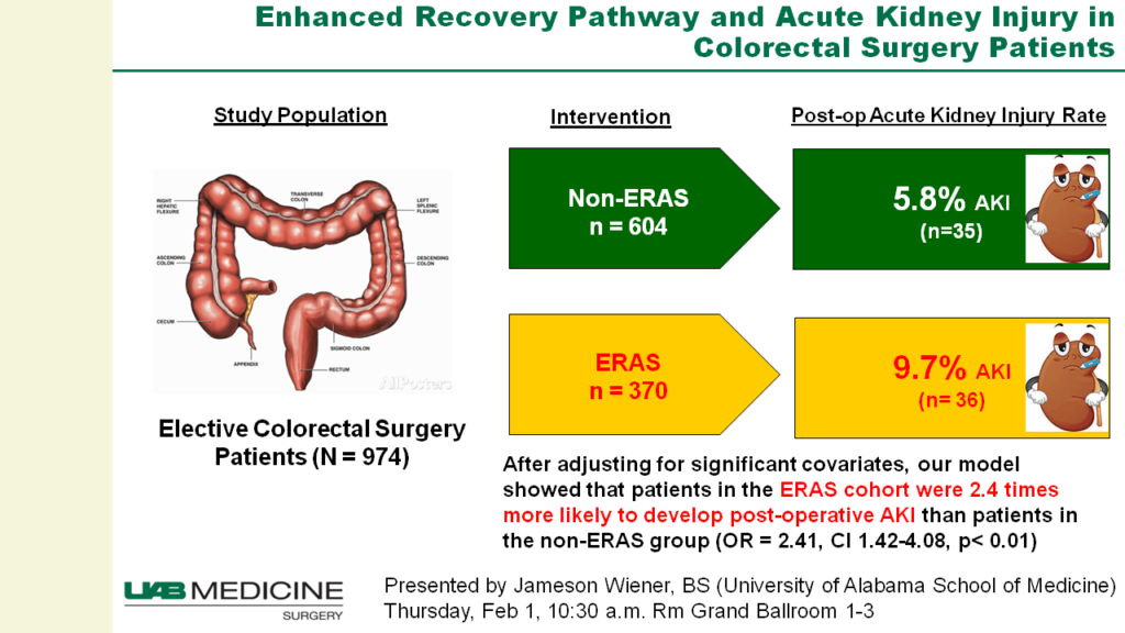
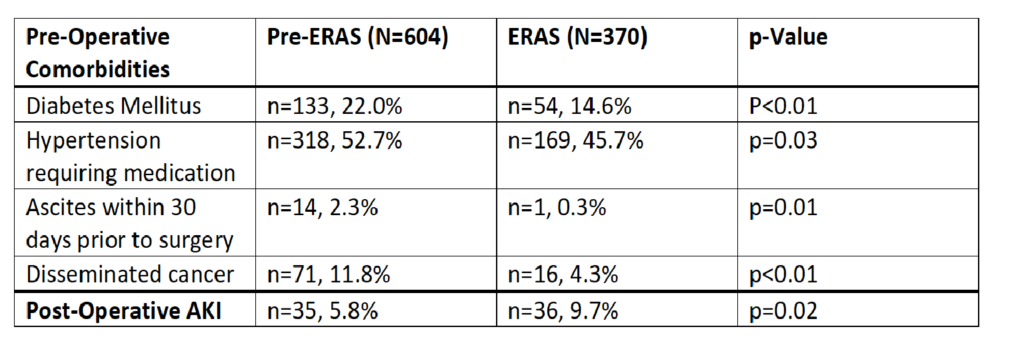
Our study cohort included 974 total patients, 604 in the pre-ERAS group and 370 patients in the ERAS group. The two groups were similar except for significantly higher incidences in the pre-ERAS group of diabetes mellitus, hypertension requiring medication, ascites within 30 days prior to surgery, disseminated cancer, and contaminated or dirty wounds in the pre-ERAS group compared to the ERAS group (Table). There was no significant difference in age or BMI at the time of surgery between the two groups. Postoperatively, 9.7% of the ERAS group developed AKI compared to 5.8% of the pre-ERAS group (p=0.02).  After adjusting for significant covariates, our model showed that patients in the ERAS group were 2.4 times more likely to develop post-op AKI than patients in the pre-ERAS group (OR=2.41, CI 1.42-4.08, p < 0.01).

Conclusion:
Implementation of an Enhanced Recovery Protocol is associated with higher levels of acute kidney injury following elective colorectal surgery. Future studies will determine which aspects of the ERAS protocol, such as NSAID use in the multi-modal pain management or intraoperative goal directed fluid delivery, may be associated with this increased incidence of AKI.

72.03 Clinical Significance of BRAF Non-V600E Mutations in Colorectal Cancer

Y. Shimada1, Y. Tajima1, M. Nagahashi1, H. Ichikawa1, M. Nakano1, H. Kameyama1, J. Sakata1, T. Kobayashi1, Y. Takii2, S. Okuda3, K. Takabe4,5, T. Wakai1  1Niigata University Graduate School Of Medical And Dental Sciences,Division Of Digestive And General Surgery,Niigata, , Japan 2Niigata Cancer Center Hospital,Department Of Surgery,Niigata, , Japan 3Niigata University Graduate School Of Medical And Dental Sciences,Division Of Bioinformatics,Niigata, , Japan 4Roswell Park Cancer Institute,Breast Surgery,Buffalo, NY, USA 5University At Buffalo Jacobs School Of Medicine And Biomedical Sciences,Department Of Surgery,Buffalo, NY, USA

Introduction: Recent advances of comprehensive genomic sequencing (CGS) enables to detect not only BRAF V600E mutation but also BRAF non-V600E mutations in a single assay. While it has been proved that BRAF V600E mutation in colorectal cancer shows poor prognosis and poor response to anti-EGFR therapy, clinical significance of BRAF non-V600E mutation has not been fully investigated. The present work aimed to describe clinicopathological characteristics and clinical outcome of BRAF non-V600E mutant type compared with BRAF wild-type and BRAF V600E mutant-type.

Methods:  One-hundred-eleven Stage IV CRC patients were analyzed. We investigated genetic alterations using 415-gene panel, which includes BRAF V600E and non-V600E mutations. The differences of clinicopathological characteristics and genetic alterations were analyzed among BRAF wild-type, BRAF V600E mutant-type, and BRAF non-V600E mutant-type using Fisher’s exact test. Overall survival (OS) and Progression-free survival (PFS) in response to targeted therapies were analyzed among the 3 groups using log-rank test. 

Results: CGS revealed that 98 patients (88%), 7 patients (6%), and 6 patients (6%) were BRAF wild-type, BRAF V600E mutant-type, and BRAF non-V600E mutant-type, respectively. The variants of BRAF non-V600E in each 6 patients were as follows: G469A, G469A and V502I, D594G, I326V, N581Y, D594G. BRAF V600E mutant-type were more frequently right-sided, histopathological grade 3, mucinous type, and with multiple peritoneal metastases distant from primary lesion. BRAF non-V600E mutant-type were more frequently left-sided, non-mucinous type, and with bilateral multiple lung metastases. While BRAF V600E mutant-type showed significantly worse OS than BRAF wild-type and non-V600E mutant-type (P < 0.001 and P = 0.038, respectively), BRAF non-V600E mutant-type showed no significant difference compared with BRAF wild-type. Two of 6 patients with BRAF non-V600E mutation underwent R0 resection and showed no evidence of disease at final follow-up. In 47 patients with anti-EGFR therapy, while BRAF V600E mutant-type showed significantly worse PFS than BRAF wild-type (P = 0.013), BRAF non-V600E mutant-type showed no significant difference compared with BRAF wild-type. In 73 patients with anti-VEGF therapy, there was no significant difference on PFS among the 3 groups.

Conclusion: BRAF non-V600E mutant-type demonstrates different clinicopathological characteristics and clinical outcome from BRAF V600E mutant-type. Further preclinical and clinical investigations are needed to clarify the role of BRAF non-V600E mutation in colorectal cancer.