24.03 IL-1 Signaling Induces Stemness To Mediate Chemoresistance in Pancreatic Ductal Adenocarcinoma

A. R. Dosch1, C. Roberts1, M. VanSaun1, S. Banerjee1, P. Lamichhane1, A. Gaidarski1, N. Nagathihalli1, D. Dai1, F. Messaggio1, N. B. Merchant1  1University Of Miami,Department Of Surgical Oncology,Miami, FL, USA

Introduction:
Pancreatic cancer (PDAC) remains a major therapeutic challenge due to its innate and acquired chemoresistance. PDAC tumors are heterogeneous entities which containing tumor cells, immune cells, cancer associated fibroblasts (CAF) and cancer stem cells (CSC). Cytokines produced within the tumor microenvironment (TME) are a prominent mechanism for the activation and maintenance of the CSC phenotype. IL-1α and IL-1β are produced by a variety of cells in the TME, including CAFs and inflammatory cells. These molecules are potent upstream mediators of the transcription factor NF-κB which has been shown to increase PDAC CD133+ overexpression, a marker of CSC differentiation. CD133+ cells confer chemoresistance and promote tumor progression through multiple downstream targets implicated in self-renewal, pluripotency, and epithelial-to-mesenchymal transition (EMT). The purpose of this study is to elucidate the molecular regulation of IL-1 signaling that contributes to the CSC phenotype and mediates therapeutic resistance in PDAC.

Methods:
The role of IL-1α and IL-1β signaling was determined in human PDAC cells lines Capan1, MiaPaCa-2, and Panc-1. Total level of IL-1α/β and expression of IL-1 receptor was determined in vivo in Ptf1acre/+;LSL-KrasG12D/+; Tgfbr2flox/flox (PKT) mice and compared with control tissue using cytokine array kit and immunohistochemistry, respectively. ELISA was used to quantify IL-1α/β expression in PDAC cells, inflammatory cells, and CAFs. Expression of target genes related to pluripotency and EMT were quantified using qPCR. Flow cytometry and immunofluorescence were used to delineate changes in CD133 expression in response to IL-1 treatment.

Results:
Our results demonstrate that CAFs secrete greater amounts of IL-1β compared with tumor cells or tumor associated macrophages. Expression of IL-1R was significantly increased in PDAC tumor cells when compared with normal ductal cells in PKT mice. Exogenous IL-1α and IL-1β stimulation activated the ERK1/2 and NF-κB pathways, which correlated with upregulation of pluripotent gene expression including Sox2, Nanog, Oct4, as well as enhanced transcription of the metastatic gene Snai1. Additionally, IL-1 significantly increased percentage of CD133+ cells in PDAC cell lines. Inhibition of the NF-κB or MEK pathway decreased expression of EMT-related genes, CD133+ cells, and decreased cell invasion.

Conclusion:
The inflammatory cytokines IL-1α and IL-1β regulate pluripotent genes essential for EMT and CSC differentiation in the TME. These data show a novel therapeutic potential of targeting IL-1 signaling through MEK inhibition and/or NF-κB in effectively reversing EMT and reducing the CSC population to enhance therapeutic response in PDAC. These results may broadly applicable to many types of cancers due to the commonality of CSCs and the mechanisms regulating pluripotent gene expression.
 

24.04 Adenosquamous Carcinoma of the Pancreas: A Translational Approach

M. C. Hernandez1, J. Leiting1, L. Yang2, J. R. Bergquist1, M. J. Truty1  1Mayo Clinic,Department Of Surgery,Rochester, MN, USA 2Center For Individualized Medicine,Biomarker Discovery Program,Rochester, MN, USA

Introduction:

Adenosquamous carcinoma of the pancreas (ASCP) is a rare and lethal histologic subtype of pancreatic cancer. ASCPs are defined by a mix of at least 30% malignant squamous cell carcinoma and coexisting ductal adenocarcinoma. Normal pancreas tissue has no benign squamous epithelial components. Thus the origin of this tumor is uncertain. Postoperative recurrence rates are high and ASCP demonstrates significantly worse overall survival, even compared to ductal adenocarcinoma. The low prevalence of ASCPs makes research studies and clinical trials exploiting unique features of this tumor difficult. We aimed to generate and amplify patient-derived ASCP malignant tissue in order to (1) genomically characterize  (2) functionally assess for sensitivity to MTH1 inhibition and (3) correlate functional assay sensitivity with therapeutic response using tumor bearing ASCP mice.

Methods:

Patient derived xenografts (PDX) were generated from surgical resection of patient tumor tissue in NOD SCID mice. All patient and derived PDX tumors were histologically (H&E and IHC) confirmed. We performed whole genome mate pair sequencing (MPseq) on PDX tissues. Western blot and immunohistochemistry for the presence of MTH1 enzyme was performed to identify possible sensitivity to MTH1 inhibition. Cells were cultured using a hanging drop technique and treated with cytotoxic and targeted therapies. Cell viability was assessed using daily cell counts and Prestoblue dye.

Results:

Five ASCP PDX models were created with 100% initial engraftment rate and >90% engraftment ratio. Immunohistochemistry for p63 (squamous) and mucin components demonstrated the ASCP phenotype. MPseq revealed distinct patterns of aneuploidy and all losses in 17p, 18q and 21q. Each predicted homozygous loss of p16 (CDKN2A) (9p21.3) and heterozygous losses of both TP53 and SMAD4. Two models also predicted double and single losses of PTEN.

Western blot and immunohistochemistry revealed variable MTH1 expression. Cellular spheroids and 2D cultures demonstrated cytostatic sensitivity to the combination of gemcitabine and oxaliplatin as well as the sensitivity to high MTH1 expression tumors and insensitivity to low MTH1 expression tumors. These findings were confirmed with in-vivo treatment studies in tumor bearing PDX models of ASCP.

Conclusion:

ASCP is a rare but more malignant phenotype compared to pancreatic adenocarcinoma. We have generated the world’s first successful models of ASCP and demonstrate variable expression of MTH1. Whole genome sequencing reveals common genomic aberrations. Functional assays using three dimensional organoids demonstrate cytotoxic as well as targeted monotherapy responses. This correlated with therapeutic response in tumor bearing PDX models.
 

24.02 Active K-RAS Mutant Gene detected in Pancreatic Juice from Patients with Peri-Ampullary Neoplasms

J. A. Reza1, S. Litherland2, X. Zhu5, P. Veldhuis4, A. J. Almodovar6, N. Fanaian3, J. P. Arnoletti1,2,4  1Florida Hospital,Center For Specialized Surgery,Orlando, FLORIDA, USA 2Florida Hospital Cancer Institute,Translational Research,Orlando, FLORIDA, USA 3Florida Hospital,Center For Diagnostic Pathology,Orlando, FLORIDA, USA 4Florida Hospital,Institute For Surgical Advancement,Orlando, FLORIDA, USA 5Florida Hospital,Center For Interventional Endoscopy,Orlando, FLORIDA, USA 6Florida Hospital,Translational Research Core,Orlando, FLORIDA, USA

Introduction:
Mutations in the KRAS oncogene (K-RASmut) have been associated with pancreatic ductal adenocarcinoma (PDAC) progression and aggressive behavior. We have previously reported the presence of K-RASmut transcriptionally active circulating tumor cells (CTC) in the portal circulation of PDAC patients. We hypothesized that active chromatin K-RASmut DNA from tumor cells may be detected in pancreatic juice following pancreatico-duodenectomy and indicative of risk for progression among surgically treated patients with peri-ampullary neoplasms.

Methods:
We collected portal venous blood and pancreatic juice samples from 17 patients who underwent pancreatico-duodenectomy for surgical treatment of various peri-ampullary neoplasms (PDAC=6, ampullary cancer=5, neuroendocrine tumor=3, cholangiocarcinoma=1, IPMN=1), and chronic pancreatitis (n=1). Portal vein circulating tumor cells (CTC) were isolated by high-speed FACS and analyzed by quantitative RT-PCR for K-RAS exon 12 mutant gene expression (K-RASmut). DNA, chromatin, and histone acetylated active chromatin were isolated from pancreatic juice samples (collected post-operatively from temporary external pancreatic duct stents) by chromatin immunoprecipitation (ChIP) and analyzed for the presence K-RASmut and other cancer related gene sequences by quantitative PCR and ChIP SEQ.

Results:
DNA, chromatin, and acetylated chromatin containing mutated K-RAS gene were detectable in pancreatic juice secreted after surgical resection of pancreatic, ampullary and bile duct carcinomas. The detection of pancreatic juice K-RASmut in chromatin and acetylated chromatin directly correlated with the number of CTC found in the portal venous blood (p=0.0108, p=0.0405, respectively). ChIP SEQ analyses indicated the presence of acetylated chromatin in pancreatic juice from PDAC patients, including RET and two other candidate chromatin loci, not found in similar analysis of non-malignant pancreatic juice from pancreatitis. PDAC patients with K-RASmut+ DNA in their portal blood CTC exhibited K-RASmut mRNA expression, indicative of transcriptionally active CTC surviving after primary tumor resection. K-RASmut gene activation in CTC correlated positively with progression free survival (p=0.0267, r2=0.4827, n=13, median follow-up of 4.5 months). 

Conclusion:
Detectable K-RASmut chromatin in pancreatic juice suggests that viable cancer cells remain in the pancreatic duct following surgical resection. Analysis of pancreatic juice and portal venous blood CTC may be useful for stratification of tumor recurrence potential and identification of molecular therapeutic targets in peri-ampullary neoplasms.
 

23.09 Interrogation of Immune Checkpoints in Pancreatic Cancer Organoids Reveal Novel Cytotoxic Therapies

K. Hirai1, M. Lin1, S. Hoque1, M. Choi1, Y. Zhang1, G. V. Georgakis1, A. R. Sasson1, M. Gao1, J. Kim1  1Stony Brook University Medical Center,Stony Brook, NY, USA

Introduction:  We have detected the immune checkpoints programmed cell death ligand 1 (PD-L1) and programmed cell death 1 (PD-1) on pancreatic cancer cells and tissues, even in the absence of immune cells.  In this study we sought to determine the functional role of PD-L1/PD-1 in pancreatic cancer cells and tissues and to determine the cytotoxic efficacy when targeting the PD-L1/PD-1 axis with current FDA approved drugs. 

Methods:  To evaluate the function of immune checkpoints in pancreatic cancer, we used the established human pancreatic cancer cell lines AsPC-1, MIAPaCa-2, and PANC-1. With IRB approval and written informed consent, we established pancreatic adenocarcinoma patient derived organoids (PDO) from operative tissues. Initially, we exposed all pancreatic cancer cells to exogenous PD-L1 or PD-1 (1μg/mL) and examined the activation of the mitogen activated protein kinase pathway. To assess the specificity of the PD-L1-PD-1 interaction, we stably transfected PD-1 short hairpin RNA into PANC-1 cells and also used pembrolizumab, a clinically active anti-PD-1 monoclonal antibody. We utilized pancreatic cancer cells and PDOs to assess the cytotoxic efficacy of current FDA approved inhibitors of the PD-L1/PD-1 axis.

Results: Exposure of exogenously administered PD-L1 or PD-1 increased levels of ERK phosphorylation in all 3 pancreatic cancer cell lines. Using stably transfected PD-1 shRNA in PANC-1 cells, we observed that ERK phosphorylation was attenuated when cells were exposed to PD-L1. Additionally, pretreatment of the 3 pancreatic cancer cell lines to pembrolizumab (anti-PD-1) inhibited ERK phosphorylation. Altogether, these studies indicate that the specific PD-L1-PD-1 interaction results in activation of MAPK signaling. Using pancreatic cancer cells and successfully created PDOs, we sought to determine treatment efficacy using FDA approved immune checkpoint inhibitors [nivolumab, NIV (anti-PD-1); pembrolizumab, PEM (anti-PD-1); and atezolizumab, ATE (anti-PD-L1)]. We utilized daratumumab (DARA) as a negative monoclonal antibody control. In PDOs, we also assessed trametinib (TRAM), a small molecule MAPK inhibitor. We completed a cytotoxicity assay (CellTiter-Glo) in triplicate and observed considerable PDO cytotoxicity with NIV (34.1%), PEM (52.0%), and ATE (39.4%). However, greatest PDO death was observed when anti-PD1 drug (PEM) was combined with TRAM (83.2%).

Conclusion: The immune checkpoints PD-L1 and PD-1 are expressed on pancreatic cancer cells and activate oncogenic signaling pathways. Drug therapies combining immune checkpoint inhibitors with MAPK antagonists have tremendous potential for novel therapeutic advances in pancreatic cancer.


23.06 Blocking NETs Reduces Macrophage Infiltration and Progression of Steatohepatitis to Liver Cancer

D. J. Van Der Windt1, V. Sud1, P. Varley1, J. Goswami1, H. Zhang1, H. Yazdani1, P. Loughran2, M. I. Minervini4, H. Huang1, R. L. Simmons1, A. Tsung1  1University Of Pittsburgh,Surgical Oncology,Pittsburgh, PENNSYLVANIA, USA 2University Of Pittsburgh,Center For Biologic Imaging,Pittsburgh, PENNSYLVANIA, USA 3University Of Pittsburgh,Department Of Medicine,Pittsburgh, PENNSYLVANIA, USA 4University Of Pittsburgh,Department Of Pathology,Pittsburgh, PENNSYLVANIA, USA

Introduction:
Nonalcoholic steatohepatitis (NASH) is a rapidly increasing precursor of hepatocellular carcinoma (HCC) whether or not cirrhosis is evident, underlining the importance of inflammation as a hallmark of cancer development. Neutrophils are increasingly recognized as regulators of the protumorigenic inflammatory environment. Neutrophils can expel their chromatin structures along with pro-inflammatory proteins into the extracelluar environment leading to the formation of neutrophil extracellular traps (NETs). We have previously reported that blocking NETs can reduce the development of HCC in an experimental model of NASH. Here we sought to further characterize the role of NETs in the inflammatory environment in NASH that can give rise to HCC.

Methods:
NASH mice were created by exposing C57Bl/6 mice to streptozotocin (200ug I.P. <5d from birth) and high fat diet. The development of NASH was evaluated over time by serum and tissue levels of inflammatory cytokines, flow cytometry of liver non-parenchymal cells, and histology. NET blockade was achieved with injections of DNase1 (100U I.P. 3x/wk), or by using peptidylarginine deiminase 4 knockout (PAD4 KO) mice (that are genetically unable to form NETs) 

Results:
Neutrophil infiltration was seen in NASH mice as early as 5 weeks by flow cytometry (6.6±0.8% of CD45+ non-parenchymal cells in NASH vs. 3.6±0.5% in control mice, p<0.05). NASH mouse livers exhibited NET formation by immunofluorescence and western blot for citrullinated histone, a specific NET marker. In vitro, the common free fatty acids, palmitic and linoleic acid (but not oleic acid), were able to stimulate neutrophils to form NETs, suggesting a mechanism for NASH to stimulate NETs. In NASH mice in vivo, neutrophil infiltration was followed by an influx of infiltrating macrophages at 8 weeks with increased levels of IL-6 and TNFa. NET blockade with DNase1 reduced macrophage infiltration from 6.2±1.3% to 3.2±0.7% (p<0.05), and reduced inflammation in the liver (IL-6 was reduced from 22.2±6.7 to 5.8±1.9 pg/mL, TNFa expression was reduces 4-fold, all p<0.05), resulting in a decrease in NASH activity scores (p<0.05). In the long term, the alterations in the liver inflammatory environment resulted in significantly fewer tumors in DNase1 treated NASH mice and PAD4KO NASH mice, compared to wild type NASH mice (1.3±1.1 and 2.2±1.8, vs. 8.7±3.4, p<0.05). 

Conclusion:
In steatohepatitis, neutrophil extracellular traps (NETs) attract macrophages, which are a known effector cell in NASH. NET blockade reduces macrophage infiltration and significantly alters the chronic inflammation, thereby reduces the risks of developing HCC.
 

22.08 Chloroquine Ameliorates Pancreatitis through Inhibition of Neutrophil Extracellular Traps

B. A. Boone1, P. Murthy1, X. Liang1, A. D. Singhi1, R. Kang1, D. Tang1, H. J. Zeh1  1University Of Pittsburgh,Surgical Oncology,Pittsburgh, PA, USA

Introduction:  Neutrophil extracellular traps (NETs) occur when activated neutrophils release their intracellular contents, including histones, DNA, elastase and other proteins, into the extracellular space, tissues or circulation.  Recently, NETs have been implicated in acute pancreatitis, worsening pancreatic inflammation, and promoting pancreatic duct obstruction.  We have previously shown that the autophagy inhibitor chloroquine (CQ) inhibits NET formation; therefore we sought to determine if CQ could improve severity and outcome in murine pancreatitis through NET inhibition.  

Methods:  Acute pancreatitis was induced in C57/Bl6 mice through two hourly injections of L arginine (4g/kg) into the peritoneal cavity.  Sham control mice were injected with saline.  Mice were sacrificed 48 hours after injection to collect serum.  Neutrophils were harvested from bone marrow using density gradient centrifugation and stimulated with platelet activating factor to induce NET formation.  Hoechst staining of DNA was then utilized to visualize NETs using fluorescence microscopy.  Citrullinated histone H3 (Cit H3), which allows for unwinding and expulsion of neutrophil DNA during NET formation and is a critical marker of NETs, was measured in murine serum using ELISA.  Serum DNA was measured using Quant-It picogreen.  For survival experiments, mice were injected with L arginine weekly x 3 weeks.  Animals were treated with CQ in the drinking water (100 mg/kg/day PO), beginning 1 hour after induction of pancreatitis. 

Results:  Injection of L arginine resulted in increased serum amylase and trypsin compared with sham controls, consistent with induction of pancreatitis. Neutrophils harvested from mice with pancreatitis were more prone to NET formation than sham injected controls.  Induction of pancreatitis resulted in a significant increase in serum DNA and citrullinated histone H3, suggesting upregulated in vivo NET formation.  CQ treatment decreased the propensity to form NETs from neutrophils harvested in mice with pancreatitis.  Both serum DNA and Cit H3 were significantly decreased with CQ treatment, suggesting a decrease in NETs in response to CQ.  CQ lessened the severity of acute pancreatitis, resulting in a reduction in serum amylase and trypsin with CQ treatment.  CQ treatment improved survival from pancreatitis (Figure 1, median survival 15 days vs. not reached, p<0.05). 

Conclusion:  CQ treatment decreases the severity of L arginine induced murine pancreatitis through inhibition of neutrophil extracellular traps, resulting in improved survival.  NET inhibition represents a novel treatment strategy in acute pancreatitis.  Further study to translate these findings into treatment of patients with severe acute pancreatitis is warranted.
 

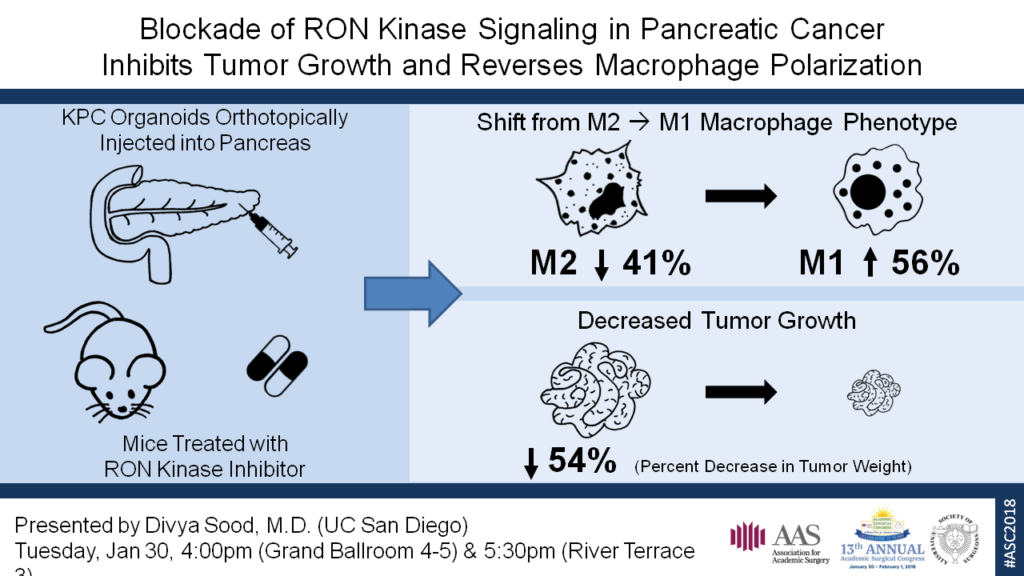
21.10 RON Kinase Inhibition Modulates the Pancreatic Cancer Microenvironment to Promote an Antitumor State

D. Sood1, A. Cazes1, D. Jaquish1, E. Mose1, R. French1, A. M. Lowy1  1University Of California – San Diego,Department Of Surgery, Division Of Surgical Oncology, Moores Cancer Center,La Jolla, CA, USA

Introduction:
RON, a receptor tyrosine kinase and c-Met proto-oncogene homolog, is over-expressed in pancreatic cancer relative to normal tissue. RON’s function in normal biology is to curtail the inflammatory response, such as in the terminal stages of wound healing. It does this in large part by regulating the transition of macrophages from a pro-inflammatory, tumor-suppressive (M1) state to an immunosuppressive, tumor-promoting (M2) state. In cancer, alternatively polarized macrophages secrete pro-tumorigenic cyto- and chemokines that suppress effector T cell activation and may prevent their access to the tumor microenvironment. In this study, we aim to determine if RON kinase inhibition can effectively modulate the pancreatic cancer microenvironment to promote antitumor activation states in macrophages.

Methods:
KPC (Pdx1-Cre; K-Ras+/LSLG12D; p53R172H/+) organoids were orthotopically injected into the pancreas of F1 hybrid B6-129S mice. LY2801653 is an orally bioavailable small molecule RON inhibitor in Phase II trials for advanced solid cancers. IPI549, a PI3Kγ  inhibitor in Phase I trials, shown to have potent preclinical antitumor activity and promote classical activation of macrophages to the M1 state, was selected as a positive control. Treatment was started one week after injection for the following groups: A) vehicle control (10% acacia gum/5% NMP), B) LY2801653 12 mg/kg, C) LY2801653 24 mg/kg, D) IPI549 15 mg/kg, and E) LY2801653 24 mg/kg + IPI549 15 mg/kg. Mice were treated daily via orogavage for 2 weeks. The tumors were harvested and dissociated into a single cell suspension. The cells were labeled with fluorescent-conjugated antibodies to markers of leukocytes and macrophage polarization for flow cytometry. One way ANOVA with a Bonferroni correction for multiple comparisons was used to analyze the results. 

Results:
Macrophages were defined as cells that labeled positive for CD45, CD11b, MHC II, and F4/80. Within that population, cells that labeled positive for CD80 and CD86, but negative for MRC1 were considered M1 macrophages, whereas those that were MRC1 positive, but negative for CD80 and CD86 were considered M2 macrophages. Flow cytometry demonstrated a significant decrease in the M2 macrophage population in each of the four treatment groups when compared to the vehicle control (p < 0.001). There was also a corresponding increase in the M1 macrophage population; this was significant (p < 0.001) for all except group E. Finally, mice in all four treatment groups exhibited significantly suppressed tumor growth by weight, relative to the vehicle control-treated mice (p < 0.005).

Conclusion:
In this study, we demonstrate that inhibition of RON kinase signaling alters the immunosuppressive microenvironment of pancreatic cancer and can be effectively targeted to shift that environment toward an antitumor state. Further studies are underway to better understand RON’s potential as a therapeutic target in pancreatic cancer.

19.03 RON Kinase Inhibition Modulates the Pancreatic Cancer Microenvironment to Promote an Antitumor State

D. Sood1, A. Cazes1, D. Jaquish1, E. Mose1, R. French1, A. M. Lowy1  1University Of California – San Diego,Department Of Surgery, Division Of Surgical Oncology, Moores Cancer Center, La Jolla, CA, USA

Introduction:
RON, a receptor tyrosine kinase and c-Met proto-oncogene homolog, is over-expressed in pancreatic cancer relative to normal tissue. RON’s function in normal biology is to curtail the inflammatory response, such as in the terminal stages of wound healing. It does this in large part by regulating the transition of macrophages from a pro-inflammatory, tumor-suppressive (M1) state to an immunosuppressive, tumor-promoting (M2) state. In cancer, alternatively polarized macrophages secrete pro-tumorigenic cyto- and chemokines that suppress effector T cell activation and may prevent their access to the tumor microenvironment. In this study, we aim to determine if RON kinase inhibition can effectively modulate the pancreatic cancer microenvironment to promote antitumor activation states in macrophages.

Methods:
KPC (Pdx1-Cre; K-Ras+/LSLG12D; p53R172H/+) organoids were orthotopically injected into the pancreas of F1 hybrid B6-129S mice. LY2801653 is an orally bioavailable small molecule RON inhibitor in Phase II trials for advanced solid cancers. IPI549, a PI3Kγ  inhibitor in Phase I trials, shown to have potent preclinical antitumor activity and promote classical activation of macrophages to the M1 state, was selected as a positive control. Treatment was started one week after injection for the following groups: A) vehicle control (10% acacia gum/5% NMP), B) LY2801653 12 mg/kg, C) LY2801653 24 mg/kg, D) IPI549 15 mg/kg, and E) LY2801653 24 mg/kg + IPI549 15 mg/kg. Mice were treated daily via orogavage for 2 weeks. The tumors were harvested and dissociated into a single cell suspension. The cells were labeled with fluorescent-conjugated antibodies to markers of leukocytes and macrophage polarization for flow cytometry. One way ANOVA with a Bonferroni correction for multiple comparisons was used to analyze the results.

Results:
Macrophages were defined as cells that labeled positive for CD45, CD11b, MHC II, and F4/80. Within that population, cells that labeled positive for CD80 and CD86, but negative for MRC1 were considered M1 macrophages, whereas those that were MRC1 positive, but negative for CD80 and CD86 were considered M2 macrophages. Flow cytometry demonstrated a significant decrease in the M2 macrophage population in each of the four treatment groups when compared to the vehicle control (p < 0.001). There was also a corresponding increase in the M1 macrophage population; this was significant (p < 0.001) for all except group E. Finally, mice in all four treatment groups exhibited significantly suppressed tumor growth by weight, relative to the vehicle control-treated mice (p < 0.005).

Conclusion:
In this study, we demonstrate that inhibition of RON kinase signaling alters the immunosuppressive microenvironment of pancreatic cancer and can be effectively targeted to shift that environment toward an antitumor state. Further studies are underway to better understand RON’s potential as a therapeutic target in pancreatic cancer.

17.20 Multidisciplinary Approach For Management Of Necrotizing Pancreatitis: A Case Series

P. SENTHIL-KUMAR1, W. Alswealmeen1, Q. Yan1, P. O’Moore1, T. Braun1, D. Ringold1, O. Kirton1, T. Vu1  1Abington Memorial Hospital,Surgery,Abington, PA, USA

Introduction:

 Necrotizing pancreatitis is often a devastating sequelae of acute pancreatitis. Historically several approaches have been described with variable outcome. Open necrosectomy is associated with higher morbidity (95%) and mortality (25%). Endoscopic necrosectomy often is tolerated well but associated with stent migration and multiple procedures. Video-assisted retroperitoneal debridement is tolerated well but associated with severe bleeding if adjacent blood vessels are injured during the procedure leading to severe complications

Methods:
In our series. We perform a step up approach by Involvement of a multidisciplinary group consisting of general surgeons, gastroenterologists, Infectious disease physicians, critical care internalist, interventional radiologist and nutritional services to formulate a management plan. The necrotized pancreas is initially drained with an IR guided drain, fluid cultures sent for microbiology and treatment with appropriate antibiotics if deemed necessary. The drain is gradually upsized to a 24 Fr sized drain to form a well-defined tract for surgical debridement; A pre-operative CT scan of the abdomen with IV contrast to access the location and proximity of the vasculature around the necrotized pancreas. A collaboration with the interventional radiologist to discuss possible IR embolization of splenic artery prior to surgical debridement. The patient would then undergo video assisted retroperitoneal pancreatic necrosectomy and a sump drain left in-situ at the pancreatic fossa. Post-operative management in the surgical ICU would be lead by the critical care internalist.

Results:
Three patients were managed by this multidisciplinary approach with excellent outcomes. One patient underwent preoperative IR embolization followed by surgical debridement; second patient underwent embolization immediately following debridement; one patient did not require any embolization but had IR on standby if needed to intervene. Post-operatively all three patients recovered well. They all were tolerating good oral intake and were discharged to rehabilitation facilities.

Conclusion:

As this series show an early plan and collaboration with various subspecialities will produce an optimal outcome. It will lead us to a pragmatic and successful approach to this potentially catastrophic condition.