4.08 Notch Activator Jagged1 Results in Increased Closure Rates in an Ex Vivo Murine Skin Wound Model 1

Z. Aburjania1, T. W. King1  1University Of Alabama at Birmingham,Plastic Surgery,Birmingham, Alabama, USA

Introduction:
Decreased rates of wound healing affect millions of patients annually. We are interested in discovering novel strategies to enhance the wound healing process in diabetic patients.  We have previously shown that inhibiting Notch inhibits wound healing. Based upon our previous work, we propose that upregulation of Notch would increase rates of wound healing.  JAG1 is a known activator of Notch. Therefore, we hypothesized that applying topical JAG1 to ex vivo excisional wounds on the backs of mice would result in increased Notch activity, and thus an increased wound healing rate as compared to untreated wounds.

Methods:
Skin biopsies from 12-week old, healthy mice, (1-cm2 full-thickness) were cultured ex vivo. A 4-mm wound was created in the center of the skin biopsy. A topical application onto the open wound bed of JAG1 (10 nM) or vehicle (PBS) was applied daily for 14 days. Digital photographs were taken daily and the skin was processed for histological and protein analysis on days 3, 7, 10, and 14. The wounds were analyzed using ImageJ software. Wound area was calculated as a percent area of the original wound size. Statistical significance was defined as p<0.05 using the students’ t-test.

Results:
Partial to complete re-epithelialization was seen in the wounded tissues over the experimental period in both the control & JAG1 treated groups. The mouse skin treated with topical JAG1 had an increased rate of wound closure when compared to wounds treated with PBS.

Conclusion:
JAG1 increases the rate of re-epithelialization of cutaneous wounds in an ex vivo murine wound-healing model, indicating that Notch signaling plays a crucial role in wound healing in mice. Based upon our findings, further study of Notch in wound healing should be conducted which may then lead to better therapeutics for the wound healing process in patients. 

1

4.07 A Novel Human Xenograft Model to study Strategies in Articular Cartilage Regeneration.

M. P. Murphy1, M. Lopez1, R. C. Ransom1, O. Marecic1, R. E. Brewer1, L. S. Koepke1, S. Mascharak1, C. F. Chan1, M. T. Longaker1  1Stanford University,Surgery,Palo Alto, CA, USA

Introduction:  Currently there are no effective strategies for regenerating articular cartilage in diseases such as osteoarthritis (OA). Our group has utilized the mouse model to study the effects of surgical, chemical and cellular manipulation in articular cartilage regeneration. We aim to understand the effects of surgical, chemical and cellular manipulation in articular cartilage regeneration in a novel xenograft human model.

Methods: We transplanted the phalanges of 18-week old fetal specimens subcutaneously in the dorsum of immuno-compromised NSG P3 mice. After we confirmed viabiliy of the human xenograft using MRI, microCT and on gross inspection we performed microfracture (MF) surgery on the articular joints. Histological composition was assessed using Movat’s Pentachrome stain and Safranin O/Fast green stain. Immunohistochemistry (IHC) was performed assessing levels of Col 1, 2, 10 and MMP13. Proliferation was assessed with EdU labelling in histology and intracellular FACS. 

Results: We successfully developed a model of investigating the effects of surgical manipulation on human articular cartilage regeneration. (Figure 1) Following 6 weeks of implantation the xenograft had grown in size. The microCT images show calcification and MRI show viable articular cartilage. We have found that similar to our mouse model, fibrocartilage forms after MF. On histology and IHC we have determined the difference between normal articular cartilage and MF tissue. Cellular proliferation increases following injury.

Conclusion: We believe that our surgical technique and topical factors including BMP2 and Avastin will provide regenerative surgeons with a new approach to treating OA. We will further augment these factors with induced SSC from human Adipose-derived Stromal Cells. Our findings provide us with a "preclinical" human model that can be utilised to effectively investigate strategies for articular cartilage regeneration. 


4.06 Cellular and Mechanical Mechanisms Underlying Regeneration in Mandibular Distraction Osteogenesis

R. C. Ransom1,2, A. C. Carter3, A. Salhotra1,2, T. Leavitt1, O. C. Marecic1,2, M. Lopez1,2, M. Murphy1,2, C. K. Chan1,2, D. C. Wan1, H. Y. Chang3, M. T. Longaker1,2  1Hagey Laboratory For Pediatric Regenerative Medicine,Department Of Surgery, Division Of Plastic And Reconstructive Surgery, Stanford University School Of Medicine,Stanford, CA, USA 2Institute For Stem Cell Biology And Regenerative Medicine,Stanford University,Stanford, CA, USA 3Center For Personal Dynamic Regulomes,Stanford University,Stanford, CA, USA

Introduction:  Accumulated evidence indicates that mechanical cues, which include physical forces, alterations in extracellular matrix mechanics and changes in cell shape, are transmitted to the nucleus directly or indirectly to orchestrate transcriptional activities that are crucial for tissue regeneration. Although mechanotransduction is thought to occur via integration of multiple signaling pathways, the precise mechanism leading to downstream cellular responses is not well understood. We have developed a mouse model of mandibular distraction osteogenesis (DO) which allows for tracing of cell fate and genetic dissection of mechanotransduction during bone formation.

Methods:  We examined cell-type-specific responses to mechanical force within distinct subpopulations of the mouse skeletal stem cell (mSSC) hierarchy. After determining that bone, cartilage, and stromal tissue are clonally derived in mice from lineage-restricted stem and progenitor cells in vivo, we employed this strategy to purify specific skeletogenic populations during mandibular distraction osteogenesis by prospective isolation using FACS. We employed the assay for transposase-accessible chromatin with high-throughput sequencing (ATAC-seq) to profile open chromatin landscapes in these cell populations to understand the epigenetic changes in response to distraction. To investigate the role of mechanotransduction via focal adhesion kinase (FAK) in distraction osteogenesis, we inhibited FAK using the small molecule PF57223, a specific and potent inhibitor of FAK signaling. Three-dimensional reconstruction of μCT images of gradually distracted specimens revealed disrupted bone formation under conditions of FAK inhibition. ATAC-seq was employed for determination of FAK-responsive regions of the epigenome within each subpopulation of the skeletal stem cell hierarchy.

Results: We show that mechanical force augments the numbers and function of multiple cell populations across the skeletal hierarchy, including mouse skeletal stem and progenitor cells and their differentiated subsets. Mechanistically, distraction induces robust cell-matrix interactions that are coupled to cell-specific transcriptional responses via epigenomic pathways and pharmacological inactivation of this pathway disrupts bone formation. 

Conclusion: Here we employ a rigorous mandibular DO model in mice that is genetically dissectable, allowing for detailed examination of the fundamental principles regulating de novo bone formation. The identification of the cellular source of regeneration, the timeline for progenitor cell response, and determination of how these cells transduce physical stimuli to enact a regenerative response may provide new and effective strategies for reconstruction of the craniofacial skeleton.


4.02 Automated Quantification of Soft Endpoints in Wound Healing Analysis

S. Mascharak1, A. L. Moore1,2, B. Duoto1, D. S. Foster1, R. E. Jones1,3, G. Wernig4, M. T. Longaker1  1Stanford University,Dept. Surgery,Palo Alto, CA, USA 2Brigham And Women’s Hospital,Dept. Surgery,Boston, MA, USA 3University Of Texas Southwestern Medical Center,Dept. Surgery,Dallas, TX, USA 4Stanford University,Dept. Pathology,Palo Alto, CA, USA

Introduction:  Studies on dermal wound healing and scarring commonly rely on qualitative assessments of morphological and histological characteristics. These can be time consuming and prone to individual observer bias. Furthermore, it is not always feasible to visually ascertain metrics of scar organization on the micron scale. We have developed a package of image processing methods to automate quantification of morphological and histological dermal scar features. Using this tool, we elucidate the effects of an anti-scarring treatment on collagen content, fiber organization, and hair follicle neogenesis after dermal wounding. 

Methods:  Two stented dermal wounds were made on the dorsa of C57BL/6 mice and injected with various concentrations (PBS, 0.1, 2, 20 mg/mL) of a potential anti-scarring drug (Doxycycline). Histology (Masson’s Trichrome, Picrosirius Red) and hair follicle neogenesis were assessed at 14 and 28 days, respectively, and analyzed with Matlab 2017a. Histological features were measured using a combination of color deconvolution, adaptive filtering, and skeletonization of individual collagen fibers. Hair follicles were segmented using top-hat and shape filters.

Results: Automated quantification of color-deconvoluted histology images showed a significant decrease in collagen deposition for wounds treated with anti-scarring drug (*p = 0.036, 2 mg/mL vs PBS, Fig. 1A-C). Color deconvolution also segmented mature (red) and immature (green) collagen fibers in birefringent Picrosirius Red-stained samples, revealing a concomitant increase in immature collagen deposition (*p = 0.018, 2 mg/mL vs PBS, Fig. 1D Picro Green). Next, images of collagen fibers were skeletonized to trace individual tracks, revealing significant decreases in mature fiber length (*p = 0.042, 2 mg/mL vs PBS), number (*p = 0.036), and branching (*p = 0.038) after treatment (Fig. 1E-F). Strikingly, alignment of mature collagen fibers decreased after treatment (*p = 0.048, 2 mg/mL vs PBS), indicating basket-weave organization reminiscent of unwounded skin. Shape filtration segmented hair follicles, dramatically speeding up the counting process (Fig. 1G). A significant decrease in follicles was observed after treatment (*p = 0.025, 2 mg/mL vs PBS), though no change in morphology was observed (Fig. 1H). 

Conclusion: Automated image processing facilitates histological and morphological study of scars. Additionally, analysis of collagen fibers reveals differences in scar organization after treatment with Doxycycline, that would not be apparent to the naked eye. Based on these findings, we will expand the use of image processing tools to models of hypertrophic scarring and fetal wound healing, towards the goal of new quantitative benchmarks for scar analysis. 

32.03 Operating Room Teams: Does Familiarity Make a Difference?

S. Fitzgibbons1,2, S. Kaplan3, X. Lei3, S. Safford5, S. Parker4  1MedStar Georgetown University Hospital,Surgery,Washington, DC, USA 2Georgetown Univeristy Medical Center,Washington, DC, USA 3George Mason University,Psychology,Fairfax, VA, USA 4Virginia Tech Carilion School Of Medicine And Research Institute,Human Factors,Roanoke, VA, USA 5Carillion Clinic,Pediatric Surgery,Lynchburg, VA, USA

Introduction: The composition of any given operating room team may vary procedure-to-procedure. Studies in healthcare have documented that greater familiarity between certain team member pairs or dyads (ex. surgeon and scrub) corresponds to improved effectiveness, with outcomes ranging from shorter cross clamp times during cardiopulmonary bypass to shorter operative times during mammoplasty. We sought to further our understanding of this effect beyond simple dyads by developing an OR team familiarity score reflective of the larger and more complex group, and determining the impact of the larger group familiarity on surgical processes and clinical outcomes.  

Methods: Data from a diverse, primarily urban healthcare system including 6 acute-care hospitals was extracted from a system-wide electronic medical record.  All knee arthroplasty cases performed between 2013 and 2016 were included in the data set.  Information regarding individual OR team participants and their roles in the surgery were collected, in addition to patient demographics (ASA class, age, gender, race, ethnicity), case information (surgical procedure, date and time of the operation)  and outcome variables (length of procedure, length of hospital stay).  Team familiarity was calculated using a previously published formula from Huckman, Staats, and Upton (2009).  A multilevel regression (i.e., random coefficient modeling) framework was applied to examine the impact of a team’s familiarity score on case length and post-op length of stay. In addition, specific familiarity scores for each possible dyad on the team was calculated and analyzed. Dyads were defined as pairs of core team members: surgeon, scrub, circulator, anesthesiologist.

Results:A total of 4546 knee arthroplasty cases were included in the data set with an average case length of 92.68 minutes and an average length of hospital stay of 3.22 days.  When controlling for patient age, gender, hospital, and ASA class, a team’s familiarity score during a case was significantly associated with a shorter case length, with 10 previous team member interactions predicting a decreased case length of approximately 1.1 minutes (p=.012).  Similarly, an increased team familiarity score predicted a decreased length of stay, with 10 previous team member interactions predicting a decrease in hospital length of stay of 0.1 days.  With respect to the impact of specific dyad familiarity, all dyads involving the circulator predicted a shorter length of hospital stay, while all three dyads between the surgeon, scrub and circulator predicted a shorter case length.

Conclusion:Overall team member familiarity in the operating room is associated with a small but significant decrease in the case length and hospital length of stay for patients undergoing total knee arthroplasty.


28.01 The Impact of ERAS protocol on Urinary Tract Infections after Free Flap Breast Reconstruction.

B. Sharif-Askary1, R. Zhao1, S. Hollenbeck1  1Duke University Medical Center,Division Of Plastic And Reconstructive Surgery,Durham, NC, USA

Introduction:  Hospitals are evaluated for quality based on a number of metrics including the occurrence of complications. Recently, our hospital instituted the Enhanced Recovery After Surgery (ERAS) protocol for patients undergoing free flap breast reconstruction. Urinary tract infections are among the most common healthcare-associated infections, with the majority seen after prolonged urinary catheterization. The ERAS protocol calls for early removal of urinary catheters. In this study, we compare the rate of UTI in patients who have undergone traditional recovery after surgery (pre-ERAS) to those who were enrolled in the ERAS protocol. We hypothesized that early catheter removal would decrease the rate of UTI in patients undergoing breast reconstruction with free flaps.

Methods:  We retrospectively reviewed the charts of 238 patients who underwent free flap breast reconstruction. We initiated the ERAS protocol in May of 2015. This study includes patients seen between March 2012 and June 2017 to capture both pre- and post-ERAS cohorts. UTI was defined using the American College of Surgeons NSQIP definition. Statistical analyses were conducted using SPSS software (Version 24.0, IBM Corp). We compared the incidence of UTI before and after ERAS initiation using a logistic regression while controlling for age, BMI, rate of diabetes and length of surgery.

Results: There were 160 patients evaluated prior to ERAS implementation and 78 patients evaluated in the post-ERAS group. The overall incidence of UTI for all patients who underwent free flap reconstruction was 4.6%. Next, we compared patients from the pre-ERAS group to the post-ERAS group. There were no significant differences with regards to mean age, BMI, or length of surgery. However, the rate of diabetes was higher in the pre-ERAS group compared to the post-ERAS group (11% vs. 4%, p=0.04, t-test). Post-ERAS patients had a significantly higher rate of UTI than pre-ERAS patients when controlling for age, BMI, rate of diabetes and length of surgery (1.9% vs. 10.3% p=0.008, OR=6.72). Of post-ERAS patients who were found to have post-op UTI, 25% were found to have bacteria on a pre-operative urinalysis.

Conclusion: In contrast to our hypothesis, we found that the rate of UTI was significantly higher in the post-ERAS patients. Further analysis is needed to determine the cause of this finding but may include the need for re-catheterization after early catheter removal. Based on these findings, we suggest individualized decision-making within the ERAS protocol in regards to timing of urinary catheter removal.


21.09 Scholarly Activity in Academic Plastic Surgery: The Gender Difference

S. E. Sasor1, J. A. Cook1, S. P. Duquette1, T. A. Evans1, S. S. Tholpady1, M. W. Chu1, L. G. Koniaris1  1Indiana University,Plastic Surgery,Indianapolis, IN, USA

Purpose:

The number of women in medicine has grown rapidly in recent years. Women constitute over 50% of medical school graduates and hold 38% of faculty positions at U.S. medical schools. Despite this, gender disparities remain prevalent in most surgical subspecialties, including plastic surgery. The purpose of this study is to analyze female authorship trends to identify factors that affect scholarly output and professional advancement in academic plastic surgery.


Methods:

A cross-sectional study of all academic plastic surgeons was performed. Data was collected on gender, degrees, titles, and affiliation residency programs from department websites and online resources. National Institute of Health (NIH) funding was determined using the Research Portfolio Online Reporting Tools database. Number of published manuscripts, citations, and h-index were obtained from Scopus (Elsevier Inc., New York, NY). Statistical analysis was performed in SPSS.


Results:

A total of 814 plastic surgeons were identified from 91 training programs in the US. Mean years in practice was 15.3 and the majority were male (83.2%). Average number of published manuscripts, citations, and h-index were 45.5, 974.9, and 11.6, respectively.


Compared to male surgeons, female surgeons had significantly fewer years in practice (9.9 vs. 16.4, p<0.001), held lower academic ranks (57.3% vs. 33.9% assistant professor, p<0.001 compared to 6.6% vs. 29.0% full professor, p<0.001), and published less (20.0 vs. 50.2 manuscripts, p<0.001, and 7.3 vs 12.6 h-index, p<0.001). Females with titles of assistant or associate professor had significantly fewer years in practice than males with the same title (6.1 vs. 9.0 years, p<0.001 assistant and 13.5 vs. 15.8 years p=0.01 associate). The trend continued for full professors but was not significant. Males published more manuscripts, had more citations and higher h-indexes than female surgeons at each academic rank but the findings were not statistically significant beyond the level of assistant professor.


Seven of 136 female surgeons (5.1%) and 46 of 678 male surgeons (6.9%) received an NIH grant during their career (p=0.57). Mean number of awards and total amount of funding was 10.3/$2,996,734 for women and 8.0/$1,853,345 for men (p=0.469 and p=0.57, respectively). NIH-funded surgeons of both sexes published more than non-funded surgeons (119.5 vs. 40.3 publications, p<0.001 and 24.7 vs. 10.7 h-index, p<0.001). There was no gender difference in scholarly output amongst NIH-funded surgeons.


Conclusion:

Research productivity is a metric for promotion in academic medicine. This study identifies significant gender disparities in scholarly productivity amongst plastic surgeons in academia. Differences are most apparent at junior ranks, suggesting that women who remain in academic medicine may later overcome publishing barriers faced earlier in their careers. 


21.05 ATAC-seq and Reciprocal Transplantation Prove Scar-Forming Behavior is Cell Intrinsic

A. L. Moore1,3, C. Marshall1, U. Litzenburger4, R. C. Ransom4, L. Barnes4, B. Duoto4, D. Foster1, R. E. Jones2,4, S. Mascharak4, M. Hu4, H. Y. Chang4, H. P. Lorenz1, M. T. Longaker1  1Stanford University,Department Of Surgery,Palo Alto, CA, USA 2University Of Texas Southwestern Medical Center,Department Of Surgery,Dallas, TX, USA 3Brigham And Women’s Hospital,Department Of Surgery,Boston, MA, USA 4Stanford University,Palo Alto, CA, USA

Introduction:

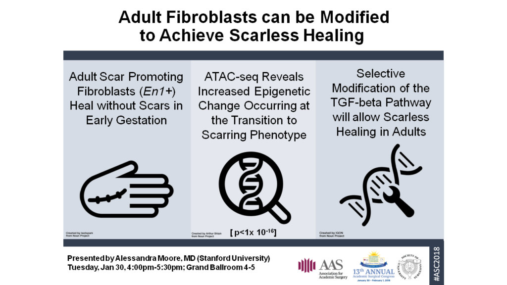
Skin scarring poses a major financial and physiologic burden on patients and the US healthcare system. In 2015, our group discovered a subset of fibroblasts in the dorsal dermis identified by embryonic expression of the homeobox transcription factor Engrailed-1 that deposit the bulk of scar collagen in adult mice. These cells are also present in embryonic development around the time of phenotypic transition from regenerative scarless healing to fibrotic scarring. Given Engrailed-1 positive fibroblasts (EPFs) share a common precursor cell, we hypothesized that the EPFs accumulate epigenetic changes over time that result in their phenotypic change and result in a permanent cellular phenotype.

Methods:

Fibroblasts were isolated from En1Cre; R26mTmG mice using fluorescence-activated cell sorting (FACS) at gestational ages embryonic day (e)10, e16, e18, postnatal day (p)1, p30, and p30 wounded skin. Epigenetic regulation was analyzed using the assay for transposase accessible chromatin using sequencing (ATAC-seq). Fibroblasts were then transplanted from e16 embryos to p1 mouse pups, and vice versa, to observe scar forming behavior in vivo.

Results:

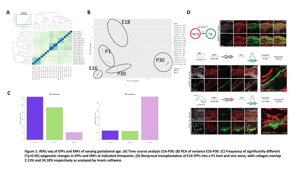
E10 fibroblasts are of a single lineage and were excluded from analysis. Time course analysis of e16-p30 EPFs and Engrailed-1 negative fibroblasts (ENFs) shows appropriate correlation between samples (Figure 1A). Grouping analysis of variance shows p30 EPFs and ENFs being the most dissimilar, and EPFs from p30 are most like e16 EPFs (Figure 1B). Most epigenetic changes in the EPF lineage occur in embryonic development between e16 and e18 with fewer epigenetic changes occurring postnatally between p1 and p30 (significant peaks = 124 vs. 20, Figure 1C). In contrast, the ENF lineage accumulates more epigenetic changes between p1 and p30 (significant peaks = 62 vs. >250, Figure 1C). Lastly, reciprocal transplantation of e16 fibroblasts into a p1 host and vice versa reveal a significant difference in collagen overlap (2.13% versus 24.18%) and morphologic changes suggestive of quiescence versus reactivity (Figure 1D).

Conclusions:

ATAC-seq analysis of EPF lineage cells reveals most epigenetic changes occurring at the time of phenotypic change from scarless to scarring (between e16 to e18). In future experiments, we will identify transcription factors to perturb using CRISPR-Cas9. Manipulated cells will be transplanted into fetal and postnatal hosts. These experiments will reveal if master regulators are involved in the scar forming phenotype of EPFs.

20.05 Rapamycin Inhibits Primary and Recurrent Heterotopic Ossification in Genetically Susceptible FOP Mice

C. Hwang1, S. Ucer1, C. Pagani1, N. Patel1, A. Vaishampayan1, M. Sorkin1, M. T. Chung1, J. Li1, C. Breuler1, C. Priest1, A. N. Economides3, S. Agarwal1, Y. Mishina2, B. Levi1  1University Of Michigan,Section Of Plastic Surgery,Ann Arbor, MI, USA 2University Of Michigan,Department Of Biologic And Material Sciences, School Of Dentistry,Ann Arbor, MI, USA 3Regeneron Pharmaceuticals And Genetics Center,Tarrytown, NY, USA

Introduction: Heterotopic ossification (HO) is a debilitating formation of ectopic bone restricting joint mobility and causing chronic pain. Fibrodysplasia Ossificans Progressiva (FOP) is a congenital variant of HO caused by a genetic mutation in a bone morphogenetic receptor that causes severe, progressive lesions resulting in immobility and fatal mechanical respiratory failure at a premature age. With increased proclivity for osteogenesis at baseline, surgical extirpation is contraindicated in FOP patients due to universal recurrence. This disease presents a unique clinical challenge with no current treatments available. Previous research has shown mTOR inhibition by rapamycin to be a viable therapy for reducing trauma-induced HO. Mice of an FOP mouse model with a hyperactivating mutation in the ACVR1 receptor (ACVR1R206H/+) were examined to determine the effects of rapamycin on both primary and post-surgical HO.

Methods: Mice were designated for 2 distinct study arms. In the primary HO cohort, ACVR1R206H/+ P21 mice received bilateral hindlimb cardiotoxin (CTX) with Ad.cre injection and stratification to daily i.p. rapamycin (n=10 hindlimbs) v. PBS control (n=12). 3 weeks later, mice were scanned with in vivo µCT. Mice within the separate recurrence cohort received initial Ad.cre/CTX followed by baseline CT scans, Ad.cre reinjection bilaterally, and surgical removal of HO at the 3 week timepoint. After surgery, mice were randomized to daily rapamycin (5 mg/kg, n=12) or PBS (n=8) for 3 weeks with endpoint µCT. Contours were drawn manually around HO to compute total volumes at 800HU. Primary and recurrence cohorts were analyzed by Student’s t test and log-transform/ANOVA/Hochberg post-hoc respectively.

Results: Mice treated with rapamycin for 21 days showed 261-fold less ectopic bone when compared to PBS vehicle injection (p=0.002). In post-surgical mice, PBS injection showed statistically similar HO volumes to baseline volumes of pre-excision mice (p=0.054). However, rapamycin treatment reduced the recurrence of HO volume 11-fold (p=.044, Figure). Geometric means from de-transformed data for pre-excision v. PBS, pre-excision v. rapamycin, and PBS v rapamycin were 0.061, 1.398, and 22.961 respectively.

Conclusions: These studies demonstrate that rapamycin prevents primary development of HO and is also effective in preventing recurrence following surgical excision in a FOP mouse model. This study further corroborates rapamycin as a promising candidate for primary and post-surgical HO prophylaxis in children with FOP. Existing literature implicates similar molecular mechanisms among various etiologies of HO, suggesting a putative role for rapamycin even beyond FOP in post-traumatic and post-surgical HO patients.

20.02 The Impact of ERAS protocol on Urinary Tract Infections after Free Flap Breast Reconstruction.

B. Sharif-Askary1, R. Zhao1, S. Hollenbeck1  1Duke University Medical Center,Division Of Plastic And Reconstructive Surgery,Durham, NC, USA

Introduction:  Hospitals are evaluated for quality based on a number of metrics including the occurrence of complications. Recently, our hospital instituted the Enhanced Recovery After Surgery (ERAS) protocol for patients undergoing free flap breast reconstruction. Urinary tract infections are among the most common healthcare-associated infections, with the majority seen after prolonged urinary catheterization. The ERAS protocol calls for early removal of urinary catheters. In this study, we compare the rate of UTI in patients who have undergone traditional recovery after surgery (pre-ERAS) to those who were enrolled in the ERAS protocol. We hypothesized that early catheter removal would decrease the rate of UTI in patients undergoing breast reconstruction with free flaps.

Methods:  We retrospectively reviewed the charts of 238 patients who underwent free flap breast reconstruction. We initiated the ERAS protocol in May of 2015. This study includes patients seen between March 2012 and June 2017 to capture both pre- and post-ERAS cohorts. UTI was defined using the American College of Surgeons NSQIP definition. Statistical analyses were conducted using SPSS software (Version 24.0, IBM Corp). We compared the incidence of UTI before and after ERAS initiation using a logistic regression while controlling for age, BMI, rate of diabetes and length of surgery.

Results: There were 160 patients evaluated prior to ERAS implementation and 78 patients evaluated in the post-ERAS group. The overall incidence of UTI for all patients who underwent free flap reconstruction was 4.6%. Next, we compared patients from the pre-ERAS group to the post-ERAS group. There were no significant differences with regards to mean age, BMI, or length of surgery. However, the rate of diabetes was higher in the pre-ERAS group compared to the post-ERAS group (11% vs. 4%, p=0.04, t-test). Post-ERAS patients had a significantly higher rate of UTI than pre-ERAS patients when controlling for age, BMI, rate of diabetes and length of surgery (1.9% vs. 10.3% p=0.008, OR=6.72). Of post-ERAS patients who were found to have post-op UTI, 25% were found to have bacteria on a pre-operative urinalysis.

Conclusion: In contrast to our hypothesis, we found that the rate of UTI was significantly higher in the post-ERAS patients. Further analysis is needed to determine the cause of this finding but may include the need for re-catheterization after early catheter removal. Based on these findings, we suggest individualized decision-making within the ERAS protocol in regards to timing of urinary catheter removal.


19.05 ATAC-seq and Reciprocal Transplantation Prove Scar-Forming Behavior is Cell Intrinsic

A. L. Moore1,3, C. Marshall1, U. Litzenburger4, R. C. Ransom4, L. Barnes4, B. Duoto4, D. Foster1, R. E. Jones2,4, S. Mascharak4, M. Hu4, H. Y. Chang4, H. P. Lorenz1, M. T. Longaker1  1Stanford University,Department Of Surgery,Palo Alto, CA, USA 2University Of Texas Southwestern Medical Center,Department Of Surgery,Dallas, TX, USA 3Brigham And Women’s Hospital,Department Of Surgery,Boston, MA, USA 4Stanford University,Palo Alto, CA, USA

Introduction:

Skin scarring poses a major financial and physiologic burden on patients and the US healthcare system. In 2015, our group discovered a subset of fibroblasts in the dorsal dermis identified by embryonic expression of the homeobox transcription factor Engrailed-1 that deposit the bulk of scar collagen in adult mice. These cells are also present in embryonic development around the time of phenotypic transition from regenerative scarless healing to fibrotic scarring. Given Engrailed-1 positive fibroblasts (EPFs) share a common precursor cell, we hypothesized that the EPFs accumulate epigenetic changes over time that result in their phenotypic change and result in a permanent cellular phenotype.

Methods:

Fibroblasts were isolated from En1Cre; R26mTmG mice using fluorescence-activated cell sorting (FACS) at gestational ages embryonic day (e)10, e16, e18, postnatal day (p)1, p30, and p30 wounded skin. Epigenetic regulation was analyzed using the assay for transposase accessible chromatin using sequencing (ATAC-seq). Fibroblasts were then transplanted from e16 embryos to p1 mouse pups, and vice versa, to observe scar forming behavior in vivo.

Results:

E10 fibroblasts are of a single lineage and were excluded from analysis. Time course analysis of e16-p30 EPFs and Engrailed-1 negative fibroblasts (ENFs) shows appropriate correlation between samples (Figure 1A). Grouping analysis of variance shows p30 EPFs and ENFs being the most dissimilar, and EPFs from p30 are most like e16 EPFs (Figure 1B). Most epigenetic changes in the EPF lineage occur in embryonic development between e16 and e18 with fewer epigenetic changes occurring postnatally between p1 and p30 (significant peaks = 124 vs. 20, Figure 1C). In contrast, the ENF lineage accumulates more epigenetic changes between p1 and p30 (significant peaks = 62 vs. >250, Figure 1C). Lastly, reciprocal transplantation of e16 fibroblasts into a p1 host and vice versa reveal a significant difference in collagen overlap (2.13% versus 24.18%) and morphologic changes suggestive of quiescence versus reactivity (Figure 1D).

Conclusions:

ATAC-seq analysis of EPF lineage cells reveals most epigenetic changes occurring at the time of phenotypic change from scarless to scarring (between e16 to e18). In future experiments, we will identify transcription factors to perturb using CRISPR-Cas9. Manipulated cells will be transplanted into fetal and postnatal hosts. These experiments will reveal if master regulators are involved in the scar forming phenotype of EPFs.