71.10 Chest Wall Ewing Sarcoma: The Results of a Population Based Analysis

A. J. Jacobs2, J. Fishbein3, C. Fein Levy4, R. D. Glick1 1North Shore University And Long Island Jewish Medical Center,CCMC, Division Of Pediatric Surgery,Manhasset, NY, USA 2Hofstra North Shore-LIJ School Of Medicine,Hempstead, NY, USA 3Feinstein Institute For Medical Research,Biostatistics Unit,Manhasset, NY, USA 4North Shore University And Long Island Jewish Medical Center,CCMC, Department Of Pediatric Hematology/Oncology,Manhasset, NY, USA

Introduction:
The globally low incidence of pediatric chest wall Ewing sarcoma (CWES) has limited prior studies of this disease to mostly small, single institution reviews. Our objective was to assess incidence, demographics, treatment patterns, and long-term survival of this disease through a population based analysis.

Methods:
The Surveillance, Epidemiology, and End Results (SEER) database was used to identify patients aged 0-21 diagnosed with CWES from 1973-2011. Patients were grouped by decade to assess changes in treatment patterns and outcomes. The effects of clinical, demographic, and treatment variables on overall survival were assessed by the computation of Kaplan-Meier curves and the log-rank test, with Cox proportional hazard regression used for multivariate analysis.

Results:
A total of 193 pediatric patients with histologically-confirmed CWES were identified. The disease was more common in males (60.6%), whites (92.2%), and 11-17 year-olds (48.7%). It was metastatic at presentation in 36.8% of patients. When grouped approximately by decade, 10-year overall survival improved progressively from 38.2% in 1973-1979 to 65.4% in 2000-2011 (p=0.033). The use of radiation decreased from 84.2% in the earliest time period to 40.0% in the most recent, while the proportion of patients receiving surgery increased from 75.0% to 84.9%. When controlling for covariates on multivariate analysis, male patients were found to have a higher mortality than female patients (HR: 2.4; CI: 1.4, 4.4; p=0.0028).

Conclusion:
This population-based analysis of CWES demonstrated an impressive trend of improving overall survival, with increasing use of surgery and decreasing use of radiation therapy. As has been previously noted for Ewing sarcoma in general, our study demonstrated a gender difference in survival of CWES, with girls having a better prognosis.
71.11 Volumetric Liver Analysis Guides Determination of Resectability

G. J. Bundley1, S. K. Geevarghese2 1Meharry Medical College,School Of Medicine,Nashville, TN, USA 2Vanderbilt University Medical Center,Liver Transplant,Nashville, TN, USA

Introduction:
Resection is an important therapeutic modality for benign and malignant disease of the liver. Volumetric analysis of the liver in addition to standard assessment of patient performance status and liver function can help determine the ideal treatment plan for patients with risk factors for postoperative liver insufficiency such as cirrhosis and hepatic steatosis. Analysis using Pathfinder Scout,™ which utilizes contrasted CT or MRI data, can provide accurate future liver remnant (FLR) volume and guide assessment of resectability.

Methods:
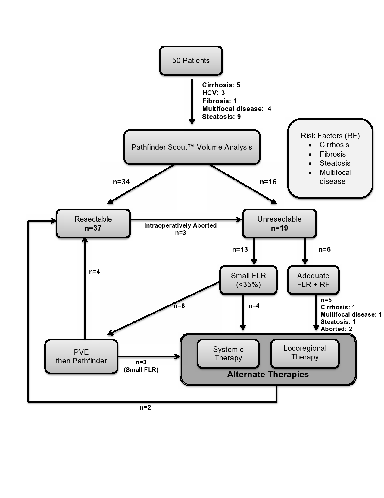
Between 2009 and 2015, 50 patients at Vanderbilt University Medical Center were evaluated for liver resection using Pathfinder Scout™. After approval by the Institutional Review Board, the medical record of each patient was reviewed for pertinent clinical information. Data extracted from the records included preoperative diagnosis, laboratory data, type of resection planned, final pathological findings, and postoperative complications such as liver insufficiency.

Results:
Optimal candidates for liver resection were defined as good performance status, FLR>35%, and limited risk factors. Risk factors for liver insufficiency were defined as hepatic steatosis by imaging or biopsy, fibrosis, or cirrhosis. Sixteen patients were deemed unresectable based on FLR < 35% (n=12) or adequate FLR but possessed one or more risk factors (n=4). An additional 3 resections were intraoperatively aborted due to vascular invasion or additional lesions. Four of the unresectable patients underwent locoregional therapy (LRT e.g. radiofrequency ablation, chemoembolization, radioembolization) and/or systemic therapy; two of the patients were rendered resectable. Eight patients underwent portal vein embolization (PVE) and 4 patients’ FLR improved above 35% leading to resection.

Conclusion:
Formal volumetric assessment prior to liver resection is an important consideration for patients with cirrhosis, steatosis, and multifocal disease. Based on volumetric data 24 % of patients in our cohort underwent LRT rather than resection, mitigating the risks of liver failure postresection in these patients. In addition to standard assessment of liver function and performance status, volumetric analysis of the FLR can determine the success of PVE as an adjunct to improve FLR.

71.07 Socioeconomic Status, Medical History, and Pathologic Findings in Breast Cancer Surgery Decisions

V. J. Tapia4, G. F. D’Souza1, F. Qiu5, G. Nguyen3, Q. Ly2 1University Of California, San Diego,Plastic Surgery,SAN DIEGO, CA, USA 2University Of Nebraska Medical Center,Surgical Oncology,Omaha, NE, USA 3Medical College Of Wisconsin,Milwaukee, WI, USA 4University Of California – San Diego,School Of Medicine,San Diego, CA, USA 5University Of Nebraska Medical Center,College Of Public Health,Omaha, NE, USA

Introduction:
Currently accepted research in oncology has demonstrated that, in early stage breast cancer (BC), lumpectomy followed by whole breast radiation, also known as breast conservation therapy (BCT), has comparable survival to that of mastectomy. Initial investigations on surgical decision-making reported that a patient’s choice was related to socioeconomic status (SES), geographic location, patient characteristics, and physician influence. However, little research has been conducted on the influence of tumor characteristics on surgical decision-making.

Methods:
A retrospective analysis of the 2002-2010 University of Nebraska Breast Cancer Collaborative Registry was performed. The demographic, medical history, and tumor characteristics of patients were compared between oncologic surgery decision groups (BCT, unilateral mastectomy, bilateral mastectomy) in a univariate analysis. A multinomial logistic regression analysis was performed on the predictor variables that were associated with the outcome variable of surgical decision to the 0.2 significance level.

Results:
320 women were included with 100 receiving BCT, 176 unilateral mastectomy, and 44 bilateral mastectomy. On univariate analysis, factors associated with surgery decision were bilateral carcinoma (p=0.0001), staging (p=0.0006), tumor metastasis (p=0.02), tumor histology (p=0.03), BRCA mutation (p=0.04), unilateral multifocal lesions (p=0.047), and lymphoma history (p=0.04). On multinomial analysis, tumor stage and bilateral carcinoma were independently associated with treatment decision. Patients with bilateral tumors had 12.1 times higher odds of choosing bilateral mastectomy (95% OR CI: 2.3-63.3, p=0.003). When compared to patients with in situ tumors, stage II patients had 8.8 times higher odds of choosing bilateral mastectomy (p=0.048) and a 2.9 times higher odds of choosing a unilateral mastectomy (p=0.02). Subjects with stage III tumors had 32.1 times higher odds of choosing bilateral mastectomy than those with in situ tumors (p=0.007), and 13.6 times higher odds of choosing a unilateral mastectomy (p=0.0003). Stage IV patients had 36.1 times higher odds of choosing a bilateral mastectomy than those with in situ tumors (p=0.009), and an 11.5 times higher odds of choosing a unilateral mastectomy (p=0.004).

Conclusion:
Our findings suggest that our patients’ decision of surgical procedure had greater association with tumor characteristics rather than demographic or medical history, as previously demonstrated in other studies. Women with stage II-IV BC and bilateral tumors are still more likely to opt for more extensive surgical interventions despite evidence to the safety, efficacy, and comparable survival rates of more conservative treatments in patients with stages I-III, and well established education on surgical options. Patients with in situ and stage I BC may be more willing to undergo BCT, underscoring the efficacy of current patient education endeavors.

71.08 Financial Implications of Routine Postoperative ICU Care after CRS/HIPEC – More is Not Always Better

H. D. Mogal1, E. A. Levine1, N. F. Fino2, T. I. Fleming1, V. Getz1, P. Shen1, J. H. Stewart1, K. I. Votanopoulos1 1Wake Forest University School Of Medicine,Department Of Surgery, Division Of Surgical Oncology,Winston-Salem, NC, USA 2Wake Forest University School Of Medicine,Department Of Biostatistics,Winston Salem, NC, USA

Introduction: The financial considerations of admitting patients undergoing Cytoreductive surgery and Heated Intraperitoneal chemotherapy (CRS/HIPEC) routinely to the ICU for postoperative care have not been elucidated. Our aim was to study cost differences between patients admitted postoperatively to the ICU and floor and to assess if avoiding routine ICU admission in selected patients can minimize costs without compromising quality.

Methods: Single index-surgical encounter costs for patients admitted directly to the floor or to the ICU for less than 48 hours were retrospectively analyzed from a prospectively maintained institutional database of CRS/HIPEC patients between April 2012 and June 2014. Comparison of clinicopathological variables, complications and average costs between the groups was performed.

Results: 65 patients were observed in the ICU for less than 48 hours, while 51 patients were sent directly to the floor. The two groups were similar for race (p = 0.87), sex (p = 0.12), number of comorbidities (p = 0.17), primary site of tumor (p = 0.37) and ECOG status (p = 0.16). PCI (Peritoneal Cancer Index) score was higher for patients in the ICU (mean 15.6 ± 7.4) compared to those on the floor (mean 10.3 ± 8.1; p = 0.0006). Estimated blood loss (OR 1.26, p = 0.0075) and PCI scores (OR 1.12, p = 0.02) were independent risk factors for admission to ICU. For patients that were observed directly on the floor, average costs were $4460 less than for patients who were observed in the ICU for less than 48-hours ($15209 and $19669 respectively; p < 0.0001). Analysis between these two groups showed no significant difference in minor complications (p = 0.23) or major morbidity (p = 0.44).

Conclusion: Selective postoperative ICU admission is associated with a substantial reduction in cost and no increase in major or minor morbidity.

71.05 Social Support for Patients undergoing Colorectal Cancer Treatment: A Diverse Population-Based Study

M. R. Kapadia1, C. M. Veenstra3, R. E. Davis4, S. T. Hawley3, A. M. Morris2 1University Of Iowa,Surgery,Iowa City, IA, USA 2University Of Michigan,Surgery,Ann Arbor, MI, USA 3University Of Michigan,Medicine,Ann Arbor, MI, USA 4University Of South Carolina,Public Health,Columbia, SC, USA

Introduction:
Social support is associated with adherence to recommended treatment, quality of life, and survival. Colorectal cancer (CRC) often requires extensive treatment, but little is known of needs, sources, and availability of social support among CRC patients.

Methods:
We surveyed Stage III CRC patients from the Detroit and Georgia SEER registries regarding socio-demographics and emotional support desired and received from spouses/partners, families, important others, and providers. We examined differences using chi-square and t- tests.

Results:
Among 1351 eligible respondents (68% response rate), 68% were white and 25% were black. Patients were evenly distributed in age, sex, and income categories. Most endorsed high support from all sources, especially family (88.6%), followed by important others (82.9%), healthcare providers (71.3%), and spouses/partners (55.6%). Only 58% of patients had spouses/partners and among these, 95% endorsed high support. Older patients, black patients, women, or those with <$20,000 annual income were less likely to have spouses/partners (p<0.001). Blacks were significantly less likely than whites to report that the support they received was ‘just right’ (vs. too little or too much, p<0.001). Increasing age and income were significantly associated with increased likelihood that the support received was just right (p<0.006).

Conclusion:
Most patients undergoing CRC treatment reported high social support from all sources. However, black and lower income patients were at risk for low support or support that did not meet their needs. Spouse/partner support was important and met needs, but was only available to 58% of respondents. In future work, we will examine how social support affects long-term outcomes for CRC patients.

71.06 Black and White Pancreatic Adenocarcinoma Patients Receiving Similar Treatment Have Similar Survival

B. J. Flink1,2, Y. Liu3,4, R. Rochat3, D. A. Kooby1,4, J. Lipscomb3,4, T. W. Gillespie1,4 1Emory University School Of Medicine,Surgery,Atlanta, GA, USA 2VA Atlanta Health System,Surgery,Atlanta, GA, USA 3Emory University Rollins School Of Public Health,Atlanta, GA, USA 4Winship Cancer Institue,Atlanta, GA, USA

Introduction: Pancreatic adenocarcinoma (PA) is a lethal cancer that newly affects over 41,000 Americans annually. Prior evidence demonstrates treatment and survival disparities between black and white patients. Our study examines receipt of surgery and overall survival by race, using novel data from a large population cancer database.

Methods: Using the National Cancer Data Base Participant User Files from 2003 to 2011, we identified PA patients with potentially resectable (T1-3M0) disease. Only patients resected with curative intent were included. Univariate, and multivariate logistic and Cox regression models were used to examine resection and survival. Multivariate models were adjusted for demographic factors, comorbidities, T stage, tumor size, facility type, and facility volume (20 or more resections/year = high volume).

Results: Of 33,255 patients with potentially resectable disease, 3,727 (11.2%) were black and 27,908 (85.2%) were white. Black patients had lower odds of undergoing resection as compared with their white patients on univariate (OR 0.69 [95% CI 0.65-0.74, p<0.0001] and multivariable logistic regression (OR 0.64 [95% CI 0.58-0.71], p<0.0001). There was no significant difference in the refusal of surgery by race. By univariate survival analyses, resected patients survived 11.2 months longer in median survival time than unresected patients and had a lower hazard of death (HR 0.39 [95% CI 0.37-0.41], p<0.0001) while across both resected and unresected, black patients had a higher hazard of death and a reduced median survival time (HR 1.10 [95% CI 1.02-1.17], p=0.007; 9 vs. 9.66 months). This persisted in a multivariable Cox regression model that did not account for treatment factors (HR 1.10 [95% CI 1.01-1.21], p=0.03). Survival by race was similar after controlling for method of treatment (Table 1).

Conclusion: Our analyses demonstrate the importance of treating patients with resectable disease the same irrespective of race. Black patients are at much lower odds of resection as compared with white patients, but they have similar survival in multivariate models when they do undergo resection. These data highlight the importance of improving access to resection for all potential resectable PA patients.

71.03 Frailty Markers and Thyroid/Parathyroid Surgical Outcomes in the Elderly

S. X. Jin1, T. W. Yen1, A. A. Carr1, B. Lalande1, K. Doffek1, D. B. Evans1, T. S. Wang1 1Medical College Of Wisconsin,Surgical Oncology/Surgery,Milwaukee, WI, USA

Introduction: Frailty, defined as decreased physiologic reserves due to decline of multiple organ systems, is a risk factor for poorer postoperative outcomes in the elderly. Cervical endocrine surgery (thyroidectomy and parathyroidectomy) is associated with low rates of morbidity and mortality, and is increasingly being performed in the elderly. This study sought to identify potential frailty biomarkers in patients undergoing cervical endocrine surgery.

Methods: A retrospective chart review of prospective databases was performed of 309 patients who underwent thyroidectomy and/or parathyroidectomy between 7/1/2012 to 6/30/2013. Demographic and clinical data were collected, including pre- and postoperative lab values, extent of surgery, length of stay (LOS) and postoperative complications within 30 days of surgery. Endocrine-specific complications were documented for 6 months in order to categorize transient vs. permanent injury; patients with <6 months of follow-up were not excluded from the cohort. Patients were divided into groups by age: <50, 50-64, and ≥65 years. Finally, to identify potential frailty markers, preoperative biochemical data were compared between patients who experienced complications and those who did not. Kruskal-Wallis test was used for continuous variables and chi-squared test was used for categorical data; a p-value <0.05 was considered statistically significant.

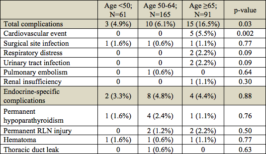
Results: Median age was 57 years (range, 19-86) and 252 (82%) patients were female. The median LOS was one day, and there was no difference in LOS among the three age groups (p=0.87). Overall, there were 28 complications; 14 (50%) were endocrine-specific (Table 1). Patients ≥65 years experienced more complications (n=15; p=0.03) overall and more cardiac events (n=5; p=0.002) than patients 50-64 and patients <50 years. There was no difference in endocrine-specific complication rates by age group. When analyzed by preoperative biochemical values and comorbidities, there were no biochemical factors that were associated with the development of complications. However, patients who experienced complications had a higher rate of anticoagulant use (22% vs. 5%; p=0.001) and congestive heart failure (CHF) (9% vs. 2%; p=0.03), compared to those who did not.

Conclusion: In this cohort, age correlated with higher postoperative complications but was not predictive of hospital LOS. Preoperative factors such as anticoagulant use and history of CHF may be markers for frailty in cervical endocrine patients.

71.04 Influence of HIV infection on hepatocellular carcinoma incidence and survival

A. Mokdad1, A. Singal3, J. Mansour1, H. Zhu2, A. Yopp1 1University Of Texas Southwestern Medical Center,Surgical Oncology,Dallas, TX, USA 2University Of Texas Southwestern Medical Center,Medical Oncology,Dallas, TX, USA 3University Of Texas Southwestern Medical Center,Digestive And Liver Diseases,Dallas, TX, USA

Introduction:

Liver-related complications such as hepatocellular carcinoma (HCC) are a major cause of morbidity and mortality in individuals infected with HIV, particularly among those also infected with hepatitis B or hepatitis C viruses. There is a lack of consensus regarding the clinical presentation, treatment, and outcomes in HIV-infected patients with HCC. We compared the clinical presentation, treatment, and survival of patients with HCC, with and without HIV infection.

Methods:

We linked the Texas cancer registry to the HIV/AIDS data for all years between 2001 and 2011. Patient demographics, socioeconomic status, cancer stage, and treatment were compared between patients with HCC and patients with HCC and HIV. Using a standard HIV population, we calculated annual age, sex, and race standardized incidence and all-cause mortality of HCC in patients with and without HIV. We estimated standardized incidence and mortality ratios for the entire study period. We calculated the fraction of mortality related to the following cause-of-death categories: HCC, end-stage liver disease, and HIV. We used a shared frailty model to evaluate risk-adjusted survival in patients with HCC and HIV and with HCC only. We explored the association between HIV infection and treatment of HCC using a mixed-effects logistic regression model.

Results:

18,291 patients with HCC were included in the study; 236 had HIV infection. Compared to patients with HCC only, patients with HCC and HIV were younger at the time of HCC diagnosis (63 years vs. 53 years, p-value < 0.01), male (91.3% vs. 71.9%; p-value < 0.01), African American (41.8% vs. 12.9%; p-value < 0.01), and of lower socioeconomic status (52.6% vs 41.9%; p-value = 0.02). Overall cancer stage and treatment provision were similar. The unadjusted median survival was 6.1 and 6.4 months (log-rank test p-value = 0.38) in the HCC and the HCC and HIV groups, respectively. Age, sex, and race standardized incidence increased and mortality decreased in both groups over the study period. The mean standardized incidence ratio for patients with HCC and HIV was 2.4 ± 0.35; the mean standardized mortality ratio was 2.7 ± 0.36. The most common cause of death was liver cancer, 70 percent and 54 percent, in patients with HCC only and with HCC and HIV, respectively. In the HCC and HIV group, 23 percent died from HIV sequelae. Adjusted survival was worse in patients with concurrent HIV infection (hazard ratio = 1.23, 95% confidence interval: 1.04 – 1.46). After accounting for facility effect, patient demographics, socioeconomic status, and HCC characteristics, there was no difference in provision of resection, ablation procedure, or chemotherapy between both groups.

Conclusion:

Patients with HIV are associated with a higher risk of developing HCC. HCC and concurrent HIV infection is associated with worse survival. It is imperative to improve screening, diagnosis, and management of HCC in patients with HIV.

70.20 Neutrophil-to-Lymphocyte Ratio Predicts Outcomes of Pancreaticoduodenectomy for Pancreatic Cancer

A. I. Salem1, E. R. Winslow1, C. S. Cho1, S. M. Weber1 1University Of Wisconsin School Of Medicine And Public Health,General Surgery,Madison, WI, USA

Introduction:

Neutrophil-to-lymphocyte ratio (NLR) has been introduced as a serological marker with a potential prognostic role for many cancer types. The role of NLR in predicting pancreas cancer outcome is understudied. Previous reports have suggested that higher NLRs are associated with worse survival. We sought to investigate the relation between NLR and both short and long term outcomes after pancreatic ductal adenocarcinoma resection in our institution.

Methods:

Patients with pancreas cancer who underwent pancreaticoduodenectomy for pancreatic ductal adenocarcinoma between 1999 and 2012 were evaluated. NLR was calculated by dividing the absolute neutrophilic count value by the absolute lymphocytic count value. We identified 216 patients from our prospectively maintained database with 104 patients excluded for lack of data on neutrophilic or lymphocytic counts within 30 days prior to surgery. Out of 112 eligible patients analyzed, 33 (29.5%) had NLR ≥ 4.5.

Results:

There was no difference in 30-day mortality between patients with NLR ≥ 4.5 and those with NLR < 4.5 (0 (0%) vs 1 (1.3%), p=0.5) and no differences in overall 30-day morbidity (16 (49%) vs 45 (57%), p=0.4). Patients with NLR < 4.5 were more likely to have nodal metastases than their counterpart group (69 (87%) vs 16 (49%), p<0.001), while patients with NLR ≥ 4.5 had a higher median estimated blood loss (EBL) (550 mL (150-12,500) vs 500 mL (150-1,400), p=0.02), a higher median number of intraoperatively-transfused packed red cell units (2 units (0-23) vs 0 units (0-9), p=0.01), and a longer hospital stay (LOS) (10 days (5-39) vs 8 days (4-35), p=0.04). Kaplan-Meier survival analysis showed improved median overall survival in the NLR ≥ 4.5 group (21 months vs 18 months, p=0.02). On multivariable analysis, after adjusting for nodal status, EBL, intraoperative packed red cell transfusion, NLRs ≥ 4.5 were found to be the only predictor of improved overall survival (HR=0.54 , CI=0.30 – 0.97, p=0.04) (Figure.1).

Conclusion:

In our experience of pancreas cancer patients undergoing pancreaticoduodenectomy, elevated NLR was associated with an increased risk of EBL, need for transfusion, and longer LOS, but improved long-term survival. This is in contrast to previous reports describing elevated NLR as a negative prognostic variable. Further studies on larger numbers of patients are required to better assess the prognostic role of preoperative NLR for both short and long-term outcomes after curative resection of pancreas cancer.

70.21 Murine Breast cancer cells eliminated in non-derived strain mice; using an improved breast cancer model

E. Katsuta1, S. DeMasi1, K. P. Terracina1, H. Aoki1, M. Aoki1, P. Mukhopadhyay1, K. Takabe1 1Virginia Commonwealth University School Of Medicine And Massey Cancer Center,Division Of Surgical Oncology, Department Of Surgery,Richmond, VA, USA

Introduction: We have previously established a murine syngeneic breast cancer model utilizing cell implantation under direct vision technique, which mimic human cancer progression (Rashid, Takabe et al. Breast Cancer Research and Treatment 2014). Other groups have reported that cell implantation using Matrigel produced stable results in xenograft models. Here, we report the establishment of improved syngeneic orthotopic murine breast cancer model using Matrigel. In this study, we determined the maximum amount of Matrigel to be implanted without spillage, the tumor growth with various number of cells, and utilizing this new model, we investigated the growth of murine cancer cell derived from different strain mice.

Methods: Matrigel was injected to #2 and #4 mammary glands. Various number of murine breast cancer E0771cells in Matrigel were implanted into bilateral #2 and #4 mammary gland of C57Blk6 mice. Three weeks after inoculation, tumorigenesis were compared. 1 x 10⁴ of murine breast cancer 4T1-luc2 cells, derived from Balb/C mice, were implanted into the Right side #2 gland of Balb/C or C57Blk6 mice. Tumor growth was monitored by bioluminescence (IVIS) imaging.

Results: We found that implantation of the cells will be more efficient with less variability when the cells are suspended in Matrigel compared with PBS, which was the technique we reported previously. Maximum volume of Matrigel inoculated without spillage was 20 μl in #2 gland, 30 μl in #4 gland, respectively. Therefore, we implanted 20 μl of Matrigel in #2 gland, and 30 μl in #4 gland in the subsequent experiments. In order to determine the difference of tumor development, 504, 105, 505, 106 E0771 cells suspended in 20µl Matrigel were inoculated. Three weeks after inoculation, ‘the take rates’ (tumorigenesis) were 0%, 12.5%, 75%, 75%, 100%, respectively. Utilizing 4T1-luc2 cells in Matrigel suspended cell implantation method, we investigated how long the mouse-derived cancer cells survive in mice from a different background. The fold increase in tumor growth in both backgrounds were nearly identical 24 h after inoculation at 5-fold increase measured by bioluminescence imaging. By 7 days after inoculation, tumor in C57Blk6 reached a maximum increase of approximately 720-fold their Day 0 size, whereas the tumor in Balb/C had almost a 2000-fold increase in tumor size. The Balb/C tumor continued to increase rapidly to reach an almost 3000-fold increase in size, while the C57Blk/6 mice tumors swiftly decreased from Day 7 and was eliminated by Day 14.

Conclusion: We identified the maximum amount of Matrigel that can be implanted into #2 or #4 mammary gland without spillage, and the difference in take rates with various number of cells for murine orthotopic breast cancer model. Utilizing Matrigel implantation method, we found that cancer cells will continue to grow until one week, then it will eliminated by 2 weeks when implanted into different background strain mice.

71.01 Preoperative Platelet to Albumin Ratio is a Prognostic Factor for Pancreatic Cancer.

Y. Shirai1, H. Shiba1, N. Saito1, T. Horiuchi1, K. Haruki1, Y. Nakaseko1, Y. Takano1, K. Furukawa1, M. Kanehira1, S. Onda1, T. Sakamoto1, T. Gocho1, Y. Ishida1, K. Yanaga1 1The Jikei University School Of Medicine,Surgery,Minato-ku, TOKYO, Japan

Introduction:
Pancreatic cancer is one of the most aggressive digestive cancers. Because pancreatic cancer recurs after pancreatic resection in as many as 70-80%, it is important to predict tumor recurrence and prognosis in regard to decision making of additional adjuvant therapy. There are several inflammation based prognostic index such as Glasgow prognostic score (GPS), mGPS, neutrophil to lymphocyte ratio, platelet to lymphocyte ratio, CRP/Albumin, and prognostic nutrition index. However, preoperative estimation of oncological prognosis remains to be established. The aim of this study is to evaluate the prognostic value of preoperative platelet to albumin ratio (PAR) in pancreatic ductal adenocarcinoma after pancreatic resection.

Methods:
A total of 115 patients who underwent pancreatic resection for pancreatic invasive ductal adenocarcinoma were available from prospectively maintained database. The patients were divided into two groups as PAR ≥ 53.1 x 10³ or < 53.1 x 10³ on the basis of ROC curve analysis (2 years survival, AUC=0.640, p=0.011). Survival data were analyzed using the Log-rank test for univariate analysis and Cox proportional hazards for multivariate analysis. P value <0.05 was judged as significant.

Results:
The preoperative PAR was significant prognostic index on univariate analysis for disease-free and overall survivals. The median overall survival in patients with PAR ≥ 53.1 x 10³ was 17.6 months, which was poorer than 36.1 months for patients with PAR < 53.1 x 10³ (p=0.0039). The PAR retained its significance on multivariate analysis for overall survival (HR 1.666, 95%CI 1.021-2.717, p=0.041) along with tumor stage (p=0.047) and serum CA19-9 (p=0.010). PAR ≥ 53.1 x 10³ was also a significant independent prognostic index for poor disease-free survival on multivariate analysis (HR 1.771, 95%CI 1.055-2.973, p=0.031).

Conclusion:
The preoperative PAR is a novel significant independent prognostic index for disease-free and overall survival in resected pancreatic invasive ductal adenocarcinoma.

71.02 Conservative Management of Desmoid Tumors is Safe and Effective

J. S. Park1, Y. Nakache4, J. Katz3, R. D. Boutin3, A. Monjazeb2, R. J. Canter1 1University Of California – Davis,Surgical Oncology,Sacramento, CA, USA 2University Of California – Davis,Radiation Oncology,Sacramento, CA, USA 3University Of California – Davis,Radiology,Sacramento, CA, USA 4University Of California – Davis,Medical School,Sacramento, CA, USA

Introduction:

Desmoid tumors are locally aggressive neoplasms without metastatic potential. Although surgical resection was once thought to be the mainstay of therapy, this is a potentially morbid approach associated with a high risk of local recurrence. There is an increasing role for watchful waiting and conservative management for these tumors.

Methods:

We identified 36 desmoid tumor patients who were categorized by the nature of treatment rendered (surgical resection versus observation). Data were abstracted on clinical and pathological factors. Disease stability or progression was determined radiographically. Univariate and Kaplan-Meier analysis was used to determine predictors of recurrence/progression of disease. Main outcome measurements were tumor recurrence following surgical resection versus tumor progression with conservative management.

Results:

Of the 36 patients, 58% were female and average age was 44 years old. The tumors were primarily located in the extremities including hip and shoulder girdle at 58%. 33% were located in the trunk and 8% in the retoperitoneum. Median tumor size was 9.2 cm (range 2.7-24 cm). Of the patients that had beta catenin staining, 96% were positive. The patients were categorized into surgical versus nonsurgical therapy. 18 patients underwent operation either prior to referral, due to refractory symptoms or due to patient preference. 17 patients were observed without surgical resection, including 3 patients who received nonsurgical therapy such as chemoradiation and/or medical therapy. One patient was lost to follow up. Median follow up time was 23 months.

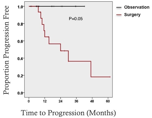
Among 17 of the patients who underwent surgical resection, ten patients developed recurrent disease (59%). Among those who developed recurrent disease, 8 (80%) patients were stable or partially responding to subsequent observation. Of the 17 patients who underwent conservative management, one patient experienced disease progression (5%) and 94% of patients had no disease progression, including one patient with complete response and three patients with partial responses.

Conclusion:

This retrospective analysis adds to growing data that observation of both primary and recurrent desmoid tumors is safe and effective with higher rates of stable disease than other published series. Conservative management of desmoid tumors appears to be safe and effective, sparing patients the morbidity and risk of recurrence that is associated with extensive operations.

70.17 Limited Utility of Gene Expression Classifier in Surgical Patients with Bethesda III Thyroid Nodules

T. M. Vaghaiwalla1, G. A. Rubio1, M. LoPinto1, Z. F. Khan1, A. R. Marcadis1, J. I. Lew1 1University Of Miami,Division Of Endocrine Surgery, The DeWitt Daughtry Family Department Of Surgery,Miami, FL, USA

Introduction: With implementation of the Bethesda System for Reporting Thyroid Cytopathology (BSRTC), Bethesda III FNA results remain a clinical dilemma for clinicians and surgeons alike. Gene expression classifier (GEC) testing was developed to further stratify patients with Bethesda III nodules as benign or suspicious for cancer. Given the known variability of GEC testing between institutions, this study evaluates the utility of this genetic testing in patients with Bethesda III nodules at a single institution.

Methods: A retrospective review of prospectively collected data of 663 consecutive patients with index thyroid nodules who underwent FNA and thyroidectomy was performed. FNA results were based on the BSRTC system, and GEC testing was later utilized in Bethesda III patients as benign or suspicious for malignancy. Patients with Bethesda III nodules underwent initial thyroid lobectomy for definitive diagnosis unless there was a history of radiation exposure, familial thyroid cancer, obstructive symptoms, bilateral nodules and/or patient preference for which total thyroidectomy was performed. Bethesda III nodules were subdivided into malignant or benign groups based on final pathology. Among patients who underwent GEC testing, final pathology was compared to initial GEC results.

Results: Of 663 patients who underwent FNA, 129 patients had Bethesda III nodules of which 66 (51.2%) had malignancy (Papillary thyroid cancer, n=54; Follicular thyroid cancer (FTC), n=10; Medullary carcinoma, n=1; Lymphoma, n=1) on final pathology. Of the remaining Bethesda III patients, 63 (48.8%) had benign pathology (Adenomatoid nodule, n=18; Hürthle Cell adenoma, n=2; chronic lymphocytic thyroiditis, n= 6; Follicular adenoma n= 10; Multinodular hyperplasia (MNH), n=26; Cyst, n=1). A total of 108 patients with Bethesda III nodules without GEC testing had a malignancy rate of 52.8% (57/108) and benignity rate of 47.2% (51/108). Of 21 patients with Bethesda III nodules who underwent GEC testing, 38.9% (7/18) had suspicious results and malignancy on final pathology with a sensitivity of 77.8% (7/9). Of 3 patients with benign GEC results, 2 (66.7%) had malignancy (FTC, n=2; MNH, n=1) on final pathology. Overall, patients with Bethesda III nodules and suspicious GEC results had a malignancy rate of 38.9% compared to a rate of 52.8% in patients with Bethesda III nodules without GEC testing.

Conclusion: In surgical patients with Bethesda III nodules, there was a greater than expected malignancy rate. However, in patients with Bethesda III nodules and GEC testing, malignancy rates were as predicted. GEC testing may have limited utility in surgical decision making, as this patient population is already highly selected for malignancy due to other factors. Surgeons should assess their local institutional experience to determine whether there is added utility of GEC testing for Bethesda III nodules in their everyday clinical practice.

70.19 A Novel Magnetic Resonance Imaging Protocol for Pancreatic Cystic Lesions

R. K. Schmocker1, S. B. Reeder2, E. R. Winslow1, S. M. Weber1 1University Of Wisconsin,Surgery,Madison, WI, USA 2University Of Wisconsin,Radiology,Madison, WI, USA

Introduction: Cystic pancreatic tumors present a diagnostic dilemma, as initial imaging often fails to provide a definitive diagnosis; therefore serial imaging is needed to follow the lesion over time. CT scan has traditionally been used to follow these lesions, however, given its excellent depiction of fluid containing lesions, and its lack of ionizing radiation, MRI has been increasingly utilized to assess these lesions. We sought to evaluate an initial experience and costs utilizing a novel pancreatic cyst-specific protocol for MRI.

Methods: A single center retrospective study was performed by searching an electronic imaging database for MRI pancreas studies from 2006-2014. After identification of the studies, the reports were examined to confirm that the indication for imaging was pancreatic cystic lesions. Retrospective chart review was used to assess demographics, imaging profiles (presence of other serial imaging – ex: CT), and the presence of interventions. Reimbursed costs of the studies were determined using the CMS physician fee data for the imaging procedures including both the professional and technical components, however contrast cost was not included.

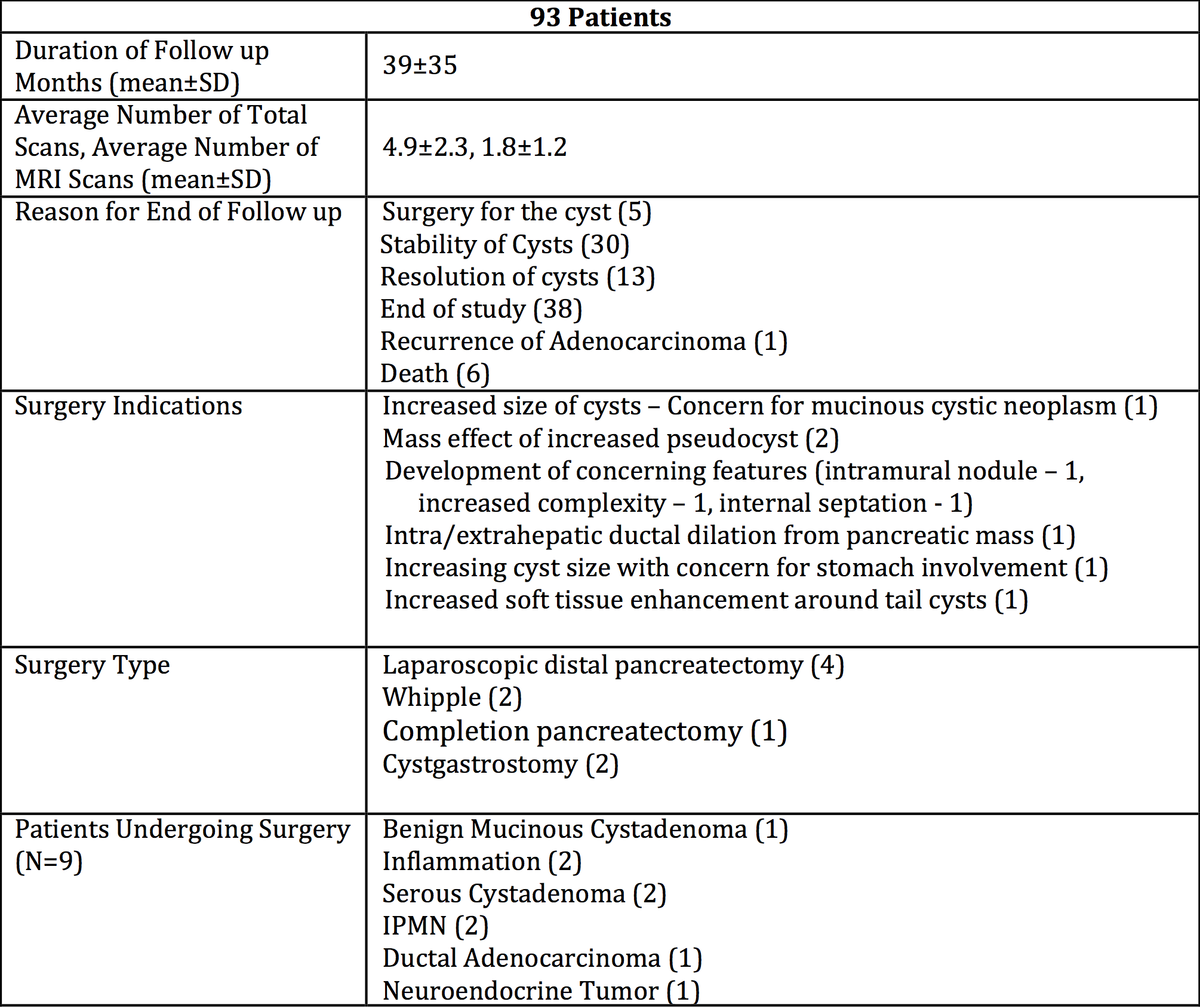
Results: 93 patients underwent an MRI pancreas protocol (average age: 65±13, % female: 61.5%). All patients had one contrasted imaging scan (MRI or CT) during the study period. 9 of these patients underwent a surgical intervention for a pancreatic cyst – 5 with a definitive operation, while 4 underwent surgery, but required surveillance of cysts in the remaining pancreas. Surgery was most often indicated due to concerning features or size (n=6). An MRI pancreas was used to diagnose cyst change for 3/5 patients that had a definitive operation, while CT scan identified a change in imaging characteristics for the other 2 patients. 43 patients underwent EUS/FNA during the study period. The average follow-up was 39 months, with the time between studies being 8.2 months. Patients had, on average, a total of 4.9 scans over the 39 months (1.7 CT scans, 1.3 MRI w/contrast, and 1.8 MRI pancreas protocol scans) or 1.5 scans per year. MRI pancreas was more expensive than CT w/Contrast ($333 vs. $230), but with the absence of radiation and contrast related complications. Additionally, if the average patient were exclusively followed with CT scans; the overall cost savings would be minimal ($189).

Conclusion: Our initial experience with the use of a novel pancreas protocol MRI is favorable, with minimal increase in cost. The lack of ionizing radiation and intravenous contrast with the MRI pancreas potocol, is an important consideration for patients undergoing serial imaging. The use of MRI for patients with pancreatic cystic disease shold be investigated further.

70.16 Causes and Predictors of Early Mortality Following Pancreatic Resection, Recognize Then Prevent

C. Mosquera1, T. L. Fitzgerald1, E. E. Zervos1 1East Carolina University Brody School Of Medicine,Division Of Surgical Oncology,Greenville, NC, USA

Introduction: Pancreas resection is among the highest risk procedures in terms of post-operative mortality. The purpose of the study was to determine predictors and causes of early and late post-operative mortality in patients undergoing pancreatectomy.

Methods: A prospective pancreas surgery registry at a very-large volume hospital was queried to identify early (30-day) and late (90-day) postoperative mortality between 2008 and 2015. Logistic regression was undertaken to determine predictors of each. Causes of death were categorized as: hemorrhagic, cardiorespiratory, sepsis and disease progression. Sepsis was sub-categorized as related (presence of infected intra-abdominal collections, abscess, fistula, and surgical site infection) or unrelated to surgery (pneumonia, CLABSI, CAUTI). Disease progression signified withdrawal of care in an otherwise viable patient due to perceived poor prognosis as a result of documented recurrent or persistent disease.

Results: A total of 312 patients underwent resection (68% Whipple, 32% Distal Pancreatectomy). Early postoperative mortality occurred in 12 (3.8%) patients and late in 24 (7.7%) patients with a total postoperative mortality of 11.5%. Early deaths occurred during index admission in 83% while 95.5% of late deaths occurred outside of the hospital or in patients that were discharged and returned to hospital and died. (Table)

On univariate analysis, age, smoking history, intensive care unit requirement (ICU), presence of complications, operative estimated blood loss, body mass index, discharge destination, presence of tachycardia, elevated WBC at discharge and type of insurance predicted any postoperative mortality. On multivariate analysis, only ICU (OR 20.3 p 0.0003) and discharge destination of home and SNF compared to rehabilitation facility remained significant (p<0.05). Private insurance was a protective factor compared to Medicare, Medicaid and uninsured (p<0.05). Sepsis arising from surgical complications was the primary source of early postoperative mortality while disease progression was the most common cause of late mortality.

Conclusion: Surgical complications remain the primary source of early postoperative mortality, which are largely unavoidable. Late postoperative mortality may be mitigated by minimization and early detection of hospital-acquired infection through adherence to best care guidelines and neo-adjuvant strategies to identify patients with low risk of disease progression in which further treatment is non futile.

70.11 Hepato-Pancreatectomy: Outcomes of Synchronous Hepatic and Pancreatic Resection

T. B. Tran1, J. N. Leal1, M. M. Dua1, B. C. Visser1, J. A. Norton1, G. A. Poultsides1 1Stanford University,Surgery,Stanford, CA, USA

Introduction: Simultaneous resection of both the liver and the pancreas remains controversial due to the high morbidity and questionable oncologic benefit. The purpose of this study is to evaluate the short-term morbidity and mortality after combined resection of the liver and pancreas, as well as to determine whether or not these aggressive operations are associated with long-term survival.

Methods: Consecutive patients who underwent synchronous hepatectomy and pancreatectomy (SHP) at a single instution were retrospectively evaluated. Extent of liver resection was categorized into major hepatectomy (defined as lobectomy or trisectionectomy) and minor hepatectomy (less than 3 liver segments). Preoperative patient features, perioperative outcomes, and long-term survival following synchronous hepatectomy and pancreatectomy (SHP) were evaluated.

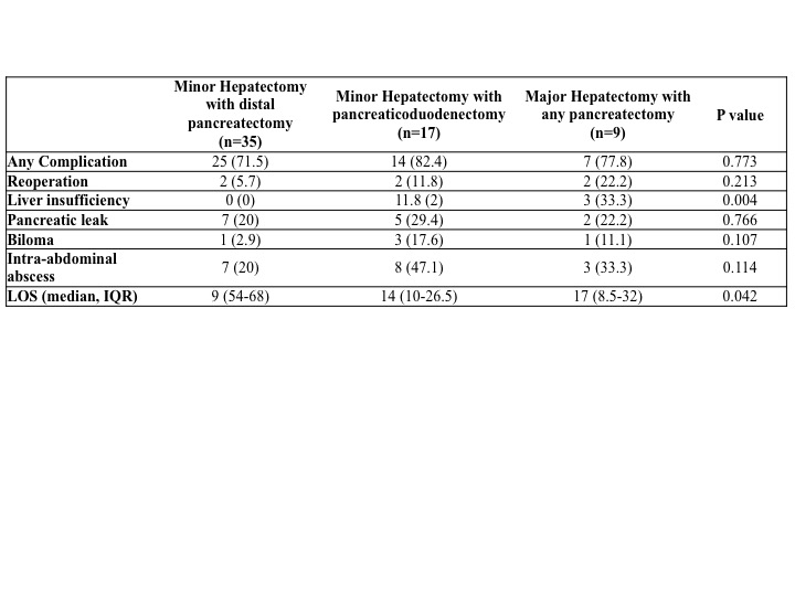
Results: From 2005 to 2014, 61 patients underwent SHP. Diagnoses include 34 pancreatic neuroendocrine tumors (PNET), 6 sarcomas, 3 cholangiocarcinomas, 3 adrenocortical carcinomas, 3 cystic pancreatic neoplasms, 2 renal cell carcinomas, 2 colon cancers, 2 recurrent gastric cancers, 2 gastrointestinal stromal tumors, 2 peritoneal mesotheliomas, 1 appendiceal cancer, and 1 ovarian cancer. Patients were further categorized into 3 groups based on the extent of SHP (Table). There were no differences in patient demographics, comorbidities, ASA status, estimated blood loss, need for blood transfusion, 30 or 90-day mortality. Furthermore, the incidence of major complications was similar between the three groups, except for liver insufficiency (defined as peak postoperative bilirubin of 7 or more), which affected one-third of patients who underwent major hepatectomy with any pancreatectomy (p=0.004). Length of stay correlated with extent of SHP (p=0.042). Patients with PNET had more favorable prognosis compared to all other malignant histologies combined (5-year overall survival 47% vs. 26%; p=0.002).

Conclusion: Combined hepatectomy with pancreatectomy is a technically challenging operation. In carefully selected patients, combined hepatectomy and pancreatectomy can be associated with long-term survival.

70.12 Incidence and Pattern of Port-Site Metastasis Following Cholecystectomy in GBCA: A Systematic Review

D. Berger-Richardson1, T. Chesney1, M. Englesakis3, S. Cleary1,3, A. Govindarajan1,2, C. Swallow1,2 1University Of Toronto,Division Of General Surgery,Toronto, ONTARIO, Canada 2Mount Sinai Hospital,Toronto, ONTARIO, Canada 3University Health Network,Toronto, Ontario, Canada

Introduction: Laparoscopic cholecystectomy for the treatment of symptomatic cholelithiasis was rapidly adopted in the early 1990’s. However, there were early reports of port-site metastasis (PSM) when incidental gallbladder adenocarcinoma (GBCA) was discovered in the specimen after surgery. In a review of cases up until 1999, the estimated incidence of PSM in incidental GBCA was reported to be 14%. Recognition of this phenomenon may have prompted changes in surgical technique, and we questioned whether the incidence of port-site metastasis has changed since then. Furthermore, direct contact between the specimen and wound edges during extraction is one of the possible mechanisms of PSM. Determining the incidence of both extraction and non-extraction port PSMs may provide insight into this hypothesis. Hypothesis: 1) The risk of PSM following resection of GBCA has decreased over the past fifteen years. 2) Extraction ports are more likely to harbour recurrence than non-extraction ports.

Methods: A systematic review of articles related to wound recurrence of GBCA was conducted by two independent reviewers. Inclusion criteria for our first objective of determining the modern incidence of PSM were English language papers reporting the presence or absence of PSM following a minimum of 5 cholecystectomies harbouring GBCA published from 2000 – 2014, the 15 year period following the previous review. Inclusion criteria for our second objective of determining the location of the port site involved include English language papers reporting the presence of PSM following laparoscopic cholecystectomy in the setting of GBCA, without limiting this search temporally or by study size.

Results:5448 abstracts were screened. 23 published case series met criteria for estimating the modern incidence of PSM in GBCA. PSM were found in 73 of 728 patients (incidence=10%). Results from the two largest studies using prospectively collected databases (n= 114 and n= 96) had PSM incidences of 14% and 15% respectively. With respect to the pattern of PSM, data was extracted from 98 papers meeting inclusion criteria. More than 305 individual port sites have been reported to harbour recurrence in 236 patients with GBCA (number of PSM/ person = 1-4). Of those with decipherable location (extraction vs non-extraction) (n=190) based on description within the paper, 53% occurred at extraction ports compared to 47% at non-extraction ports (p=0.526).

Conclusion:Despite the implementation of techniques for risk reduction, the incidence of PSM following gallbladder cancer resection is stable at 10-15%. Recurrence at non-extraction ports may indicate mechanism(s) other than direct contact with the wound during extraction. Studies into mechanisms of PSM are warranted. Preoperative imaging studies should be carefully reviewed to minimize inappropriate laparoscopic resection of gallbladder cancer.

70.14 Gender Differences in Length of Hospital Stay After Elective Major Operations for Colon Cancer

C. E. Cauley1,2, D. Chang1,3, L. Bordeianou1, A. B. Haynes1,2,3 1Massachusetts General Hospital,Department Of Surgery,Boston, MA, USA 2Ariadne Labs At Brigham And Women’s Hospital And The Harvard T.H. Chan School Of Public Health,Boston, MA, USA 3Codman Center Center For Clinical Effectiveness In Surgery,Boston, MA, USA

Introduction:
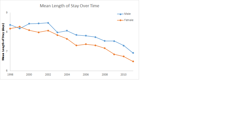
Length of stay (LOS) after elective colectomy has markedly decreased with the advent of enhanced recovery pathways, yet there remains substantial variability in postoperative stays. This study aims to assess the influence of gender on LOS after elective colectomy for colon cancer.

Methods:
Patients undergoing elective colectomy for colon cancer between 1998 and 2011 were identified from the Nationwide Inpatient Sample. Multivariate logistic regression modeling was used to determine if gender was a significant predictor of extended postoperative LOS (> 8 days) controlling for potential confounders, including year of operation, race, age, comorbidity, complications, and hospital factors.

Results:
The cohort consisted of 88,778 patients, 40,526 (45.65%) men and 48,252 (54.35%) women. The mean LOS was 7.7 days (SE = 0.020), which decreased from 8.3 to 6.6 days over the study period, with a mean stay of 7.9 days (SE = 0.031) versus 7.5 days (SE = 0.025) for men and women, respectively (p<0.001). Multivariate logistic regression analysis revealed lower odds of extended LOS in women (OR 0.90; 95% CI: 0.87-0.94). This gender difference was greater in patients over 60 years old, and has continued despite decreasing LOS in more recent years.

Conclusion:
Men have a longer length of stay after elective colectomy for colon cancer, controlling for patient and operative characteristics. This difference in healthcare utilization between genders despite increasing standardization of clinical care should be explored further to determine if factors such as social support influence this phenomenon.

70.15 Very Long Acyl Chain (C24:0 and C22:0) Ceramides are Associated with Obesity and Breast Cancer Progression

K. Moro1, M. Nagahashi1, J. Tsuchida1, T. Niwano1, K. Tatsuda1, C. Toshikawa1, M. Hasegawa1, Y. Koyama1, T. Kobayashi1, S. Kosugi2, H. Kameyama1, H. Aoki3, K. Takabe3, T. Wakai1 1Niigata University Graduate School Of Medical And Dental Sciences,Digestive And General Surgery Niigata University,NIigata, NIIGATA, Japan 2Uonuma Kikan Hospital,Division Of Digestive And General Surgery,Minami-Uonuma, NIIGATA, Japan 3Virginia Commonwealth University School Of Medicine And The Massey Cancer Center,Surgical Oncology,Richmond, VA, USA

Introduction:
It is well established that obesity is associated with poor prognosis of breast cancer patients. Obesity evokes chronic inflammation, which stimulates cancer progression. Ceramide is a key metabolite in both anabolic and catabolic pathways of sphingolipids, and the very long fatty acyl chain (C24:0 and C22:0) ceramides are elevated both in the tissues and in the circulation of obesity, insulin resistance and diabetes. One of the mechanisms of obesity-mediated inflammation is due to release of ceramide from adipocytes after lipolysis. It has been reported that the ceramides and other obesity-related factors trigger activation of inflammasome that result in the secretion of inflammatory cytokines such as IL-1 beta, IL-6, and TNF-alpha under the obese condition. However, serum levels of ceramides in breast cancer patients associated with obesity have not been elucidated to date. In this study, we examine the levels of ceramides in breast cancer patients to reveal the association of the sphingolipids with obesity and breast cancer progression.

Methods:
A retrospective analysis was conducted of 59 patients with breast cancer, in whom the disease was diagnosed as stage I, II or III pathologically. The serum from the patients were obtained when the diagnosis was made and any treatment had been done before the blood collection. Ceramides (C14:0, C16:0, C18:1, C18:0, C20:0, C22:0, C24:1, C24:0, C26:0) were measured by liquid chromatography-electrospray ionization-tandem mass spectrometry . The levels of ceramides were analyzed with clinicopathological data of the patients.

Results:

Body mass index (BMI) was significantly associated with the pathological stage (P < 0.05). The levels of ceramides were detected successfully in the serum from 59 breast cancer patients. The levels of ceramide were not associated with clinical demographics of the patients including age, hormone receptors (ER, PgR) and HER2 status, Ki-67 index, nuclear grade, lymphatic and vascular invasion of the tumor. Interestingly, however, levels of C24:0 ceramide in patients with high BMI (> 25) was significantly higher than that in patients with normal BMI (< 22) (P < 0.05). Further, levels of C24:0 ceramide were significantly elevated with pathological stage (P < 0.05). C22:0 ceramide also showed trends of similar associations with BMI and pathological stage, albeit they were not statistically significant.

Conclusion:

Our results show an elevation of very long fatty acyl chain (C24:0 and C22:0) ceramides in serum of obesity-related breast cancer. Further studies are needed to elucidate the mechanism, and possible association with the prognosis. This work was supported by the Japan Society for the Promotion of Science Grant-in-Aid for Scientific Research Grant Number 15H05676 and 15K15471 for M.N and 15H04927 for W.T. M.N. is supported by the Uehara Memorial Foundation, Nakayama Cancer Research Institute, Takeda Science Foundation, and Tsukada Memorial Foundation.

70.08 Outcomes of Non-elective Gastric Cancer Surgery Following Admission Through the Emergency Department

I. Solsky1,2, P. Friedmann1,2, P. Muscarella1,2, H. In1,2 1Montefiore Medical Center,Department Of Surgery,Bronx, NY, USA 2Albert Einstein College Of Medicine,Department Of Surgery,Bronx, NY, USA

Introduction: Outcomes following non-elective surgery for gastric cancer are poorly defined. Gastric cancers are mostly asymptomatic and the presence of symptoms generally signals more advanced disease. Studies suggest that emergent cancer surgery for gastrointestinal (GI) cancers are associated with later cancer stages and worse outcomes. Our objective was to compare outcomes of patients who underwent non-elective gastric cancer surgery following an admission through the emergency department (ED) with patients receiving elective surgery or surgery after planned admission using a US representative database.

Methods: Nationwide Inpatient Sample (NIS) was used to examine adults admitted with gastric cancer who underwent gastric cancer surgery over five years (2007-2011). NIS is an all-payer database designed to yield national estimates of hospital inpatient stays. Demographics and outcomes were compared between those who had their surgery performed non-electively after an ED admission with those who did not. Multivariable logistic regression was used to examine predictors of being discharged to home.

Results: 9,279 patients who underwent gastric cancer surgery were included for analysis. 1,143 (12%) underwent non-elective surgery following an ED admission. These patients were more likely to be female (42% vs. 35%), non-white (61% vs. 43%), elderly ≥75 years (40% vs. 26%), admitted to an urban non-teaching hospital (46% vs. 25%), in the lowest quartile for median household income (31% vs. 25%), and have one or more comorbidities (87% vs. 70%). They were less likely to have private insurance (19% vs. 37%). They had a longer median length of stay (16 vs. 9 days), number of days to surgery (5 vs. 0), were more likely to die during their hospitalization (8% vs. 3%) and less likely to be discharged home (63% vs. 82%). On multivariable logistic regression analysis, we found that having non-elective surgery following an ED admission was independently associated with a lower likelihood of being discharged to home [OR: 0.49 (95% CI: 0.42 – 0.57)]. On sensitivity analysis, our findings remained unchanged regardless of whether specific complications were included in the model.

Conclusion: Nationally, 12% of all gastric cancer surgeries are done following an admission through the ED. This tends to occur more frequently in vulnerable populations. Our finding that patients undergoing non-elective surgery following an ED admission had worse outcomes than those of patients getting surgery electively has implications towards the design of future studies to help improve outcomes for these patients. Outcomes after gastric cancer surgery may be improved by a) developing programs to detect gastric cancer in patients prior to the development of symptoms prompting presentation to the ED, b) improving access to health care in vulnerable populations, and c) encouraging elective scheduling of surgery for stable gastric cancer patients.