76.09 Incidence and Outcomes of Extremity Compartment Syndrome After Blunt Trauma

R. S. Jawa1, J. E. McCormack1, E. C. Huang1, M. J. Shapiro1, J. A. Vosswinkel1 1Stony Brook University Medical Center,Stony Brook, NY, USA

Introduction: Extremity compartment syndrome is a recognized complication of blunt trauma, especially with underlying fractures, where rates in excess of 10% have been reported. We sought to evaluate the prevalence and outcomes of extremity compartment syndrome at a suburban regional trauma center.

Methods: The trauma registry was retrospectively reviewed for all admitted adult (age>18) blunt trauma patients admitted from 2010 to 2014. Deaths in the emergency department and burns were excluded.

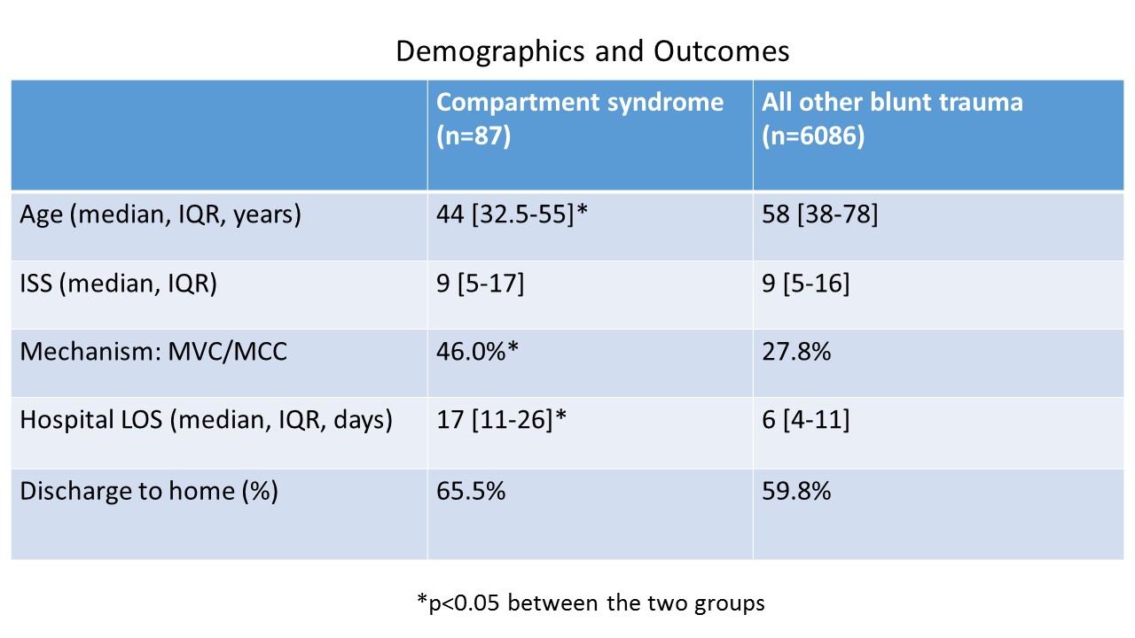
Results: During this time period, there 6173 adult blunt trauma admissions, of which 87 developed extremity compartment syndrome and were further evaluated (table). Their median age was 44 years [IQR 32.5-55] and the vast majority (90.8%) were male. While 76.9% had a National Trauma Data Standard (NTDS) defined comorbidity, 36.8% had ≥ 2 NTDS defined comorbidities. The most common mechanism was MVC (46.0%) followed by fall (23.0%). The median ISS was 9 [IQR 5-17]. The primary severe injury (AIS >3) locations in order of frequency were extremity (63.2%), followed by chest (16.1%), and abdomen (12.6%). Further 96.5% of these patients underwent major surgery, with the overwhelming majority being major orthopedic surgery (96.5%), followed by major abdominal surgery (8.0%). The median hospital LOS was 17 days [IQR 11-26], 34.5% required ICU admission with a median ICU LOS of 6 days [IQR 3.4-19.5], and 34.5% required mechanical ventilation. Further 44.8% had a complication. While 65.5% of admissions were discharged to home, 32.2% did require acute rehabilitation, and 2.3% died during hospitalization. This mortality rate was not significantly different from the rate in other blunt trauma patients (4.0%).

The vast majority (82.7%) of patients had lower extremity compartment syndrome. The most commonly injured bone associated with a compartment syndrome was the tibia (61.5%, n=48); in these 48 patients, it was associated with fibula fractures in 32 patients and with acetabular or other lower extremity fractures in 18 patients. Finally, 10.3% of patients did not have any fracture: 3 had contusions, 1 contusion with venous injury, 1 contusion with arterial injury, 1 sciatic nerve injury, and 3 crush injuries.

Conclusions: The prevalence of extremity compartment syndrome was quite low during this time period at 1.4%. Compartment syndrome most often (82.7%) occurred in the lower extremity, with the tibia being the most frequent fractured bone. However, 37.8% had minor extremity injuries (AIS ≤ 2); 10.3% of patients without a fracture had compartment syndrome. Vigilance is warranted in evaluating the compartments of patients with and without severe extremity injuries, even in the absence of underlying fracture.

76.06 Effects of Mechanism of Injury and Patient Age on Outcomes in Geriatric Rib Fracture Patients

H. Shi2, M. Esquivel1, K. Staudenmayer1, D. Spain1 1Stanford University,Department Of Surgery,Palo Alto, CA, USA 2Case Western Reserve University School Of Medicine,Cleveland, OH, USA

Introduction: Rib fractures are the most common type of chest trauma sustained by elderly patients and are often associated with significant morbidity and mortality in this patient population. Based on studies that show 2-5 times the mortality in patients ≥ 65 with 2 or more rib fractures, our academic institution and Level I trauma center recommends intensive care unit (ICU) admission for the first 24 hours. In this study, we aim to evaluate the utilization of the protocol and outcomes of this patient population.

Methods: A retrospective review was completed of all patients ≥ 65 years of age in our Trauma Registry who sustained isolated rib fractures from January, 2008 to March, 2015 at an urban Level I Trauma center. Data included basic demographics, comorbidities, injuries, length of intensive care unit and hospital stay (LOS), ventilator days, analgesic used, morbidity, mortality and disposition.

Results: Over the 7 year period, a total of 97 patients ≥ 65 years of age with at least one rib fracture and an Abbreviated Injury Score (AIS) of < 2 for other regions were admitted. The median age of these patients was 80 years, 55% were female, and 90% had two or more rib fractures. Fall was the cause of 58% of injuries, while motor vehicle collision (MVC) accounted for 33%. Of the 87 patients with more than one rib fracture, 69 (71%) were not admitted directly to the ICU and 6 (9%) of those were subsequently transferred to the ICU with a mortality of 2.9% for patients with ≥2 rib fractures not admitted directly to the ICU. Of the 4 (4%) mortalities, 3 were caused by falls and 1 by MVC. Patients who fell had a median hospital LOS that was 0.5 to 1 day longer than patients who suffered other mechanisms of injury or were involved in a MVC respectively. Patients aged 70 or older had a median length of stay of 4 days, twice the median length of stay for those between 65 to 69 years old.

Conclusion: With an aging population, trauma in the elderly will continue to increase, likely with a high volume of rib fractures. It is critical to understand the best course of triage and management for this high risk population. We found that falls were the most common mechanism of injury and had the highest median LOS and median number of rib fractures. We also found that patients over the age of 70 had a longer median LOS than those younger and admitting all patients ≥ 65 years of age is likely not necessary, though further research is needed to determine more discrete age and fracture cut-offs.

76.07 Newly Diagnosed Swallowing Dysfunction in Elderly Trauma Patients

D. Laan1, T. Pandian1, D. Morris1 1Mayo Clinic,Trauma Critical Care And General Surgery,Rochester, MN, USA

Introduction: We noticed that some of our elderly patients developed swallowing dysfunction after suffering an injury. We aimed to determine the incidence of swallowing dysfunction in our elderly trauma patient population. Additionally, we wished to evaluate the clinical impact of newly diagnosed swallowing dysfunction in this group and determine risk factors for dysphagia to identify patients that might benefit from screening.

Methods: A retrospective review of our trauma database from 2009 – 2012 was conducted. Injured patients ≥75 years who had newly diagnosed swallowing dysfunction by video swallow study were identified. A comparison group without dysphagia was also identified from this time period that was frequency matched by age, gender, injury mechanism, and injury severity score (ISS). Relevant demographics and injury characteristics were collected along with patient comorbidities, complications (pneumonia, urinary tract infection, stroke, etc.), hospital length of stay (LOS), and discharge disposition. The groups were compared using Fisher’s exact tests for categorical variables and Wilcoxon rank sum tests for interval or continuous variables. Factors which reached statistical significance (p<0.05) on univariable analysis were then used to create a multivariable logistic regression model to determine independent risk factors for dysphagia.

Results: In total, 1323 patients met age and injury criteria. Of these, 56 (4.2%) had newly identified swallowing dysfunction; the majority (n=38, 68%) were male. The average age and ISS were 84.38 (SD=4.54) and 11.29 (SD=6.26), respectively. The majority (85%) of patients were injured due to a fall. Comorbidities were similar between cases and controls. Regional injury patterns were also similar. On univariate analysis, patients with dysphagia had higher rates of pneumonia (15 % vs. 4 %, p<0.05), longer ICU LOS (5.6 vs. 1.9 days, p < 0.01), and longer hospital LOS (11.4 vs. 5.8 days, p < 0.01). In-hospital complication rates were higher in patients with dysphagia (42% vs. 14%, p < 0.01), and these patients had lower rate of discharge home (7% vs. 24%, p<0.01). On multivariable regression, ICU LOS was independently associated with a diagnosis of dysphagia (OR 1.46, p<0.05). All patients who had a tracheostomy were diagnosed with dysphagia.

Conclusion: Newly diagnosed swallowing dysfunction is relatively uncommon in elderly trauma patients, but the clinical implications of newly diagnosed dysphagia are important. Screening for dysphagia with swallowing study should be considered for patients with ICU LOS > 2 days or who had a tracheostomy.

76.08 Pharmacologic stress ulcer prophylaxis may not be needed in ICU patients tolerating enteral nutrition

N. M. Grimmer1, B. McKinzie1, P. L. Ferguson1, E. Chapman1, M. Dorlon1, E. A. Eriksson1, B. Jewett1, S. M. Leon1, A. R. Privette1, S. M. Fakhry1 1Medical University Of South Carolina,Charleston, Sc, USA

Introduction: Stress gastropathy is a complication of ICU stay with high morbidity and mortality, but a low incidence of occurrence. Prophylaxis against stress gastropathy is recommended in national guidelines, and the standard of care is pharmacologic acid suppressive therapy. There are data that support the concept that patients tolerating enteral nutrition have sufficient gut blood flow to obviate the need for prophylaxis; however, no robust studies exist. The current standard in our academic level 1 trauma center is to discontinue pharmacologic prophylaxis once enteral nutrition is providing full caloric requirements. This retrospective cohort study assesses the incidence of clinically significant gastrointestinal bleeding in surgical-trauma intensive care unit (STICU) patients at risk of stress gastropathy secondary to mechanical ventilation in this setting.

Methods: A retrospective chart review was performed of adult patients admitted to the STICU between 2008 and 2013. The primary objective was to assess the incidence of clinically significant gastrointestinal bleeding. Secondary objectives include the rates of ventilator-associated pneumonia (VAP), Clostridium difficile infection (CDI), and mortality. Patients were included if they received full enteral nutrition while on mechanical ventilation. Exclusion criteria included any coagulopathy, glucocorticoid use, prior-to-admission acid suppressive therapy use for other indications, direct trauma or surgery to the stomach, failure to tolerate enteral nutrition goal, and orders to allow natural death. Patients were excluded if pharmacologic stress ulcer prophylaxis was not discontinued during intensive care unit stay.

Results: A total of 239 patients were included. The median age was 42 years, 81.2% were male, and 96.7% were trauma patients. The incidence of clinically significant gastrointestinal bleeding was 0.42%, with a subset analysis of traumatic brain injury patients yielding an incidence of 0.57%. Rates of VAP and CDI were low at 1.24 cases/1000 vent days and 0.71 events/1000 patient days, respectively. Hospital all-cause mortality was 1.7%. An average $242 per patient was saved by discontinuing medication. The incidence of clinically significant gastrointestinal bleeding was comparable to historic rates of bleeding with provision of pharmacologic stress ulcer prophylaxis. The patient population studied is limited to predominantly trauma patients with mechanical ventilation and/or traumatic brain injury as risks for stress gastropathy.

Conclusion: We conclude that stress gastropathy is rare in this population. Surgical trauma patients who are at risk for stress gastropathy may not benefit from pharmacologic prophylaxis once they tolerate enteral nutrition. Pharmacologic prophylaxis was safely discontinued in this patient population. Further investigation is warranted to determine whether continued prophylaxis after attaining enteral feeding goals is detrimental.

76.03 Outcomes after Inpatient Rehabilitation for Trauma Patients

C. W. Lancaster1, P. Ayoung-Chee2 1Emory University School Of Medicine,Atlanta, GA, USA 2New York University School Of Medicine,Department Of Surgery, Division Of Trauma, Emergency Surgery And Surgical Critical Care,New York, NY, USA

Introduction:

Traumatic injury can result in substantial loss of physical function. Of the 18.1% of trauma patients discharged to post-acute care, 19.5% are discharged to an inpatient rehabilitation facility (IRF). Admission to IRF is based on expected functional improvement, but there is little data that documents benefit in injured patients. The purpose of this study was to quantify functional improvements experienced by trauma patients after discharge from IRF and identify predictors of functional improvement.

Methods:

Data were retrospectively collected on patients ≥18 yrs old admitted after injury to a Level 1 trauma center from January, 2012, to March, 2013, and discharged to IRF. Data included demographics, injury characteristics, hospital course (e.g. procedures, length of stay (LOS)), and IRF course (e.g. LOS). The functional independence measure (FIM) was used to measure change in physical and cognitive function from IRF admission to discharge. The FIM score is the sum of scores (range 1 (total assistance) to 7 (total independence)) from 18 domains (13 motor, 5 cognitive).

Results:

There were 136 patients with a mean (SD) age of 56 yrs (21.3). Most (99.3%) were living at home, alone or with support, at time of injury. Mean Charlson Comorbidity Score (CCS) was 0.17 (0.46). Fall was the leading mechanism of injury (44.9%). Median (range) injury severity score (ISS) was 9 (0-45). Median hospital LOS was 8 days (2-89). Mean IRF LOS was 14.3 days (8.07). Due to extenuating circumstances, 11 were transferred to other IRFs and not included. Of the remainder, FIM scores were available for 124. On IRF admission, 43.6% required moderate assistance or greater. From IRF admission to discharge, the mean intra-individual change in FIM score was 28.3 (18.0); 84.7% improved ≥1 levels of independence. On discharge from IRF, 53.2% were at a modified independent level of independence or better; 49.6% were discharged home (with family or agency support); 20.8% were discharged to skilled nursing facility; 21.6% were readmitted to acute care; 8.0% were discharged home independently. Using multivariable analysis, for every 1 yr increase in age, improvement in FIM scores decreased by 0.15 (p=0.05) when adjusted for CCS, primary injury (PI), ISS, intensive care unit (ICU) stay, and IRF LOS. For chest PI, improvement in FIM scores decreased by 17.2 (p=0.04) when adjusted for age, CCS, ISS, ICU stay, and IRF LOS. For every 1 day increase in IRF LOS, improvement in FIM scores increased by 0.65 (p=0.002) when adjusted for age, CCS, PI, ISS, and ICU stay.

Conclusion:

Trauma patients experienced improved functionality after admission to IRF: 80.7% of patients experienced an increase in level of independence. However, despite this improvement, 42.4% of patients were unable to be discharged home. While significant predictors of functional improvement are non-modifiable, they are known at time of acute hospital admission and can help with early resource planning.

76.04 An Examination of Expected and Observed Mortality in the Setting of Mono- And Polytrauma

E. Eklund1, O. Kassar1, N. Napoli2, W. Barnhardt3, L. Barnes2, J. Young1 1University Of Virginia,Department Of Surgery,Charlottesville, VA, USA 2University Of Virginia,Department Of Systems & Information Engineering,Charlottesville, VA, USA 3University Of Virginia,Emergency Services,Charlottesville, VA, USA

Introduction: During mortality gap analysis using our Relative Mortality Metric (RMM) of patient populations separated by Abbreviated Injury Scale-coded regions obtained from our institutional trauma registry, it could be seen that there was little difference in relative mortality between single and multiple injury. Therefore we examined trauma populations to determine whether patients demonstrated statistically significant differences in mortality under mono- and polytrauma conditions.

Methods: We examined data from an initial population of 38000 patient encounters from 1994 to present, yielding a final sample size of approximately 25000. Raw data were cleaned and analyzed using our RMM that describes the overall performance in relation to the anticipated mortality (benchmark TRISS threshold) and observed mortality, where RMM has a range from -1 to 1. Monotrauma (n=8606) and polytrauma (n=16250) were analyzed (defined as a single or multiple AIS-coded injuries in the registry). The relation was further evaluated by stratifying patients into subpopulations based on their overall number of injuries (1, 2-5, 6-10, and 11+) and evaluating mortality differences. Additionally, a second polytrauma definition (>2 injuries with AIS score >2) was used to assess whether patient group mortality differences held. Finally, head monotrauma was separated from the rest of the monotrauma patient population in order to determine if single high mortality head injuries could be driving monotrauma toward greater expected mortality.

Results: When each population was plotted across all AIS values and regions, no significant difference in RMM value between mono- and polytrauma was found (RMM values of 0.3392 and 0.3576, respectively) and each curve fell within the 95% confidence interval of the other. Furthermore, when data was divided by AIS region, the two largest groups (head+face and extremity) mirrored the overall trends. Subsequent analysis of injury number-stratified polytrauma subpopulations again showed no significant difference between mono- and polytrauma. Lastly, when head monotrauma was separated from the rest of the monotrauma population, there was again no significant shift of the curve toward increased patient survival relative to the polytrauma population.

Conclusion: We hypothesized that polytrauma would result in increased mortality due to the compounding effects of multiple sites of injury. However, it was seen that there is no notable difference in the mortality gap as defined by RMM value between different anatomical sites when comparing mono- and polytrauma. This trend held even when confounding factors such as differing levels of polytrauma and the sometimes more severe nature of singular head injuries were controlled for.

76.05 Assessing the Safety and Efficacy of EMS Tourniquet Application: Too Often or Just Right?

E. M. Campion1, V. Orr2, A. Conroy2, J. Gurney2, M. J. Cohen2, R. Callcut2 1Denver Health Medical Center,Surgery,Denver, CO, USA 2University Of California – San Francisco,Surgery,San Francisco, CA, USA

Introduction: Recent military experience with tourniquets has demonstrated safety and efficacy with few complications. These studies have increased enthusiasm for civilian use. Civilian data has lacked injury specifics and many remain concerned about the potential downside to overuse. Using a standard protocol for documenting duration of tourniquet use, this study investigates outcomes following prehospital tourniquet application.

Methods: A retrospective review was performed for prehospital tourniquet application in a metropolitan trauma system from 2011 to 2014. Demographics, injury details, sensorimotor exam, transfusion data, operative treatment, complications and outcome were analyzed. The need for a tourniquet was deemed appropriate if a major artery was injured or the attending trauma surgeon reapplied the tourniquet in the emergency room for significant bleeding.

Results: 38 patients with a total of 39 extremities had tourniquets applied. 1 patient died (3%). Median transport time was 8 minutes. Median ischemia time from placement to removal in ED or OR was 49 minutes. 3 (8%) patients had a prehospital amputation and 4 (11%) had partial amputations. A systolic blood pressure<90 occurred in 9 (26%) prehospital and 12 (32%) in the ED. 5 (14%) required a massive transfusion. 18 (47%) had major arterial injury and 15 (40%) major venous injury. 30 (79%) required emergency surgery with 2 (5%) undergoing fasciotomy. Tourniquets were deemed appropriate in 16 (42%), likely inappropriate in 14 (37%) and questionable in 8 (21%). 17 (47%) patients survived with intact sensation. 11 (31%) survived with a sensation or motor deficit, and 7 (19%) required amputation. No complication could be definitively linked to tourniquet use.

Conclusion: Tourniquet placement in the civilian setting is associated with a high rate of major arterial or venous injury, with a significant majority of patients requiring emergency surgery. Tourniquet application was safe, as we did not identify a single case in which harm could be attributed to tourniquet use. However, further education of prehospital providers is warranted as we identified a number of patients with tourniquets in which there was no clear indication for their use.

75.21 MORTALITY AFTER ANGIOEMBOLIZATION IN THE SETTING OF traumatic hemorrhagic shock

C. E. Nembhard1, J. O. Hwabejire1, W. R. Greene1 1Howard University College Of Medicine,Washington, DC, USA

Introduction: Angioembolization is a life-saving intervention for hemorrhage control after traumatic hemorrhage and is frequently employed in selective non-operative management of solid organ injuries. We examined the risk factors of for mortality after angioembolization in traumatic hemorrhagic shock.

Methods: The Glue Grant database was analyzed. Patients who had angioembolization were included. Survivors were compared with non-survivors. Univariate and multivariable analyses were used to determine predictors or mortality.

Results: A total of 258 patients were included with a mean age of 44 years. Compared to non-survivors, survivors were slightly younger (42±19 vs. 51±20, p=0.004), had lower multiple organ dysfunction score (6±2 vs. 9±3, p<0.0001), lower Injury Severity Score (38±13 vs. 44±16, p=0.003), lower ER lactate (4.5±2.8 vs. 6.6±3.5, p<0.001), and received less blood (2889±2357 vs. 8232±6619, p<0.001) and crystalloids (10756±5272 vs. 16561±10083, p<0.001). Predictors of mortality include age (OR: 1.045, CI: 1.022-1.070, p<0.001), multiple organ dysfunction score (OR: 1.584, CI: 1.302-1.927, p<0.001), ER lactate (OR: 1.177 , CI: 1.031-1.343, p=0.016) and cardiac arrest (OR: 37.185, CI: 7.948-173.976, p<0.001). p=0.002)

Conclusion: Age. Multiple organ dysfunction, ER lactate level, and cardiac arrest are independently associated with mortality following traumatic hemorrhagic shock.

76.01 Three Potential Methods for Prehospital Treatment of Abdominal Hemorrhage

M. J. Hurley3, J. B. Holcomb2,3 2Memorial Hermann Hospital,Department Of Surgery,Houston, TX, USA 3University Of Texas Health Science Center At Houston,Center For Translational Injury Research,Houston, TX, USA

Introduction: Uncontrolled intra-abdominal hemorrhage after injury is associated with increased mortality. Current treatment consists of rapid transport to a trauma center and emergent laparotomy. Three potential prehospital hemorrhage control interventions that could lead to improved outcomes are resuscitative endovascular balloon occlusion of the aorta (REBOA), self-expanding foam and the abdominal junctional tourniquet (AJT). The purpose of this retrospective study was to assess the number of patients who had intra-abdominal injury and received a laparotomy that could have potentially benefited from one of these three new interventions.

Methods: This retrospective study included patients who received an emergent trauma laparotomy between 1/1/2013 to 1/1/2015 at an urban Level 1 trauma center. We reviewed all the injuries found at laparotomy and based on the specific indications for each device determined if the patients would have been potentially helped or hurt by each of the three interventions. For instance, the AJT only controls bleeding from injuries below the aortic bifurcation (located below the umbilicus), while the foam is contraindicated in patients with large abdominal wall defects or diaphragm injuries. McNemar’s test was used with Bonferonni correction for statistical analysis (α=0.017).

Results: 402 patients met the inclusion criteria. REBOA was potentially beneficial for 384, (96%) of patients, foam was potentially beneficial for 351, (87%), while the AJT was potentially beneficial for only 35, (9%). There was no significant difference between REBOA and foam (p=0.022), while there was a difference between REBOA or foam and AJT (p<0.001). There were 170 (42%) patients with penetrating injuries, while only 9 (5%) patients with penetrating injuries would have been potentially helped by the AJT. 33 patients had cutaneous wounds located in the right lower quadrant (RLQ), while 45 patients had cutaneous wounds located in the left lower quadrant (LLQ). Diaphragm injuries occurred in 58 (14%) of patients, with REBOA potentially of benefit in 55 of those patients, while foam would have potentially helped only 36.

Conclusion: REBOA and foam both would potentially benefit the largest number of patients, (≥ 87%) who had intra-abdominal injury and laparotomy. AJT was found to be helpful in only 9% of patients in need of prehospital intervention for hemorrhage. These results suggest that the AJT should not be used for prehospital hemorrhage control unless it is absolutely sure that all injuries causing hemorrhage are below the aortic bifurcation.

76.02 The Impact of Smoking on Mortality and Failure to Rescue in Operative Trauma

T. Orouji Jokar1, P. Rhee1, A. Azim1, N. Kulvatunyou1, T. O’Keeffe1, A. Tang1, R. Latifi1, D. J. Green1, L. Gries1, R. S. Friese1, B. Joseph1 1The University Of Arizona,Trauma/Surgery/Medicine,Tucson, AZ, USA

Introduction: The role of chronic smoking has been well established with adverse outcomes in several surgical disciplines. However, its effect on outcomes after trauma operations has never been explored. The aim of this study was to assess the effect of active smoking on outcomes in patients who underwent surgery after trauma.

Methods: We did a two year (2011-2012) analysis of national trauma data bank. We included all critically injured (minimum AIS≥3, and required ICU admission during their stay) adult trauma patients who underwent surgical procedure on day 1. Patients, who died in ED, arrived with no vital signs, intoxicated, and fatally injured (AIS≥6) were excluded from the analysis. Our outcome measures were in-hospital complications, mortality, failure to rescue (FTR), hospital and ICU length of stay, and days on ventilator. Patients were divided into two groups based on their smoking status. Groups were matched using propensity score matching after adjusting for age, gender, race, injury severity, head AIS, admission vitals, and comorbidities.

Results: After propensity score matching 17,198 patients (Smokers: 8,599, Non-Smokers: 8,599) were included in the analysis. Mean age of the population was 46±18, 75% were male, median [IQR] ISS was 16 [10-25], median [IQR] AIS was 4[3-4], and overall mortality in our population was 8%. There was no difference in basic demographics of the groups. Overall rate of complications was significantly higher in smokers compared to non-smokers (52% vs 47%, p<0.001) and they were more likely to develop surgical site wound complications compared to non-smokers (4% vs 3%, p=0.02). Smokers had longer ICU length of stay (Median [IQR]: 3[2-7] vs. 3[2-6], p=0.04) and ventilator days (Median [IQR]: 0[0-3] vs. 0[0-2], p=0.02) compared to non-smokers. Overall mortality rate (6% vs 9%, p<0.001) and failure to rescue (4.6% vs 6%, p<0.001) were significantly lower in smokers compared to non-smokers.

Conclusion: Overall in-hospital and post-surgical complications were significantly higher in current smokers; however, it appears from our study that after controlling for comorbidities and other patient factors, active smokers have a survival advantage. Further investigation in elucidating the physiological and mechanistic basis of this association is required.

75.18 Surgical Site Infections in the Trauma and Acute Care Surgical Populations

J. R. Burgess1, T. J. Novosel1, B. Knuckles1, L. D. Britt1 1Eastern Virginia Medical School,Surgery,Norfolk, VA, USA

Introduction: For many years, the rates of surgical site infections (SSIs) have been stratified according to wound classification by the Centers for Disease Control (CDC) and American College of Surgery-National Surgical Quality Improvement Program (ACS-NSQIP). However, these predicted rates are used universally for all surgical procedures and may not be applicable to the emergency surgery population. Acute care surgery (ACS) and trauma patients are at an increased risk for infection for a variety of reasons. This study was designed to determine whether the rates of surgical site infections in ACS and trauma patients are higher than the accepted standards.

Methods: A retrospective review of patients admitted to the trauma and ACS services at a level I trauma center that underwent midline laparotomy between July 2011 and June 2013 was performed. Patients were excluded if death occurred within one week of admission or if the abdomen remained open for over 72 hours. Demographic data, risk factors, ASA classification, type of surgery, wound classification, length of stay and presence of either a deep organ space infection or superficial SSI were analyzed. The rates of surgical site infections were stratified by wound classification and compared to the CDC rates of superficial SSIs and the ACS-NSQIP rates of deep organ space infections. All results noted were significant if p<0.05.

Results: During the study period there were 178 patients on the ACS service and 107 patients on the trauma service that underwent midline laparotomy and met inclusion criteria. Rates of superficial SSIs were not significantly different between historic CDC rates and our trauma patient population across all wound classes. Using the ACS-NSQIP reported data, there were significantly more deep organ space infections in contaminated wounds (27.3% vs 2.6%, p<0.05). In the ACS patient population, there were significantly more superficial SSIs in the clean/contaminated wounds (27.7% vs 11%, p<0.05) when compared to CDC rates. There were also significantly more deep space organ infections in the clean/contaminated, contaminated and dirty wounds when compared to the ACS-NSQIP data (10.8% vs 1.9%, 30% vs 2.6% and 25% vs 4.5%, respectively, p<0.05).

Conclusion: Trauma and acute care surgery patients have a significantly higher rate of deep organ space infections than the standard estimates when compared to ACS-NSQIP published data. This is likely due to the emergent nature of the cases and increased comorbidities in this patient population. Larger studies should be done to determine if this is true nationwide. If so, a different standard of surgical site infections should be adopted for these patient populations to more accurately reflect the true rate of postoperative infectious complications in these patients.

75.19 Study of the Relationship between Outcomes of Severe Traumatic Brain Injury and Coagulopathy

P. Chang1, W. Chong1 1First Affiliated Hospital Of China Medical University,Emergency Department,Shenyang, LIAONING, China

Introduction: Traumatic coagulopathy usually follows severe traumatic brain injury (sTBI).The present study aimed to assess the relationship between coagulation parameters and outcomes in sTBI.

Methods: A total of 58 sTBI patients with Glasgow Coma Scale (GCS) <9, head Abbreviated Injury Scale (AIS)≥3 and all other body regions AIS<3 were recruited in this retrospective study. The age, gender, GCS score, pupil reaction, median line shifting and biochemical parameters including blood platelet (PLT), prothrombin time (PT), activated partial thromboplastin time (aPTT), fibrinogen (Fg) and blood glucose of sTBI patients on admission were retrieved from the medical record database. The patients were divided into good outcome group whose Glasgow Outcome Scale (GOS) was between 3 and 5 and poor outcome group whose GOS was between 1 to 2.In single-factor analysis, P≤0.15 was defined as statistical significance. The significance of parameters was determined with non-conditional multivariate Logistic regression analysis and those with P≤0.10 were used to generate receiver operating characteristic curves (ROCs).

Results: There were 41 males and 17 females ranging from 12 to 88 years old. The occurrence of TBI-associated coagulopathy is 31.03%. Compared with good outcome group, poor outcome group had significantly increased occurrence of abnormal pupil reaction[24(77.4%) vs 10(37.0%),P=0.002], occurrence of median line shifting[21(67.7%) vs 7(25.9%),P=0.001], blood glucose[mmol/L: 9.1(6.53-12.85)vs6.9(4.90-7.88),P=0.000], PT[second:14.5(13.18-16.53) vs 13.8(12.45-15.18),P=0.004],and aPTT[second:37.56(6.06) vs 34.32(7.98),P=0.087],lower GCS[6(1.75-3.32) vs 5(1.82-5.24),P=0.000], PLT[×109/L:167.33(48.8) vs 191.45(75.247),P=0.149]and Fg[g/L:2.55(1.75-3.32)vs2.98(1.82-5.24),P=0.021]. In addition, there was no significant difference of GCS between patients with coagulapothy and non-coagulopathy. The multivariate Logistic regression analysis demonstrated that pupil reaction (β=-3.650,OR=0.026,P=0.068), median line shifting (β=-1.990,OR=0.137,P=0.082), GCS (β=0.716,OR=0.195,P=0.081), PT (β=-1.200,OR=0.031,P=0.082), aPTT (β=0.293,OR=1.340,P=0.073), and blood glucose (β=-1.636,OR=0.195,P=0.078) were the independent risk factors for unfavorable outcome in patients with sTBI. Pairwise comparisons of the AUC were as follows: GCS versus PT, p = 0.516, blood glucose versus PT, p=0.227, GCS versus blood glucose, p=0.579, GCS versus aPTT, p=0.033?blood glucose versus aPTT?p=0.006, PT versus aPTT, p=0.142.

Conclusion: Prolonged PT and aPTT are the independent risk factors for unfavorable outcome in patients with sTBI. It suggests that there is a strong association between coagulopathy and an unfavorable outcome of sTBI.

75.20 Size Matters: Defining the Size tor Neurosurgical Intervention With Isolated Intra-Cranial Injury

V. Pandit1, P. Rhee1, N. Kulvatunyou1, A. Tang1, T. O’Keeffe1, L. Gries1, G. Vercruysse1, R. Latifi1, R. S. Friese1, B. Joseph1 1University Of Arizona,Trauma Surgery,Tucson, AZ, USA

Introduction: The size of the intracranial hemorrhage (ICH) in patients with traumatic brain injury is an important determinant for neurosurgical intervention (NI). However; the optimum size for NI remains unclear. The aim of this study was to define the optimum size of isolated ICH (epidural [EDH] or SDH [SDH]) for NI in patients with TBI.

Methods: We performed a 5 year retrospective analysis of a prospectively collected database of all patients with an isolated EDH or SDH to our level 1 trauma center. The outcome measure was need for neurosurgical intervention. . Neurosurgical intervention was defined as craniotomy and/or craniectomy. Receiver operating characteristic (ROC) curves were plotted to identify the optimal ICH size for EDH and SDH for need for neurosurgical intervention. Sub-analysis was performed for patient on antiplatelet/anti-coagulation therapy (AP/AC).

Results: A total of 905 patients (isolated SDH: 729, isolated EDH: 176) were included of which; 21.2% (n=192) patients underwent NI. In patients with SDH, size of 6 mm was the optimum threshold for NI (AUC: 0.80, p=0.01) while in patient with EDH, size of 6.5 mm optimum threshold for NI (AUC: 0.78, p=0.01).

On multivariate regression analysis after adjusting for demographics, neurological examination, AP/AC therapy, injury severity, patient with SDH≥6 were 3.9 times (OR: 3.9, CI: 2.5-5.2) more likely while patient with EDH ≥ 6.5 were 3 times (OR: 3, CI: 1.7-4.9) more likely to have neurosurgical intervention

On sub-analysis of patients on AC/AP therapy, size of 4.5 mm was the optimum threshold for NI for both patients with SDH (AUC: 0.79, p=0.001) and EDH (AUC: 0.77, p=0.02).

Conclusion:Our study defines the optimal size of ICH in patients with isolated SDH and EDH for the need for NI. In patient with SDH/EDH 6mm is the optimal threshold determining the need for NI while it is lower at 4.5mm in patient on AP/AC therapy. This will help establish guidelines based on the size of ICH for better defining the criteria for NI in patients with TBI and isolated ICH.

75.15 Age Predicts Discharge Disposition but Not Adjusted Mortality After Nonoperative Management of TBI

Q. Dang1,2, S. Moradian1,2, J. Catino1, L. Zucker1, I. Puente1, F. Habib1, M. Bukur1 1Delray Medical Center,Department Of Trauma And Surgical Critical Care,Delray Beach, FL, USA 2Larkin Community Hospital,Department Of Surgery,Miami, FL, USA

Introduction: Traumatic Brain Injury (TBI) continues to be a leading cause of death and disability particularly in the elderly population. Age is generally considered to be a risk factor for adverse outcomes after TBI. We sought to examine the impact of age on outcomes in TBI patients who do not require neurosurgical operative intervention.

Methods: This was a retrospective review of all patients with survivable head injuries undergoing nonoperative management at a Level I trauma center from 2008-2013. Patients were stratified by age into young and elderly groups using the age of 40 as a reference group based upon the current Brain Trauma Foundation guidelines. Logistic regression was used to adjust for baseline differences in demographic and injury variables to determine the effect of age on outcomes. The primary outcomes were in-hospital mortality, worsening discharge GCS, and discharge disposition.

Results: 1,869 patients met inclusion criteria with 77% of patients being older than 40 years. Elderly patients were more likely to be victims of falls and presented with a higher GCS despite having a higher Head AIS. Immediate need for non-intracranial operative intervention was greater in younger patients. After adjusting for differences in characteristics, there was no significant difference in overall mortality (Elderly 3.6% vs. young 5%, p=0.209) or worsening discharge GCS (14% vs. 11%, p=0.926). However, younger patients were more likely to be discharged to a rehabilitation facility or home (91% vs. 70%, AOR=2.4, p=0.001). Stratification of mortality by decade revealed similar results, with adjusted mortality being lower in the sixth and ninth decades of life (Figure).

Conclusion: Survival of patients sustaining TBI not requiring neurosurgical operative intervention may not be age dependent. However, age is associated with a less favorable discharge disposition that is independent of discharge GCS.

75.17 Secondary Overtriage in Level 3 and 4 Trauma Centers: Are These Transfers Necessary?

R. M. Essig1, K. T. Lynch1, D. M. Long2, U. Khan1, G. Schaefer1, J. C. Knight1, A. Wilson1, J. Con1 1West Virginia University,Surgery,Morgantown, WV, USA 2West Virginia University,Biostatistics,Morgantown, WV, USA

Introduction:
‘Secondary overtriage’ refers patients discharged home shortly after being transferred to another hospital. Although minimally injured patients could potentially be discharged home, a recent regional study showed that 9.8% of these seemingly uninjured patients seen at level 3 and 4 trauma centers were transferred to a higher level of care prior to being discharged. We analyze these occurrences at the national level to identify patient characteristics and injury patterns which may facilitate the development of strategies to reduce overtriage rates.

Methods:
Data from the National Trauma Data Bank was obtained from 2008-2012 to isolate those who were: 1) discharged home within 48h of arrival, and 2) did not undergo a surgical procedure. We then identified those who arrived as a transfer to a Level 1 or 2 Trauma Center prior to being discharged (secondary overtriage) from those who were discharged directly from a Level 3 or 4 Trauma Center. Factors associated with transfer were analyzed using a logistic regression. Injuries were classified based on the need for a specific consultant. Co-morbidities examined included: smoking status, functionally dependent status, diabetes, and history of cerebrovascular accidents.

Results:
Descriptive analysis showed a number of differences between transfers and non-transfers due to our large sample size. Those who were not transferred tended to be older than 65 years (21.3% vs. 15.5%) and female (35.3% vs. 26.9%). Rates of hypotension (SBP<90mmHg) were low (0.6% vs. 1.3%) and those with ISS>15 were more likely to be transferred (10.5% vs. 3.7%). No clinically significant differences in co-morbidities were found between groups. We examined the more siginificant variables using a logistic regression controlling for age, gender, ISS, SBP<90, and injury pattern (Table 1). Neurosurgical injuries, vertebral injuries, and facial injuries were associated to an increased risk of transfer, while orthopedic injuries were protective.

Conclusion:
Secondary overtriage results from a combination of limited hospital resources, patient characteristics, and injury pattern. Although some injuries such as mild traumatic brain injuries and stable spine fractures are managed expectantly, it seems that they may be the underlying reason behind many of these transfers. A fear of an underdiagnosed or a progressing central nervous system injury may prompt a referral to a specialist for evaluation. Orthopedic injuries may involve a mechanism isolated to a single limb and therefore be considered more stable. Further studies into how these types of injuries are treated at Level 3 and 4 trauma centers may help reduce inefficiencies stemming from unnecessary transfers.

75.13 Discharge Destination as an Independent Risk Factor for Readmission of the Older Trauma Patient

D. S. Strosberg2, B. C. Housley3, D. Vazquez1, A. Rushing1, C. Jones1 1Ohio State University,Department Of Surgery, Division Of Trauma, Critical Care, And Burn,Columbus, OH, USA 2Ohio State University,Department Of Surgery,Columbus, OH, USA 3Ohio State University,College Of Medicine,Columbus, OH, USA

Introduction: Unplanned readmissions are a major quality measure used to evaluate hospital care. Readmissions after traumatic injury are frequent, and older trauma patients are at increased risk for poor outcomes in both morbidity and mortality. Determining an appropriate destination after discharge in this population is difficult, and may impact readmission rates. Prior literature evaluating discharge destination’s impact on patient outcome is limited and conflicting; no prior study has evaluated this relationship in older trauma patients. The objective of this study was to explore the association between the discharge destination and rate of 30-day readmission in older trauma patients.

Methods: A database of all patients age 45 years or older undergoing trauma evaluation at our American College of Surgeons verified level 1 trauma center over a 1-year period was used to retrospectively compare frequency of 30-day readmission to the center between patients discharged to home, to inpatient rehabilitation facilities, and to other extended care facilities (ECFs, including long term acute care hospitals, skilled nursing facilities, and nursing homes). Further abstracted potentially confounding factors were trauma activation level, injury severity score, comorbidity-polypharmacy score, age, hospital length of stay (LOS), ICU LOS, Glasgow coma score, gender, pre-trauma functional status (independent, partially dependent, or dependent), and pre-trauma residence (home, rehab, or ECF). Inmates, patients who died during their hospitalizations, and patients who were discharged to hospice were excluded from analysis. Univariate analysis was undertaken using chi-square testing. Multiple logistic regression was performed with all the above variables to evaluate for independent contribution to readmission risk.

Results: 960 patients age 45 and older were evaluated over the study period; 81 (8.4%) were excluded and 879 patients age 45-103 were included in the analysis. Seventy-six patients (8.6%) were readmitted within 30 days of discharge, including 6% of patients discharged to home, 14% discharged to ECF, and 19% discharged to rehab (p=0.00009 on univariate analysis). 557 (63%) patients had data recorded for all variables analyzed using multiple logistic regression; among these, only discharge destination was independently associated with the rate of readmission (p=0.019).

Conclusion: Unplanned hospital readmission following traumatic injury is common in older patients and is used as a marker of hospital quality. In this first study of outcomes based on discharge destination of older trauma patients, discharge to inpatient rehabilitation or other extended care facilities was a strong independent risk factor for hospital readmissions. Though causes of this association are likely multifactorial, recognition of this risk factor may aid in the disposition planning of these patients and suggests the need for further evaluation of this correlation at other centers.

75.14 Time to Pelvic Embolization ts Increased in Those Presenting After Business Hours and on Weekends

R. J. Miskimins1, L. R. Webb1, S. D. West1, A. N. Delu2, S. W. Lu1 1University Of New Mexico HSC,Department Of Surgery,Albuquerque, NM, USA 2University Of New Mexico HSC,Department Of Radiology,Albuquerque, NM, USA

Introduction:
Multiple methods of hemorrhage control associated with pelvic fractures have been described. At our Level 1 Trauma center, the primary method used is placement of a pelvic binder followed by angioembolization. Angioembolization is performed by a interventional radiology (IR) team which is not in house at night or on weekends. We hypothesized that individuals with pelvic hemorrhage requiring embolization who present to the emergency department (ED) after business hours have an increased time to angioembolization, require more blood products, and have a higher mortality compared to those presenting during business hours.

Methods:
The IR database was used to identify individuals who underwent emergent pelvic angioembolization secondary to blunt trauma from January 2008 to December 2013. These were divided into, the business hours (BH) group, defined as those presenting to the ED between 7:30 AM and 5:30 PM Monday to Friday and the after business hours group (ABH) defined as those presenting to the ED on weekends, holidays or between the hours of 5:30 PM and 7:30 AM. A chart review was used to obtain the time of ED presentation, presence of contrast extravasation on CT, start time of angioembolization, units of packed red blood cells (PRBC) transfused and if the patient went to the operating room prior to IR. The trauma database was used to obtain initial vitals, demographics, ISS and mortality. Continuous variables were analyzed with the Mann Whitney U test and categorical data was analyzed using the Fisher exact test.

Results:
Ninety nine patients meet inclusion criteria (64 ABH vs 35 BH). There was no difference in initial vitals or demographics. The ISS was similar between the groups (median, 27 ABH vs 26 minutes BH). A blush was present on CT in 63% of the ABH vs 57% in the BH (p=0.67). 25% of the ABH went to the OR prior to IR vs 17% in the BH (p=0.45). There was no difference in PRBCs transfused (median, 6.5 ABH vs 5 BH, p=0.27). There was a significant difference in the time to IR (median, 304 minutes ABH vs 219 minutes BH). Ten patients died within 30 days in the ABH vs 6 in the BH group (p=0.78). There were five deaths from hemorrhage in the first 24 hours in the ABH compared to one in the BH group (p=0.32).

Conclusion:
Individuals who arrive after hours, on weekends, or holidays who require angioembolization to control pelvic hemorrhage, require more time to arrive in the IR suite for management. Although they require more time to arrive in the IR suite, our data does not demonstrate an increase in 30 day mortality, the number of PRBCs transfused, or the number of patients who died from hemorrhage in the ABH group. A limitation of our study is the ability to determine the number of patients who died in both groups while waiting for IR. We have demonstrated there is an increased time to pelvic angioembolization in those presenting outside of business hours, however, there is no difference in mortality if the patient is able to arrive in IR.

75.10 Lactate Clearance as a Predictor of Mortality in Trauma Patients with and without TBI

S. E. Dekker1, H. M. de Vries1, W. D. Lubbers1, P. M. Van De Ven2, A. Toor3, F. W. Bloemers4, L. M. Geeraedts4, P. Schober1, C. Boer1 1VU University Medical Center,Anesthesiology,Amsterdam, NH, Netherlands 2VU University Medical Center,Epidemiology And Biostatistics,Amsterdam, NH, Netherlands 3VU University Medical Center,Center For Acute Care, VU Medical Center Region,Amsterdam, NH, Netherlands 4VU University Medical Center,Surgery,Amsterdam, NH, Netherlands

Introduction: Impaired lactate clearance is predictive of mortality in general trauma patients. The relationship between this biomarker and outcome in traumatic brain injury (TBI) patients, however, is still unknown. This retrospective study examined the association between lactate clearance and mortality in trauma patients with and without TBI.

Methods: Lactate values of trauma patients admitted to the emergency department between 2010-2014 were retrieved from patient files. Patients without initial lactate drawn within 30 minutes after admission, or without a second lactate measurement within 8 hours after admission, were excluded. Lactate clearance was calculated based on modified methods described by Odom et al. (2013) [(Lactateinitial – Lactatedelayed) / Lactateinitial × 100%]; Régnier et al. (2012) [(Lactateinitial – Lactatedelayed) / Lactateinitial × 100% × Delay−1]; and Billeter et al. (2009) [four groups: (1) always below 2.5mmol/L, (2) decreasing below 2.5mmol/L, (3) increasing above 2.5mmol/L, and (4) always above 2.5mmol/L]. We studied the association between lactate clearance and in-hospital mortality in patients with isolated TBI, TBI combined with extracerebral injuries, and trauma patients without TBI.

Results: Of the 3000 admitted trauma patients, 818 (27.2%) had an initial lactate measurement. 367 patients (12.2%) were eligible for lactate clearance calculations. A high initial lactate was associated with a higher in-hospital mortality [OR = 1.20, 95% CI: (1.13 – 1.27), P<0.001]. We found a significant relationship between the lactate clearance method of Billeter et al. and in-hospital mortality (Wald chi-square = 14.614, P=0.002). Post-hoc pairwise comparisons only revealed a significant difference between groups 1 and 4 [Bonferroni corrected P=0.002, OR = 3.59, 95% CI: (1.86 – 6.93)]. There was no association between lactate clearance and in-hospital mortality using the methods of Odom et al. and Régnier et al. Initial lactate value and Billeter’s lactate clearance method did not differ in their ability to predict in-hospital mortality [AUC 0.64, 95% CI: (0.56 – 0.71) vs. AUC 0.64, 95% CI: (0.57 – 0.71), P=0.71]. Neither initial lactate nor lactate clearance differed between isolated TBI, polytrauma + TBI, and general trauma without TBI patients.

Conclusion: This is the first study to investigate the relationship between lactate clearance and outcome in TBI patients, and the first to compare three previously described lactate clearance metrics. In contrast to the available literature for the general trauma patient, only lactate clearance using the method of Billeter et al. predicted mortality in trauma patients with and without TBI. However, in our patient population we found no clinical advantage of using Billeter’s lactate clearance method, as the initial lactate value was equally effective in its ability to predict in-hospital mortality.

75.11 Comparison of Average Pre-Transfusion Hemoglobin Trigger in the SICU

N. Provenzale1, M. Cripps1,2, T. Chung1, C. Townsend1, W. Huda1, C. T. Minshall1,2 1Parkland Health And Hospital System,Dallas, TX, USA 2University Of Texas Southwestern Medical Center,Division Of Burn, Trauma And Critical Care,Dallas, TX, USA

Introduction:
Adverse effects of blood transfusions on patient outcomes are well known; however, adherence to best practice by providers is often inconsistent. We assessed whether our transfusion practice varied respective of which primary service admitted the patient to the Surgical Intensive Care Unit (SICU). We hypothesized that average pre-transfusion hemoglobin levels for patients on elective surgery services, Otorhinolaryngology (ENT), Vascular, and general surgery, would be significantly higher than hemoglobin triggers for Emergency General Surgery (EGS) and Trauma services, despite the Surgical Intensive Care Unit (SICU) operating as a closed unit.

Methods:
The expected practice of the SICU team is to transfuse packed red blood cells (PRBCs) according to the standards established by the TRICC trial. We used a semantic layer construct to identify pre-transfusion hemoglobin levels for SICU patients between June 2014 and May 2015. Patients with uncontrolled hemorrhage were excluded. Self-service electronic health record (EHR) data reporting was used to aggregate patients by primary surgical service and retrieve medical history, BMI, and age for transfused patients. Independent samples T-tests and Fischer’s exact tests were used to assess differences between groups

Results:
403 surgical patients were transfused ≥ 1 units of PRBCs during the study period. Average pre-transfusion hemoglobin level was significantly lower for EGS compared to the ENT and Vascular services. The average pre-transfusion hemoglobin level for Trauma was also significantly lower compared to Vascular (see Table). Two or more units were transfused in 37% of transfusion instances. EGS was the only service found to have a significant difference in average pre-transfusion hemoglobin for patients that received one unit (7.19 ± 0.28) versus two units (6.72 ± 0.44; p=0.042). The prevalence of coronary artery disease was similar between all services. ENT patients were significantly older than EGS (p=0.016) and Trauma (p<0.01) patients, while Vascular patients were significantly older than Trauma patients (p<0.01). EGS patients were more likely to have a history of diabetes mellitus as compared to both Trauma and ENT patients (p=0.01, p=0.039) and more likely than Trauma patients to have hypertension (p=0.041).

Conclusion:
This review of the transfusion practice in our SICU demonstrates variability in transfusion practice for patients on ENT and Vascular services as compared to the other services and is not justified by an increased incidence of comorbid conditions. We also have a high incidence of multiple unit transfusions. The results of this review will allow us to focus our provider education and strive to improve our performance.

75.12 Injury Level Impacts Dysphagia Incidence in Elderly Patients with Non Operative C-Spine Fractures

J. Pattison2, U. Pandya1 1Grant Medical Center,Trauma Services,Columbus, OH, USA 2Ohio University College Of Osteopathic Medicine,Heritage College Of Osteopathic Medicine,Athens, OH, USA

Introduction: Dysphagia is a common condition in the elderly with significant complications such as aspiration, pneumonia, and potentially increased mortality. This problem is even more magnified in elderly patients with cervical spine fractures as neck positioning and altered physiology may further predispose these patients to complications. Fractures of the cervical spine can span the entire length of the neck from C1 to C7. It stands to reason that injuries at different levels of the cervical spine could influence swallowing function in unique ways. This study seeks to investigate how the level of cervical fracture in elderly patients who are non-operatively managed impacts the incidence of dysphagia.

Methods: Medical records of all trauma patients age 65 and older admitted with cervical fractures between January 2008 and April 2014 to a level 1 trauma center were retrospectively reviewed. Patients with a past medical history of dysphagia or stroke or who had operatively managed fractures were excluded, leaving 123 patients for analysis. Bedside evaluation of swallowing function was performed and any patients with evidence of swallowing dysfunction had formal speech therapy consultation. Dysphagia was defined as any restriction or diet modification as a result of swallowing dysfunction after speech therapy assessment. Demographic data, hospital length of stay, intensive care unit days, ventilator days, injury severity score, mortality and level of cervical fracture were analyzed. P values < 0.05 were considered statistically significant. Multiple regression analysis was used to control for confounding variables.

Results: A total of 123 patients met inclusion criteria and 19 (15.4%) of those patients had dysphagia Patients with dysphagia were older (86.3 ± 8.01 vs. 80.6 ± 8.76, p = 0.007), had higher hospital length of stay (7.4 ±4.23 vs 4.3 ± 4.21, p = 0.016), and were more likely to have intensive care unit days (47.4% vs 20.2%, p=0.01).. Patients with C1 fractures were more likely to have dysphagia than patients with other cervical fractures (29.2% vs 7.1%, p = 0.0008). After using regression analysis to control for total length of stay, any intensive care unit days, and age, C1 fracture increased the likelihood of dysphagia by 4 times (OR = 4.0; 95% CI 1.2-13.0)

Conclusion: Incidence of dysphagia is significant in geriatric trauma patients with cervical spine fractures and has important ramifications for patient outcomes such as hospital length of stay. Patients with C1 fractures are at increased risk for dysphagia and these patients may benefit from more vigorous surveillance to prevent subsequent complications.