75.07 Extended Venous Thromboembolism Prophylaxis After Trauma: A Disparity of Beliefs and Practices

C. McCoy1, J. H. Lawson1, M. Shapiro1, C. Sommer1 1Duke University Medical Center,Durham, NC, USA

Introduction: Multiply injured trauma patients are at high risk for venous thromboembolism (VTE) and inpatient chemoprophylaxis is standard of care. However, the time period when a patient is at risk for VTE may extend beyond the inpatient stay. The indication for and utilization of extended VTE prophylaxis following discharge have not been previously described.

Methods: A survey regarding current use of inpatient and extended VTE prophylaxis was distributed to trauma surgeons at Level 1, 2 and 3 trauma centers nationwide with the assistance of the Trauma Center Association of America. Practice patterns including indications for prophylaxis, agents used, and duration of extended VTE prophylaxis were studied.

Results: 80 surgeons, the majority (80%) of whom staff Level 1 trauma centers, responded from 56 hospitals. Nearly all (98%) respondents believed the risk of VTE was elevated following traumatic injury. All respondents utilized VTE chemoprophylaxis for trauma inpatients. Although 72 (90%) responding surgeons believed certain trauma patients should receive chemoprophylaxis following discharge, only 50 (62%) routinely discharged these patients on extended prophylaxis. The most common indications for extended VTE prophylaxis identified by respondents were prolonged immobility and orthopedic trauma. The most common agents utilized were low molecular weight heparin and aspirin. Specific drug dosage and duration were highly variable.

Conclusion: Current practice patterns surrounding extended VTE prophylaxis in trauma patients are highly variable despite widespread belief that extended prophylaxis is warranted in certain patients. Indications, efficacy, and risk stratification of extended prophylaxis regimens should be a focus of additional prospective research in the trauma population.
75.08 Management of Major Hepatic Trauma in The Era of Damage Control Resuscitation

K. M. Ibraheem1, P. Rhee1, A. A. Haider1, N. Kulvatunyou1, T. O’Keeffe1, A. Tang1, R. Latifi1, G. Vercruysse1, L. Gries1, R. Friese1, B. Joseph1 1University Of Arizona,Trauma Surgery,Tucson, AZ, USA

Introduction: Major hepatic trauma (AAST grade ≥4) classically has been associated with mortality as high as 80%. With improvements in resuscitation, increased utilization of angioembolization, and non-operative management, morality associated with these injuries is expected to change. The aim of this study was to assess our experience with the management of major hepatic injuries, to determine if there is a difference in mortality with the use of different techniques for hemorrhage control, and to determine factors associated with mortality.

Methods: We performed a 4 year (2009-2012) retrospective analysis of all patients with major hepatic injuries (AAST grade ≥4) who presented to our trauma center and abstracted information regarding patient injuries, physiological parameters, resuscitation details, radiological findings, and operative details. Our outcome measures were mortality. Multivariate regression and ROC curve analysis were performed.

Results: A total of 98 (Grade 4, 73; Grade 5, 25) patients with a mean age of 30.6 ±19.1 years and mean ISS of 26 [18 – 34] were included. 27.6% (n=27) had other solid organ injuries and 12.2% (n=12) had juxtahepatic vascular injuries. 26.5% (n=26) patients required massive transfusion; and mean pRBC and FFP packs transfused were 8±11 and 5±8 units respectively. 66.3% (n=65) patients required operative intervention and mean estimated blood loss was 2500 mL. The most common hemorrhage control techniques performed were packing (36.7%, n=36) and hepatorraphy (34.7%, n=34) followed by Pringle’s (18.4%, n=18), hepatotomy (8.2%, n=8), non-anatomical resection (4.1%, n=4), omental packing (3.1%, n=3), total hepatic isolation (3.1%, n=3), atriocaval shunt (1.0%, n=1), and lobectomy (1.0%, n=1). Angioembolization was performed in (8.2%, n=8) patients. Overall mortality rate was 28.6% (n=28). Mortality rate with packing (70.6%) was significantly higher as compared to hepatorraphy (18.8%) and angioembolization (0.0%; p<0.001). The strongest predictor of mortality was 24hours pRBC transfusion (OR [CI]: 1.8 [1.3-2.6]; p=001). ROC curve analysis revealed >4.5units of pRBC transfusion as the most sensitive cutoff associated with mortality.

Conclusion: The use of angioembolization and hepatorraphy for hemorrhage control are associated with significantly lower mortality compared to packing after major hepatic trauma. Greater than 4.5 units of pRBC requirement is the most sensitive cutoff associated with mortality in these patients.

75.09 A Nursing Team Trained in Ultrasound-Guided IV Placement Improves Infection Rates in the SICU

J. R. MacDermott1, C. H. Cook3, J. Flaherty1, C. Jones2 1Ohio State University,Wexner Medical Center,Columbus, OH, USA 2Ohio State University,Department Of Surgery, Division Of Trauma, Critical Care, And Burn,Columbus, OH, USA 3Beth Israel Deaconess Medical Center,Department Of Surgery,Boston, MA, USA

Introduction: Central venous catheters (CVCs) are often necessary during critical illness, but are associated with higher rates of blood stream infections than peripheral intravenous catheters (PIVs). To augment ongoing efforts to reduce central line-associated bloodstream infections (CLABSIs), we sought to limit durations that CVCs are in place by placing PIVs and removing CVC as quickly as possible. Because PIV placement in critically ill patients is often difficult, we developed a team of surgical intensive care unit (SICU) nurses trained to use ultrasound (US) to place PIVs in SICU patients. We hypothesized that training SICU nurses to use US guidance for PIV placement in critically ill patients would reduce CVC catheter days and thereby reduce the rate of CLABSI.

Methods: We retrospectively reviewed data gathered prospectively from 2011 through 2014 regarding individual attempts by the nursing team at US-guided PIV (US-PIV) placement and further evaluated pre-existing SICU quality databases of central line usage, patient census, and incidence of CLABSI from 2009 to 2014. Monthly rates of central line usage were calculated from central line days and patient days. Rates of CLABSI were evaluated quarterly as infections per 1000 central line days. Differences between rates before and after implementation of the US-PIV team were analyzed via Kruskal-Wallis testing.

Results: From 2011 to 2014, 2748 attempts at US-PIV placement were recorded, with placement of 2440 PIVs in 1646 patients (success rate 89%). Rates of central line usage trended down from 61.8 central line days per 100 patient days before US-PIV team implementation (monthly range 49.6-76.0) to 58.7 central line days per 100 patient days after its implementation (38.6-81.5, p=0.208). CLABSI incidence decreased substantially from a rate of 2.40 per 1000 patient days prior to US-PIV team creation (quarterly range 1.48-3.46) to 0.85 per 1000 patient days thereafter (0-2.43, p=0.0009).

Conclusion: Properly trained SICU nurses can be highly successful at US guided PIV placement. Implementation of this technology was associated with decreased central line usage and CLABSI when used in conjunction with other infection-reducing efforts. Effective CLABSI prevention requires a multifaceted approach; creation of a nursing team trained to place PIVs using ultrasound may be a useful adjunct to standard practices in improving these quality measures.

75.04 Implementation of a Trauma Activation Checklist at an Academic Trauma Center

D. Ruter1, J. Um1, D. Evans1, C. Boulger2, C. Jones1 1The Ohio State University College Of Medicine,Department Of Surgery, Division Of Trauma, Critical Care And Burn,Columbus, OH, USA 2The Ohio State University College Of Medicine,Department Of Emergency Medicine,Columbus, OH, USA

Introduction: The unpredictable nature of trauma evaluation makes it vulnerable to human error, often communication related and occurring at times of handoff. In 2014, our university-based American College of Surgeons verified Level 1 trauma center developed a ‘Trauma Activation Checklist’ as a joint project between the Departments of Emergency Medicine and Surgery. The checklist included three sections: ‘sign in’ for introductions and preparation prior to patient arrival; ‘EMS timeout’ for attention to handoff by prehospital providers; and ‘sign out’ for planning imaging, consults, and patient disposition upon leaving the trauma bay. The checklist was posted prominently in each trauma bay and discussed during annual and monthly trauma team orientations. We sought to evaluate checklist use during the transition to new teams in July 2015.

Methods: During July 2015, trained observers monitored arrivals and evaluations of a convenience sample of patients seen as Level 1 (severely injured) and Level 2 (moderately injured) trauma activations. For each patient, they recorded the level of activation, time of day, and use of each checklist component as ‘checked’ or ‘not checked’. Failure to verbally acknowledge a checklist component led to its recording as ‘not checked’. No personally identifying data regarding patients were recorded. Descriptive statistics were performed to analyze use of the checklist.

Results: Our center evaluated 130 patients via trauma activation in July 2015. 46 evaluations (35%) were observed for checklist adherence, divided between daytime (7am-5pm, 16 patients, 35%), evening (5-10pm, 23, 50%) and nighttime (10p-7a, 7, 15%). The least frequently completed section was the sign out; only 7 (15%) of the trauma activations completed this section in its entirety. All sections of the checklist were more commonly completed in Level 1 trauma activations than in Level 2 trauma activations (sign in: 41% vs 24%; EMS timeout: 65% vs 38%; sign out: 24% vs 14%).The most commonly missed individual components in the sign in section were placing pre-arrival orders and obtaining equipment access. In the EMS timeout section, obtaining referring institution records was most commonly missed, while assuring intensive care unit handoff was the component most overlooked in the sign out section.

Conclusions: Despite prominent incorporation into trauma team training and its highly visible position in the trauma bay, a trauma activation checklist is used suboptimally in organizing the arrival and initial evaluation of injured patients at our academic medical center. Teams are particularly deficient in completing the sign out section and verbalizing forthcoming plans for patient care, especially for patients deemed less critically injured. Improvements to the checklist itself and to checklist adherence and its effects on information exchange and patient care are ongoing.

75.05 Changing Lives, Changing Lanes – A New Role for Vehicle Deformity and GCS in Trauma Triage?

M. Subrize1, B. A. Asif1, M. Mukit1, H. C. Thode1, M. C. Henry1 1Stony Brook University Medical Center,Department Of Emergency Medicine,Stony Brook, NY, USA

Introduction:
In 2001, Suffolk County, New York implemented the use of the trauma prehospital care report (TPCR), which included the current American College of Surgeons (ACS) trauma triage criteria, a vehicle diagram to identify the location of damage, body diagrams to identify the location and type of bodily injury and the use of safety equipment. We sought to use the TPCR in conjunction with Trauma Registry data to examine which criteria were predictive of patient outcomes and resource use.

Methods:
We conducted a retrospective study of adult TPCR cases matched with the County’s regional Trauma Registry. All patients with a TPCR from 2003 to 2007 were eligible for inclusion. Outcomes included severe injury (Injury Severity Score > 16), need for major OR procedure (MOR), and death. Univariate analysis of triage criteria vs. outcomes was shown as relative risks. Multivariate analysis was performed using stepwise logistic regression analysis. Predictors were included if they were significant at the 0.05 level. Stepwise receiver operating characteristic (ROC) curves were also made using triage criteria as discrete (binary) predictors. At each step the sensitivity and specificity of each remaining criterion was calculated, and the criterion with the largest YI was accepted as the next predictor. This method was repeated until all remaining predictors had a YI of < 5.

Results:

17001 TPCR’s were analyzed. The most common cause of injury was due to motor vehicle collision. Nearly every trauma triage criterion was significantly associated with need for MOR, severe injury, and death. GCS <14 and flail chest were the most related to mortality (OR=12.4 CI 5.8-25.6; 11.4, 2.3-57.3 respectively).

GCS <14 and vehicle deformity were associated with every outcome and often the first criteria accepted into an outcome model. Mechanism criteria were included in 3 of the 6 outcome models. Speed over 40 mph was a useful indicator for ISS>15 or Major OR, but not death. The model with the highest sensitivity and specificity used mortality as the outcome (81% / 85%). Vehicle rollover, vehicle ejection, death of same vehicle occupant and pulse were not accepted into any models. Only death as an outcome had high sensitivity; all models had adequate specificity (78-85%).

Conclusion:
The current study explored different triage criteria and found vehicle deformity >20” and GCS <14 had the best predictive value in traumas causing death and were significant predictors in all other models as well. Vehicle rollover, vehicle ejection, death of same vehicle occupant and pulse had little impact on outcomes. The findings were discordant with other studies evaluating the 2011 trauma triage guidelines, and offer a glimpse into a unique environment, which includes a high percentage of automobile collisions.

75.06 Injury Type Predicts Extubation Failure Despite Successful Spontaneous Breathing Trials

A. Adkins2, U. Pandya1 1Grant Medical Center,Trauma Services,Columbus, OH, USA 2The Ohio State University,College Of Medicine,Columbus, OH, USA

Introduction: Extubation failure and subsequent reintubation is associated with longer intensive care unit and hospital stay and worse outcomes when compared to successfully extubated patients. For many institutions, a successful spontaneous breathing trial is the primary criteria to determine readiness for extubation. Patients with traumatic injury, however, represent a unique population that requires special consideration. It seems reasonable that injury types such as rib fractures and cervical fractures could influence success of extubation. Relatively few studies on this topic have focused on the trauma patient population and their specific injury types. The purpose of this investigation is to determine if specific types of traumatic injury predispose patients to extubation failure. We hypothesize that rib fractures and cervical spine fractures are risk factors for failed extubation despite having a successful spontaneous breathing trial.

Methods: This study was a retrospective chart review of all trauma patients admitted to Grant Medical Center over a 6 year period from January 2009 to December 2014. All injured patients on mechanical ventilation over the age of 15 were considered for the study. Patients that were endotracheally intubated and subsequently passed a spontaneous breathing trial were included. Exclusion criteria were self-extubation, tracheostomy placement prior to attempted extubation, palliative withdrawal of life support, or death prior to an extubation attempt. Patients requiring re-intubation within 72 hours of extubation were determined to have failed extubation and were compared to patients who had successful extubation. Differences between the two groups were analyzed with a p value < 0.05 signifying statistical significance. Multiple regression analysis was used to control for age, injury severity score, and vent days prior to extubation.

Results: A total of 1,613 patients met inclusion criteria with 103 (6.4%) of those in the failed extubation group. Patients who failed extubation were older, had higher injury severity score, more ventilator days prior to extubation, and more intensive care unit and hospital days. Patients who failed extubation were more likely to have cervical spine fractures (21.4% versus 8.2%, p = <0.0001) and more likely to have rib fractures (39.8% versus 23.4%, p = 0.0002).

Conclusion: Cervical spine fractures and rib fractures are risk factors predictive of failed extubation despite successful spontaneous breathing trial. These patients may require additional weaning criteria to better determine chances of successful liberation from mechanical ventilation.

75.02 Do Transferred Patients Increase the Risk of Venous Thromboembolism in Trauma Centers?

B. K. Yorkgitis1, O. Olufajo1, G. Reznor1, J. M. Havens1, D. Metcalfe1, Z. Cooper1, A. Salim1 1Brigham And Women’s Hospital,Trauma, Burns And Surgical Critical Care,Boston, MA, USA

Introduction: It is well known that trauma patients are at increased risk for venous thromboembolism (VTE). Because many patients require initial stabilization followed by transfer to a higher level of care, administration of VTE prophylaxis may often be delayed. It is unclear if these patients are actually at increased risk for VTE. As VTE has become a quality metric for trauma centers, it is important to know if there is an actual increased risk for patients transferred.

Methods: The National Trauma Database (NTDB) v6.2 (2007-2012) was used to identify patients admitted to Level I and II trauma centers. Patients receiving anticoagulants, <18 years, or pregnant were excluded. Patients transferred <24 hours were compared to non-transferred patients with respect to age, sex, race, patient-level risk factors for VTE, and VTE rates. Multivariable logistic regression models were used to identify risk factors for deep venous thrombosis (DVT), pulmonary embolism (PE), and VTE. All calculations were done with SAS 9.3 SURVEYLOGISTIC and SURVEYFREQ procedures to include NTDB weights, strata and clustering to create nationally representative estimates. Alpha was set on P= 0.05.

Results:There were 736,374 trauma patients identified for analysis during with 189,166 (25.7%) patients transferred to a level I or II trauma center within 24 hours of injury. A total of 11,619 (1.5%) developed VTE including 9,149 (78.8%) with DVT and 3,167 (21.2%) with PE. The VTE rate was significantly higher in the transferred group compared to the non-transferred group (1.73% vs. 1.42%, p=0.002). Significant differences exist with respect to DVT (1.39% vs. 1.14%, p=0.004) and PE (0.45% vs. 0.37%, p=0.003) between transferred and non-transferred patients respectively. Multivariable analyses adjusting for patient-level risk factors demonstrated that transferred patients were independently associated with a higher likelihood of VTE (OR 1.15; 95% CI: 1.06 – 1.25, P=0.001 ), PE (OR 1.18; 95% CI: 1.08 – 1.30, p<0.001), and DVT (OR 1.13; 95% CI: 1.04 – 1.25, p=0.007).

Conclusion: Adult trauma patients transferred to a level I or II center that initially presented to another facility are at increased risk for VTE. Further studies are necessary to determine the causes for this increased risk. Accepting a transferred trauma patient results in an increased VTE risk and may not be reflective of the quality of trauma care at the receiving facility.

75.03 Loss of Health Insurance after Traumatic Spinal Cord Injury

J. K. Canner1, E. R. Hammond5, E. B. Schnieder1,3,4, A. Asemota1, S. Selvarajah2 1Johns Hopkins University School Of Medicine,Surgery,Baltimore, MD, USA 2Kennedy Krieger Institute,International Center For Spinal Cord Injury,Baltimore, MD, USA 3Harvard School Of Medicine,Brookline, MA, USA 4Brigham And Women’s Hospital,Center For Surgery And Public Health,Boston, MA, USA 5Johns Hopkins University School Of Medicine,Psychiatry And Behavioral Sciences,Baltimore, MD, USA

Introduction: Individuals are at risk of losing their insurance if they are severely injured or have a disabling illness, such as following a traumatic spinal cord injury (TSCI). A factor that may affect patients’ ability to address all their healthcare needs following a TSCI, particularly the potential life-long need for neuro-rehabilitation to improve neurological and functional outcomes, is their insurance status. We sought to describe time to insurance loss after TSCI in a population with employer-sponsored insurance.

Methods: We used the 2010-2012 Truven Health Systems MarketScan® database which includes approximately 45 million patients under the age of 65 years who are covered through an employer-sponsored insurance plan. All hospitalized patients with a primary diagnosis of TSCI or a secondary diagnosis of TSCI accompanied by a primary diagnosis of other major trauma were included in the sample. Patients were followed in the database until discontinuation of enrollment in a MarketScan-affiliated insurance plan or until December 31, 2012, and the duration of insurance coverage was calculated. Log-rank tests and univariable Cox proportional hazards regression were used to compare TSCI patients with the general population and to compare the time to coverage loss for patients with and without spinal fracture and traumatic brain injury (TBI). Multivariable Cox proportional hazards regression was used to adjust for age, sex, and injury beneficiary status (e.g., employee, spouse, or child).

Results: A total of 5,231 patients were hospitalized for a TSCI during the study period. Of these, 44.8% had a primary diagnosis of TSCI, 70.5% had an associated vertebral fracture, and 21.0% had a complete TSCI. Two-thirds of the patients (67.1%) were male and the median age was 41 years (IQR: 23-54). An estimated 43.8% of the population with TSCI lost their insurance before the end of the study period, compared to 33.9% in the general population (HR=1.18, p<0.001). After adjusting for potential confounders, the hazard ratio was 1.23 (95% CI: 1.17-1.29, p<0.001). Among patients with a TSCI, there was no difference in the risk of insurance loss between those with and without a concomitant spinal fracture (HR=1.03, p=0.61), but there was a significant difference between those with and without a concomitant TBI (HR=1.17, p=0.026).

Conclusion: Approximately 4 in 10 patients with TSCI lost their employer-sponsored health insurance during the study period, a proportion significantly higher than that seen in the general population. Further research is needed to identify modifiable factors that could mitigate the extent of, and rate of loss of insurance among patients with TSCI.

74.18 Validation Of A Checklist For Postoperative Wounds In Vascular Surgery

R. Gunter1,3, J. Wiseman1, S. Fernandes-Taylor1, Y. Ma2, S. Saha2, S. Clarkson3, D. Yamanouchi4, K. C. Kent1,3 1University Of Wisconsin,Wisconsin Institute Of Surgical Outcomes Research,Madison, WI, USA 2University Of Wisconsin,Department Of Biostatistics,Madison, WI, USA 3University Of Wisconsin,Department Of Surgery,Madison, WI, USA 4University Of Wisconsin,Department Of Surgery, Division Of Vascular Surgery,Madison, WI, USA

Introduction

Surgical site infection (SSI) is the most common nosocomial infection and reason for readmission in surgical patients, particularly in vascular surgery patients who experience the highest readmission rate for surgical populations. Technology-based transitional care that allows digital image-based wound monitoring has the potential to detect and facilitate treatment of SSI at an early stage. We undertake a study to validate the use of smartphone digital photography to evaluate postoperative vascular surgery wounds.

Methods

We developed a wound assessment checklist using previously validated criteria. All patients 18 and older who underwent a vascular surgery procedure involving a surgical incision at least 3cm in size between May and December 2014 were recruited from a high-volume tertiary-care academic vascular surgery service. Vascular surgery attending surgeons, physician assistants, nurse practitioners, and registered nurses evaluated wounds using the assessment checklist in-person; a different group of providers evaluated the wound via a Smartphone digital photograph. Inter-rater reliability for (1) wound assessment and (2) treatment plan was measured using Gwet’s Agreement Coefficient (AC) for the in-person group, the photograph group, and between groups. We used 1000 bootstrap samples with replacement to calculate 95% CI of Gwet’s AC; the samples were drawn by wound.

Results

We assessed 80 wounds (23 lower extremity wounds, 20 groin wounds, 18 abdominal wounds, 10 neck wounds, 5 upper extremity wounds, and 4 amputation stumps) in-person with a median 2 raters per wound (minimum of 1 rater and maximum of 4 raters), and via photograph with 9 raters per wound. Agreement between in-person and image-based assessments was high with respect to course of treatment with substantial agreement for antibiotics (AC=0.76) and need for debridement (AC=0.89). Relative to in-person raters, raters evaluating high-quality images were more likely to detect redness (OR=2.23, p<0.001), drainage (OR=2.1, p<0.01), and indicate the need for an ED visit (OR=6.42, p<0.001). Raters evaluating low-quality images were more likely to indicate the need for an ED visit (OR=5.69, p<0.001), but less likely to detect drainage (OR 0.13, p=0.005), than in-person raters. Less experienced clinicians were more likely to indicate the presence of ecchymosis (OR=1.47, p=0.03), more likely to indicate a need for wound drainage (OR=1.89, p=0.03), and less likely to suggest surgical debridement (OR=0.51, p=0.03).

Conclusions

Smartphone digital photography is a valid method for evaluating postoperative vascular surgery wounds that is comparable to in-person evaluation across most domains of wound abnormality. The inter-rater reliability for determining treatment recommendations was universally high. Remote wound monitoring and assessment may play an integral role in future transitional care models to decrease readmission for SSI in vascular surgery patients.

74.19 Primary Fascial Closure After Damage Control Laparotomy: Factors Affecting Outcome

M. A. Khasawneh1, D. H. Jenkins1, H. J. Schiller1, D. J. Dries2, M. D. Zielinski1 1Mayo Clinic,Trauma, Critical Care, General Surgery,Rochester, MN, USA 2University Of Minnesota,General Surgery,St. Paul, MN, USA

Introduction: Our recently completed prospective, randomized, placebo controlled clinical trial studying the effects of flaccid paralysis of the lateral abdominal wall muscles after damage control laparotomy had unexpectedly high rates of primary fascial closure (PFC) whether botulinum toxin or placebo was injected (98% vs 96%). In order to ensure patient safety, numerous exclusion criteria were applied. We aim to compare PFC rates between enrolled and excluded patients, and identify factors associated with lack of PFC.

Methods: A retrospective review of all patients who were screened for enrollment into the prospective trial was performed. The principal exclusion criterion for each excluded patient was utilized for analysis. Patients who died before an attempt at fascial closure were excluded from the current analysis. Data is presented as means [standard deviation] for continuous variables and as rates for discrete variables.

Results: 153 patients were included in the current analysis, 46 of who were enrolled in the trial and 107 were excluded. Excluded patients were older, more likely to have cardiac disease, hypertension, peripheral vascular disease, and use corticosteroids. There were no differences in sex, BMI, diabetes mellitus, renal disease, connective tissue disorder, anticoagulation, malignancy, and smoking status between the groups. The rates of PFC were significantly greater in included patients (93.5% vs 76.5%, p=0.007). 30 day mortality was significantly different between the two groups (4.4 vs 27.9%, p=0.0003). Multivariate analysis demonstrated that COPD, BMI >50 and cirrhosis were independent predictors of lack of PFC.

Conclusion: The study identifies factors associated with lack of PFC. Future trials examining the effect of botulinum toxin injection in high risk patients are needed.

74.20 Alkaline Ocular Burns in the United States

R. S. Haring1,2,3,4, I. D. Sheffield5, J. K. Canner3, A. H. Haider1,2, E. B. Schneider1,2,3,4 1Harvard School Of Medicine,Brookline, MA, USA 2Brigham And Women’s Hospital,Center For Surgery And Public Health,Boston, MA, USA 3Johns Hopkins University School Of Medicine,Johns Hopkins Surgery Center For Outcomes Research,Baltimore, MD, USA 4Johns Hopkins Bloomberg School Of Public Health,Dept. Of Health Systems And Policy,Baltimore, MD, USA 5Brigham Young University,Provo, UT, USA

Introduction: Ocular chemical burns are a severe form of injury, often leading to long-term morbidity, including reduced visual acuity and quality of life. A review of the current literature on ocular burns found that chemical burns are most common among working-age men, suggesting that these types of injuries can have a substantial impact on quality-adjusted life years. Alkaline burns, which penetrate deeper and faster into the cornea, are the most severe form of chemical burn, and are associated with significantly poorer outcomes than acid burns. Currently, there are no large-scale epidemiologic studies that describe the incidence and burden of alkaline ocular burns in the United States. We sought identify and characterize trends, incidence, and characteristics related to patients presenting to Emergency Departments (EDs) in the US from 2006-2012 with alkaline ocular burns.

Methods: Using the Nationwide Emergency Department Sample (NEDS), we identified all patients who presented with an ICD-9-CM diagnosis of alkaline ocular burn from 2006-2012. We examined burden and trends related to age and sex to determine which populations were at highest risk of alkaline ocular burn injury. Age-specific population-based rates were calculated using the US Census Bureau’s intercensal and postcensal estimates. Chi square tests were used to compare subsets across time and between age and sex groupings.

Results: Patient age at ED presentation for treatment of alkaline ocular burns followed a bimodal distribution, with the highest population-based injury rates occurring among 1-year old infants (1.29 per 100,000), with a lower rate among two-year-olds (0.811 per 100,000). The second-highest injury-rates were found among adults in their twenties (e.g., 1.28 per 100,000 among 28-year olds). Overall, 55.2% of alkaline burn injuries occur among 20-45 year olds, and among this group, 67.9% of patients were male. Mean patient age at presentation was 35.5 years (95% CI: 34.7-36.3), and did not vary by sex (p=0.991).

Conclusion: Alkaline ocular burns are among the most critical and severe forms of ocular injury. Earlier studies and reviews in the literature identified working-age men as a high-risk cohort; however, we discovered that the single highest-risk age-based rate of ED presentation with alkaline ocular burns is found among one-year-old children. These findings may be used to aid in the design and implementation of appropriate prevention efforts, which may range from appropriate storage of chemicals in the home to policies on protective eyewear in the workplace. Special attention must be paid to preventing access to alkaline agents among the youngest of children and educating parents on the need for rapid appropriate response when alkaline ocular burn is suspected in an infant.

75.01 Impact and Treatment Success of New Onset Atrial Fibrillation with RVR Development in the SICU

M. Brown2, F. Luchette2, W. Chaney2, S. Nassoiy2, T. Plackett2, R. Blackwell2, M. Engoren3, J. Posluszny2 3University Of Michigan Medical Center,Ann Arbor, MI, USA 2Loyola University Medical Center,Loyola University Stritch School Of Medicine,Maywood, IL, USA

Introduction: Atrial fibrillation (AF) with rapid ventricular rate (RVR) frequently affects non-cardiac post-operative (NCPO) surgical patients. The development of post-operative AF is associated with poor cardiovascular outcomes and mortality. There are no well established guidelines for treatment of NCPO surgical patients who develop AF with RVR. The objective of this study was to evaluate the practice patterns for evaluation and treatment for NCPO surgical patients diagnosed with new onset AF with RVR in the surgical ICU (SICU).

Methods: All adult patients (≥ 18 years) admitted to the SICU from June 2014-June 2015 were retrospectively screened for the development of new onset AF with RVR (HR >100). AF with RVR was confirmed by cardiologist reviewed EKG. Patient medical records were reviewed for demographics, hospital course, evaluation and treatment of AF with RVR, and outcome (cardiology consult, rate and rhythm (R/R) control within 48 hours, and in-hospital mortality). Data are presented as mean±SD with statistical comparisons performed with student’s t-test or Fisher exact test.

Results: There were 1070 patients admitted to the SICU during the study period. Fifty-seven patients developed AF with RVR, 33 of whom were new-onset AF with RVR (3.1%). For these 33 patients, the average age was 71±11 with 19 males (58%). Cardiology consult was obtained for 18 patients (55%). Twenty-six patients (79%) had R/R control within 48 hours of AF with RVR onset. Seven patients died while hospitalized (21%) with 8 discharged patients (31%) readmitted within 30 days. β-blockers were the most commonly used initial medication (67%). When used first, β-blockers were successful at R/R control in only 27% of patients (6/22). Amiodarone had the highest rate of success if used initially (5/6, 83%), and if used as a second treatment (11/13, 85%). Cardiology consult was not associated with improved mortality (28% vs. 13%; p=0.41) or success with R/R control within 48 hours (61% vs. 100%; p=0.009). Failure to control R/R was associated with a greater number of cardiac comorbidities (100% vs 57%; p=0.06), increased BMI (31.8±3.6 vs 26.1±4.4; p=0.006), and less volume overload (3260±2484 mL vs. 6630±4131 mL; p=0.06). In-hospital mortality was associated with being a trauma/acute care surgery patient (71% vs 15%; p=0.009), need for vasopressors at AF onset (71% vs. 27%; p=0.07), and higher SAPS II score (40.7±9.5 vs. 29.7±12.2; p=0.04).

Conclusion: New onset AF with RVR was an uncommon development in the SICU but was associated with a high mortality. Treatment with amiodarone, rather than traditional β-blocker, was associated with R/R control. Failure to establish R/R control was associated with cardiac comorbidities, but not volume overload. These results will help to form our future algorithm for the treatment of AF with RVR in the SICU and will be prospectively studied.

74.16 Abdominal Wall Reconstruction in Cytoreduction and Hyperthermic Intraperitoneal Chemotherapy

J. Rosado2, J. B. Oliver2, J. Patel2, K. M. Spiegler2, K. Houck3, R. J. Chokshi1 1New Jersey Medical School,Surgical Oncology,Newark, NJ, USA 2New Jersey Medical School,Surgery,Newark, NJ, USA 3New Jersey Medical School,Obstetrics And Gynecology,Newark, NJ, USA

Introduction: Cytoreduction surgery (CRS) and Hyperthermic Intraperitoneal Chemotherapy (HIPEC) offer the best opportunity for long term survival for peritoneal metastasis for colorectal, appendicular, and ovarian cancers. There are times in which to achieve proper cytoreduction, abdominal wall resection is required. These resections in part the need for abdominal wall reconstruction while some patients require reconstruction for other reasons as well. It is known that chemotherapy and hyperthermia in part increased risks for wound infections, poor wound healing and further complications. Therefore, we looked at the outcomes in our series of CRS-HIPEC patients taking a look at the outcoomes, morbidity and mortality of those patients requiring abdominal wall reconstruction compared to those who did not.

Methods: Demographics, comorbidities, intraoperative variables, and post-operative outcomes for all patients undergoing CRS/HIPEC at a single institution from 2012 to 2015 were analyzed. Variables were examined with chi squared or Wilcoxon Ranked Sum Test where appropriate. Survival was analyzed with Kaplan Meier curves and Cox Proportional Hazards Regression..

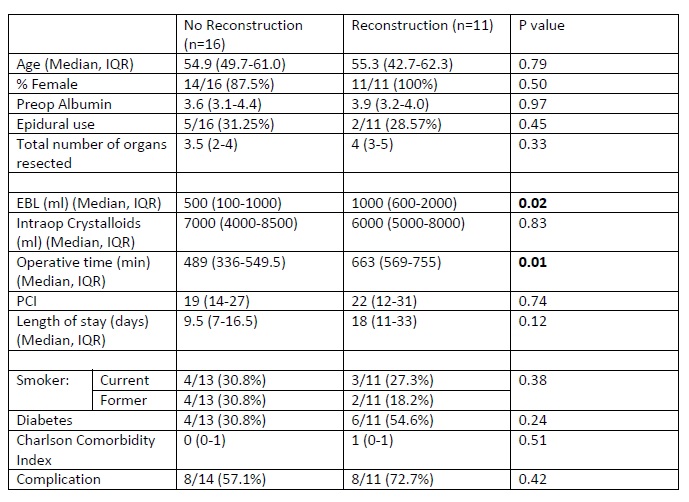
Results:During this time frame, 27 individuals underwent CRS-HIPEC. There were 11 patients which underwent reconstruction, while 16 patients had no reconstruction. These patients were similarly aged and gender distribution. They had similar epidural use and similar number of organs removed. There was no difference in Peritoneal Carcinomatosis Index for the two groups (22 vs 19 p=0.74). Patients undergoing reconstruction tended to have increased operative time (663min vs 489min p=0.01) as well as increased blood loss (1000ml vs 500ml p=0.02). Both groups had similar complication rate (72.7% vs 57.1% p=0.42). Median follow up for the no reconstruction group was 4 months compared to 1.7 months for the reconstruction group. Five of the 16 individuals within the no reconstruction group and 3 of the 11 individuals with reconstruction died during the follow up period. The survival rate for the two groups were similar (68.8% vs 72.7%, p=0.44).

Conclusion:Thus the need for abdominal wall reconstruction had no impact on patient’s mortality but did impact their operative time and blood loss. Further evaluation with larger series and longer follow up is needed to confirm these findings.

74.17 Correlation of Verified Burn Centers with CDC Burn Related Mortality: A Statewide Analysis

T. Orouji Jokarokar1, B. Joseph1, A. Hassan1, n. kulvatunyou1, E. Tran1, A. tang1, R. latifi1, d. J. Green1, l. Gries1, R. S. friese1, p. rhee1 1University Of Arizona,trauma/Surgery/Medicine,Tucson, AZ, USA

Introduction: Early referral to a verified burn center (VBC) is a critical factor in determining outcomes of a burn patient. However; variability exits in the presence of VBCs across states and the impact of VBCs on patient’s outcomes remains unclear. The aim of this study was to assess the association between VBC’s and burn related mortality across states in the United States (US).

Methods: Burn related mortality was abstracted over a one year (2013) period from the Center of Disease Control and Prevention. VBCs were obtained from the American Burn Association burn center registry. Population and area of each state were recorded from the US census data. Level of trauma center was recorded from the American College of Surgeons. States were dichotomized as: VBC and No-VBC. Linear regression analysis was performed.

Results:A total of 44 states were included of which, 50% states had a VBC. The overall burn related mortality rate was 1.47±0.5/100,000. States with VBC had lower burn related mortality rate compared to No-VBC states (1.16±0.3 vs 1.78±0.5, p=<0.001). The presence of a VBC in a state reduces the mortality rate by 0.433 per 100,000 per year.

Conclusion:Presence of a VBC significantly reduces burn related mortality within a state. Factors associated with this discrepancy need further exploration, which may help define the actual need for a verified burn center in each state.

74.12 Identification of two new mutations in BPES phenotype

B. J. Sumpio1, D. Balkin4, D. Scott2, P. Le Pabic3, T. Schilling3, D. Narayan1 1Yale University School Of Medicine,Plastic And Reconstructive Surgery,New Haven, CT, USA 2Baylor College Of Medicine,Molecular And Human Genetics,Houston, TX, USA 3University Of California – Irvine,Developmental And Cell Biology,Orange, CA, USA 4University Of California – San Francisco,Plastic And Reconstructive Surgery,San Francisco, CA, USA

Introduction:

Blepharophimosis-ptosis-epicanthus inversus syndrome (BPES) is a rare disfiguring disease that results in abnormal faces. Although originally thought to be a purely soft tissue disorder, recent evidence suggests that orbital dysmorphism is also part of the disease. This includes a more lateral orbital wall, deeper orbits and flattened projections of the orbital rims. The lateral orbital wall is vertical, the orbit is deeper than normal and there is flattened projection of the orbital rims. The orbital volume can be less than normal and the supraorbital rim can be notched. The constellation of physical features are generally isolated to the periorbital region and may have some or all of the listed traits.

The physical manifestations were originally described as the result of a mutation a transcription factor gene—FOXL2 -3q23. However, 105 mutations have been associated with BPES-like phenotypes. Here we investigate a novel, previously unreported pair of genes which result in BPES when mutated.

Methods:
A male patient with BPES was identified along with the parent and siblings who had similar facial morphology. Physical features and anthropometric measurements were recorded. Whole blood samples were obtained and genomic DNA extracted. Whole exome sequencing was performed and candidate mutations identified. Sanger sequencing was performed with appropriate primers to confirm. The entire coding region of the FOXL2 gene was resequenced via the Sanger method to confirm the absence of FOXl2 mutations.

Results:

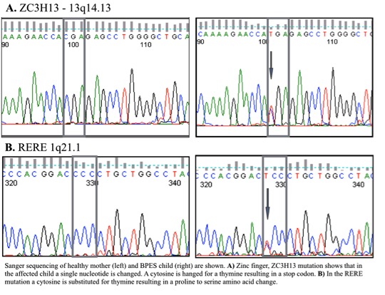
Phenotypic features of this disease were found in 2 generations of living relatives (father, 2 male children and female child) As well as documented in the grandparents as well. The inheritance demonstrated a Mendelian autosomal dominant pattern with 100% penetrance. Genetic analysis confirmed that a conserved mutation was responsible for the progression of disease, while whole exome sequencing identified candidate genes ZC3H13, and RERE with a nonsense and missense mutation, respectively.

We have identified a patient with orbitoblepharophimosis and, together with the father, the subjects were found to have a normal FOXL2 gene sequence, which was originally thought to manifest the disease. Whole exome sequencing and Sanger sequencing confirmed that FOXL2 was normal. The point mutation in ZC3H13 results in a premature stop codon of a gene which is known to be a strong transcription factor for FOXL2. Addition the single point mutation in RERE changes a cytosine for a thymine resulting in a proline to serine amino acid change.

Conclusion:

We have identified a missense and a nonsense mutation that together result in the BPES phenotype. Furthermore we have shown that FOXL2, a gene initially thought to be responsible for the mutation, to be completely normal in these patients.

74.13 Tumor Inhibition by Excisional Wounds in an Immune Competent Mouse Allograft Model

M. S. Hu1, T. Leavitt1, J. Gonzalez1, C. Marshall1, S. Malhotra1, L. Barnes1, A. T. Cheung1, G. G. Walmsley1, G. Gurtner1, A. J. Giaccia1, P. Lorenz1, M. T. Longaker1 1Stanford University,School Of Medicine,Palo Alto, CA, USA

Introduction:
We previously demonstrated that the placement of an adjacent splinted full-thickness excisional wound will inhibit human xenograft tumor growth in an immunocompromised mouse by outcompeting for neovascularization. Herein, we further explore this fascinating observation by placement of a full-thickness excisional wound both adjacent and directly over a mouse allograft tumor

Methods:
Mouse breast cancer 4T1 cells (5 x 10⁴) were injected into the left mid-dorsum of BALB/c mice. After engraftment was confirmed at 7 days, either a splinted full-thickness excisional wound was created adjacent to the tumor on the right mid-dorsum or an unsplinted full-thickness excisional wound was created directly over the engrafted tumor. Wounds were 6 mm in diameter. Tumor growth was assessed via 3-way caliper measurements taken every other day.

Results:
Tumors with an adjacent splinted full-thickness excisional wound were smaller than control tumors without a wound at 28 days post-wounding with a volume of 4639.14 mm3 versus 3742.32 mm3 and a weight of 3.27 g versus 2.47 g, respectively (*p<0.05). In addition, mice that had tumors and unsplinted full-thickness excisional wounds created directly over the tumor had inhibited growth and prolonged survival compared to mice with tumors alone (*p<0.05).

Conclusion:
These data show that placement of a full-thickness excisional wound adjacent to or directly over a tumor inhibits growth in an immune competent mouse allograft tumor model. These data have novel implications for ulcerated tumors and tumors that undergo an incomplete surgical resection. Further research promises to identify the mechanism for this inhibition.

74.14 Analysis of Gene Expression in E14 versus E18 Fetal Fibroblasts

M. S. Hu1, S. Malhotra1, W. Hong1, M. Januszyk1, G. G. Walmsley1, A. Luan1, D. Duscher1, D. Wan1, G. C. Gurtner1, M. T. Longaker1, P. Lorenz1 1Stanford University,School Of Medicine,Palo Alto, CA, USA

Introduction:
Early fetuses heal without scar formation, yet the biological mechanism behind this process is largely unknown. We aimed to examine fetal fibroblasts, which are intimately involved with the wound healing and scar formation process, in different stages of development to characterize differences that may contribute to the switch from wound regeneration to repair.

Methods:
Fetal fibroblasts were harvested and cultured from the dorsal skin of time-dated BALB/c embryos. Total RNA was isolated and microarray analysis was performed using chips with 42,000 genes. Significance analysis of microarrays (SAM) was utilized to select genes with greater than 2-fold expression differences between the wounds with a false discovery rate (FDR) of less than 2. Enrichment analysis was performed on significant genes to identify differentially expressed pathways.

Results:
Comparison of gene expression profiles revealed 275 genes that were differentially expressed between E14 and E18 fetal fibroblasts, with 30 genes significantly downregulated and 245 genes upregulated at the E18 time point compared to the E14 time point. Ingenuity pathway analysis (IPA) identified the top 20 signaling pathways that were differentially regulated between E14 and E18 fetal fibroblasts.

Conclusion:
This work represents the first instance where differentially expressed genes and signaling pathways between fetal fibroblasts at E14 and E18 have been identified. These genes and pathways drive the mechanism behind the transition from scarless fetal wound regeneration to scarring adult wound repair and may prove to be key targets for future therapeutics aimed to promote regeneration.

74.15 Variability in Didactic Hand Surgery Training for Surgical Residents

J. Silvestre1, B. Chang1, L. S. Levin1 1Perelman School Of Medicine,Division Of Plastic Surgery,Philadelphia, PA, USA

Introduction: Surgical residents in the United States receive unique hand surgery training yet these differences are poorly defined in the literaure. The purpose of this study was to compare didactic hand surgery training for orthopedic and plastic surgery trainees.

Methods: Digital syllabi of the Plastic Surgery In-Service Training Exam (PSITE) and Orthopedic In-Training Exam (OITE) were analyzed for hand surgery content (2009-2013). Content outlines for the American Board of Surgery In-Training Examination (ABSITE) were analyzed because syllabi are unpublished. Topics were categorized via the content outline for the Surgery of the Hand (SOTH) Exam. Differences were elucidated via Fisher’s test and presented as means ± standard error of mean.

Results: The ABSITE had no content specific to hand surgery. Relative to the OITE, the PSITE had greater hand representation (20.3% versus 8.1%, p < 0.001) with more yearly hand questions (40 ± 3 vs 24 ± 2, p < 0.001). PSITE questions were longer, less often level I-recall type, and less often image-based. PSITE questions focused more on finger and hand/palm anatomy, whereas OITE questions were more wrist-based. The PSITE emphasized wound management and muscle/tendon injuries, but underemphasized fractures/dislocations. References differed, but Journal of Hand Surgery (American) and Green’s Operative Hand Surgery were overwhelmingly represented on both exams. The PSITE had a greater publication lag for journal references (10.7 ± 0.5 years vs 9.0 ± 0.6, p = 0.035).

Conclusion: Hand surgery is under-represented on the ABSITE. Differences in plastic surgery and orthopedic hand training may account for the differences in passage rates of the SOTH Exam. These data may assist educators in optimizing hand surgery training in the United States.

74.11 Influence of Topical Vasodilator-Induced Pharmacological Delay on Flap Viability

Z. Wu3, M. M. Ibrahim1, R. Schweller2, B. Phillips1, B. Klitzman1,2,3 1Duke University Medical Center,Division Of Plastic, Maxillofacial And Reconstructive Surgery,Durham, NC, USA 2Duke University Medical Center,Biomedical Engineering,Durham, NC, USA 3Duke University Medical Center,School Of Medicine,Durham, NC, USA

Introduction: Surgical delay is a well-known technique that improves perfusion of random and pedicle cutaneous flaps. The aim of this study was to create a model of pharmacological delay that would induce vascular remodeling and decrease overall flap necrosis.

Methods: A modified caudally based McFarlane flap was created using a rat model. Seven groups of random flaps were created (n=8) that included application of topical minoxidil and iloprost for various durations beginning 2 weeks prior to flap elevation. A standard surgical delay group was performed for a positive control. Surgical flaps were elevated, re-inset and observed at various time points until postoperative day 7. Gross viability, histology, perfusion analysis, tissue oxygenation and vascular casting were performed for analysis.

Results:Pharmacologic delay with preoperative application of topical minoxidil and iloprost were found to have equivalent flap viability when compared to standard surgical delay. A significant increase in viability was observed when comparing these groups to a negative control using a topical vehicle. Pharmacologic delay was found to increase blood flow during the preoperative period through vascular remodeling rather than acute vasodilation. These changes were not observed in flaps that were only treated in the postoperative period.

Conclusion:Preoperative topical application of vasodilatory agents, minoxidil and iloprost, yield equivalent viability in a random cutaneous flap model compared to the gold standard surgical delay. We have created a model of non-invasive pharmacological delay that improves tissue viability and potential postoperative complications without an additional surgical procedure.

74.08 Literature Recommended as Study Aids for the Plastic Surgery In-Service Training Exam

J. Silvestre1, A. Zhang1, S. J. Lin2 1Perelman School Of Medicine,Philadelphia, PA, USA 2Harvard Beth Israel Deaconess Medical Center,Boston, MA, USA

Introduction: Each year the American Society of Plastic Surgeons administers a 200 question exam to residents and practicing surgeons in the US. The Plastic Surgery In-Service Training Exam (PSITE) offers residents and faculty an opportunity to assess plastic surgery knowledge against a national norm. Currently, however, the best resources for PSITE preparation are unknown.

Methods: Digital syllabi of 10 consecutive exams (2006-2015) were analyzed for recommended references. Each answer is accompanied by one or more references used to support the tested concept and direct interested readers for further reading. References were categorized as journal, textbook, or miscellaneous (webpages, package inserts, etc) and trends were noted over time. The most-referenced sources were noted by section (comprehensive, craniomaxillofacial, extremity, cosmetic).

Results: 2000 questions and 5385 recommended references were analyzed. The average number of references per question was 2.69 ± 1.02 (range = 0 – 11) with no differences among sections (p > 0.05). Annual PSITE journal citations increased from 63.1% of all references in 2006 to 84.7% in 2015 (r2 = 0.841). PSITE textbook references decreased from 36.5% in 2006 to 11.4% in 2015 (r2 = 0.853). Plastic and Reconstructive Surgery (PRS) comprised the plurality of journal references (38.5%) followed by Clinics in Plastic Surgery (5.6%), and Journal of Hand Surgery (American) (5.1%). PRS articles were used to support 47.0% of all PSITE questions and had highest yield in the cosmetic section (69.0%, p < 0.05). Publication lag was shortest in the cosmetic (8.0 years) versus comprehensive (9.2 years) section (p = 0.003).

Conclusion: Plastic surgery faculty and residents may utilize these data to facilitate knowledge acquisition during residency. Residency curricula focused on recent PRS articles may afford an effective means for PSITE preparation.