72.09 Factors Predictive of Complicated Appendicitis in Children

X. D. Pham1, V. F. Sullins1, D. Y. Kim1, B. Range3, A. Kaji2, C. DeVirgilio1, S. L. Lee1 1Harbor-UCLA Medical Center,General Surgery,Torrance, CA, USA 2Harbor-UCLA Medical Center,Emergency Medicine,Torrance, CA, USA 3University Of California – Los Angeles,Los Angeles, CA, USA

Introduction: The ability to predict whether a child has complicated appendicitis at initial presentation may influence clinical management. However, it is controversial whether complicated appendicitis is associated with prehospital factors (age, symptom duration, serum sodium, and WBC count) or in-hospital factors (time from admission to appendectomy). We investigate which of these factors are associated with predicting complicated appendicitis.

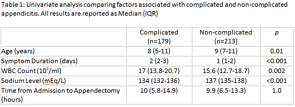
Methods: A retrospective review of all pediatric patients (≤12 years) with appendicitis treated with appendectomy between 2000-2013 was performed. The main outcome measure was intraoperative confirmation of gangrenous or perforated appendicitis. A multivariable analysis was performed and the main predictors of interest were age, symptom duration >24 hours, leukocytosis (WBC count >12 103/ml), hyponatremia (sodium <135 mEq/L), and time from admission to appendectomy.

Results: Of 392 patients, 179 (46%) had complicated appendicitis at the time of operation. Results of univariate analysis are listed in Table 1. Predictors of complicated appendicitis on multivariable analysis included symptom duration >24 hours (OR=5.1, 95% CI=3.2-8.2, p<0.001), hyponatremia (OR=3.2, 95% CI=2-5.1, p<0.001), age <5 years (OR=2.7, 95% CI=1.3-5.4, p=0.007), and leukocytosis (OR=1.9, 95%CI=1-3.4, p=0.04). Increased time from admission to appendectomy was not a predictor of complicated appendicitis (OR=0.75, 95% CI=0.5-1.1, p=0.2).

Conclusions: Complicated appendicitis is a prehospital occurrence. Hyponatremia is a novel marker associated with complicated appendicitis and may be more predictive than WBC count or age. Delaying appendectomy does not increase the risk of complicated appendicitis. This information may help guide resource/personnel allocation, timing of appendectomy, and decision for non-operative management of appendicitis in children.

72.10 Imaging Gently? Higher Rates of CT Imaging for Pediatric Appendicitis in Non-Children's Hospitals

K. T. Anderson1,2, L. R. Putnam1,2, T. Ostovar-Kermani1,2, E. Hamilton1,2, D. H. Pham1,2, M. T. Austin1,2, A. L. Kawaguchi1,2, L. S. Kao3, K. P. Lally1,2, K. Tsao1,2 2University Of Texas Health Science Center At Houston,Pediatric Surgery,Houston, TX, USA 3University Of Texas Health Science Center At Houston,General Surgery,Houston, TX, USA 1Children’s Memorial Hermann Hospital,Pediatric Surgery,Houston, TX, USA

Introduction: Growing concerns regarding radiation exposure in children has led to recommendations to minimize CT imaging, especially in pediatric appendicitis. However, these practices are not always adopted by non-pediatric specialties. We hypothesized that within a metropolitan hospital system (one children’s hospital (CH) and eight non-children’s hospitals (NCHs)), use of pre-operative CT is much higher in NCHs. We aimed to determine differences in pre-operative imaging and any associated demographic variables.

Methods: A retrospective study of patients <18 years undergoing appendectomy for acute appendicitis from April 2012 to April 2015 in a 9-hospital system was conducted. Patient demographics, pre-operative location, and imaging modality (computed tomography (CT) and ultrasound (US)) were evaluated. Patients were attributed to the hospital in which the pre-operative imaging was performed and not the location of appendectomy. Chi-squared, Student’s t-test, and multivariate regression were performed; p<0.05 was significant.

Results: 1448 pediatric patients were identified (CH=215, 15%; NCHs=1233, 85%). The median (range) number of patients triaged per NCH was 154 (85-193). Children seen at the CH were younger (mean 10.2 ± 4.0 vs 12.2 ± 3.9, p<0.01), more likely non-white Hispanic (68% vs 46%, p<0.01), and publically insured (58% vs 49%, p=0.04). CH patients underwent significantly fewer CT scans (23% vs 70%, p<0.01) and more US (75% vs 20%, p<0.01). For all patients, on multivariate regression, increased preoperative CT use was significantly associated with NCHs (OR 7.5, 95%CI 5.3-10.6), older age (age >10: OR 2.3, 95%CI 1.8-2.9), and female gender (OR 1.34, 95%CI 1.05-1.71). For patients triaged at NCHs, older age (age >10: OR 2.4, 95%CI 1.8-3.1) and female gender (OR 1.33, 95%CI 1.02-1.73) remained significantly associated with CT use. No patient characteristics were associated with increased CT use at CH. Children presenting at CH were more likely to undergo US (OR 11.8, 95%CI 8.3-16.9). Younger age (age ≤ 10: OR 1.9, 95%CI 1.5-2.5) and female gender (OR 1.5, 95%CI 1.1-1.9) were predictive of US use overall.

Conclusions: Within our hospital system, there are significant differences in utilization of imaging modalities for suspected pediatric appendicitis between CH and NCHs. Pediatric patients are 7.5 times more likely to receive a CT scan if initially evaluated in NCHs. Further investigation is needed to identify other factors contributing to imaging preference in the pediatric population in order to establish clinical practice guidelines to reduce radiation exposure in children.

72.06 Hospital Variation in Rates of Concurrent Fundoplication during Gastrostomy Enteral Access Procedures

A. M. Stey1, C. Vinocur4, B. L. Hall3,5, M. E. Cohen3, K. Kraemer3, B. Kenney2, R. Moss2, C. Ko3,6, L. Berman4 1Mount Sinai School Of Medicine,Department Of Surgery,New York, NY, USA 2Nationwide Children’s Hospital,Columbus, OH, USA 3American College Of Surgeons,Division Of Research And Optimal Patient Care,Chicago, IL, USA 4Nemours/Alfred I. DuPont Hospital For Children,Surgery,Wilmington, DELAWARE, USA 5Washington University,Surgery,St. Louis, MO, USA 6David Geffen School Of Medicine, University Of California At Los Angeles,Surgery,Los Angeles, CA, USA

Introduction: Indications for concurrent fundoplication during gastrostomy enteral access procedures are not well established. This study aimed to determine whether 1) the propensity for concurrent fundoplication during gastrostomy varies among hospitals, and 2) whether postoperative morbidities differ significantly among institutions performing fundoplication more or less frequently.

Methods: The American College of Surgeons National Surgical Quality Improvement Program Pediatric (ACS-NSQIP-P) dataset was queried to identify children who underwent gastrostomy with or without concurrent fundoplication. We created a hierarchical multivariate regression to model the propensity for concurrent fundoplication. The model included preoperative clinical variables associated with concurrent fundoplication after gastrostomy. The excess effects that hospitals exerted over the propensity of concurrent fundoplication were estimated as odds ratios. Hospitals were identified as having low, intermediate, or high odds ratios for the use of concurrent fundoplication. The risk-adjusted postoperative morbidity rates were compared among hospitals designated as low outliers, average hospitals, and high outliers in odds of concurrent fundoplication.

Results: 3,775 children underwent gastrostomy at 54 ACS-NSQIP-P participating hospitals between 2011-2013. The mean concurrent fundoplication rate was 11.7% (unadjusted range 0% to 64%). There was no significant difference in unadjusted morbidity rate in children with concurrent fundoplication, 11.0% compared to 9.7% in children without concurrent fundoplication. After controlling for clinical variables, 8 hospitals were identified as low outliers (mean observed fundoplication rate of 0.4%) and 16 hospitals were high outliers (mean observed fundoplication rate of 34.6%). The average unadjusted morbidity rate among hospitals with low, intermediate and high odds of concurrent fundoplication were 9.6%, 10.6% and 8.6% respectively (p=0.78). The added morbidity rate attributed to a concurrent fundoplication among hospitals with low, intermediate and high odds of concurrent fundoplication were 0.1%, 0.2% and 0.1% respectively (p=0.34).

Conclusion: Hospitals appear to vary significantly in propensity for concurrent fundoplication during gastrostomy in this small observational dataset yet short term morbidity did not differ significantly among the groups. These findings could demonstrate the need to clarify indications for concurrent fundoplication and the need for longer follow up to evaluate the success as well as morbidity of the procedure over time.

72.07 Safety and Cost-Effectiveness of Port Removal Outside of the Operating Room

T. F. Devji1,2, A. Madenci1,3,4, E. Carpino1, M. Samnaliev1,4, C. Weldon1, J. Cravero1 1Boston Children’s Hospital,Boston, MA, USA 2Harvard School Of Dental Medicine,Boston, MA, USA 3Brigham And Women’s Hospital,Boston, MA, USA 4Harvard Medical School,Boston, MA, USA

Introduction: Unprecedented access to health insurance coverage with the Affordable Care Act necessitates cost-conscious delivery of care. As such, health systems must optimize value without sacrificing safety or outcomes. Elective surgical procedures represent one healthcare area that may be value-enhanced by moving to a more cost-efficient setting. The purpose of this study was to assess the safety and cost of performing the removal of implantable central venous access devices (‘ports’) in settings other than the main OR.

Methods: We compared port removal in three settings, specifically: 1) Main OR, 2) Satellite OR, and 3) Clinic procedure room. This was a mixed-methods study, including a retrospective review of health records and prospective observation/interviewing. To calculate cost without the inherent biases of hospital charges, costs, and payments, we utilized the methodology of Time-Driven Activity Based Costing. Specifically, we recorded time spent by the patient in hospital facilities and with hospital personnel. This duration was weighted with the hourly cost of each professional and hospital space. Demographic information and 30-day procedure-related complications data were obtained retrospectively. Time spent by the patient was obtained retrospectively from electronic timestamps. When this information was unavailable, a proxy was obtained from the median time spent during prospective observation. The Mann-Whitney U test compared the continuous variables of time and cost across the three sites. Overall cost at each site was divided by overall cost at the referent site (Main OR) to obtain a ratio of cost savings.

Results: A total of 120 patients (40 per site) were included in the analysis. Demographic and clinical factors were not significantly different between sites. No complication occurred with port removal at any site. Patients in the Main OR spent more time in the procedure room, which was the highest-cost space (Main OR median 56 min vs. Satellite OR 42 min, P<0.001; vs. Clinic 41 min, P<0.001). Median overall cost was decreased by 25% in the Clinic (ratio=0.75, 95% confidence interval [CI]=0.66-0.87) and by 6% in the Satellite OR (ratio=0.94, 95% CI=0.89-1.02), compared to the Main OR (referent; P<0.01).

Conclusion: In this study, port removal in the Clinic procedure room was significantly more cost-effective and not significantly associated with an increased risk of negative outcomes, compared to port removal in the Main OR. Re-locating routine port removal to a non-OR setting, such as a clinic procedure room, appears to be safe and may result in substantial cost savings.

72.08 Outcome Assessment Of Renal Replacement Therapy In Newborns

I. I. Maizlin1, M. C. Shroyer1, L. Perger2, M. K. Chen1, E. A. Beierle1, C. A. Martin1, S. A. Anderson1, V. E. Mortellaro1, D. A. Rogers1, R. T. Russell1 1Children’s Hospital Of Alabama, University Of Alabama,Division Of Pediatric Surgery,Birmingham, Alabama, USA 2Scott & White Healthcare/Texas A&M Health Science Center College Of Medicine,Department Of Surgery,Temple, Texas, USA

Introduction: Recent advances in renal replacement therapy (RRT) have allowed for significant increase in rates of dialysis initiation for neonates with kidney failure or inborn errors of metabolism. Methods of RRT in neonates include peritoneal dialysis (PD), continuous renal replacement therapy (CRRT) and intermittent hemodialysis (HD). The purpose of this study was to assess morbidity and mortality following initiation of dialysis in newborn patients (<30 days) on those modalities.

Methods: Retrospective chart review was performed on all patients who had RRT initiated in the first 30 days of life between 2006 and 2014, excluding patients receiving RRT following congenital heart surgery. All charts were queried for birth history, underlying etiology for RRT, operative history, and route of RRT (HD and/or PD). We also reviewed the surgical complications associated with RRT and mortality rate.

Results: Total of 49 patients were identified, 39 boys and 10 girls. Median gestational age and birth weight were 36 (30-41) weeks and 2.85 (1.66-4.52) kg respectively. Thirty-two patients (65%) had end stage renal disease (ESRD), 11 (22%) had inborn errors of metabolism and 6 (12%) required dialysis due to other pathologies. Initial therapy was continuous veno-venous hemofiltration (CVVH) in all patients. Median age at onset of RRT was 6 (4-14) days and median weight of 3.1 (2.7-4.0) kg. Patients with ESRD spent a median of 8.5 (2-38) days on CVVH, while metabolic error and other etiology patients spent a median of 4 (3-7) days and 4.5 (1-10) days respectively. Overall mortality was 65.3% (71.9% among ESRD patients, 45.5% metabolic disease). Fifty-six percent (18 of 32 total deaths) of all deaths occurring within the 1st 30 days of life and 94% (30 of 32) occurred within the 1st year of life. Most common cause of death was sepsis (30%) among ESRD patients, and ARDS (40%) among metabolic patients. Total of 201 surgeries were performed. Excluding catheter revisions, 83 new HD lines and 28 new PD catheters placed, with a maximum of 6 HD lines and 4 PD lines placed in a single patient. Two patients died within 24 hours of catheter placement due to respiratory failure. Catheter-associated morbidities occurred in 100% of patients. Most common complications for HD included circuit clotting (87%), bleeding (68%) and bacteremia (40%), and for PD patients included peritonitis (83%), catheter complications (72%), and PD catheter leaks (55%). Among 9 survivors that required long term RRT (median follow-up of 5.3 years), 4 were severely and 2 were moderately developmentally delayed.

Conclusion: RRT is increasingly utilized for the neonatal population with renal and metabolic diseases. However, while RRT is becoming more technically feasible, it remains associated with significant morbidity and mortality. Pediatric surgeons must be aware of the challenges of neonatal dialysis and take them into account when considering the care of these critically ill children.

72.04 Institutional Experience of Ultrafiltration in Neonates with Congenital Diaphragmatic Hernia on ECMO

P. E. Lau2,4, S. M. Cruz2,4, D. L. Cass1,2,4, S. E. Horne2, T. C. Lee1,2,4, S. E. Welty3, D. Lazar2,4, J. Garcia-Prats5, O. O. Olutoye1,2,4 1Texas Children’s Hospital,Texas Children’s Fetal Center,Houston, TX, USA 2Texas Children’s Hospital,Pediatric Surgery,Houston, TX, USA 3Texas Children’s Hospital,Pediatrics,Houston, TX, USA 4Baylor College Of Medicine,Department Of Surgery,Houston, TX, USA 5Texas Children’s Hospital,Section of Neonatology,Houston, TX, USA

Introduction:
Several children with severe congenital diaphragmatic hernia (CDH) require extracorporeal membrane oxygenation (ECMO) for the management of respiratory failure. Fluid restriction is typical on ECMO, and on occasion ultrafiltration (UF) is used as a rescue measure. Since early 2012, our institution established a protocol introducing the routine use of ultrafiltration on ECMO (ECMO-UF). We hypothesized that routine ultrafiltration on ECMO (ECMO-UF) is associated with better fluid management, shorter ECMO duration, improved nutritional status and ultimately better long term outcomes without compromising renal function post-ECMO.

Methods:
A retrospective review of CDH cases from January 2004 to January 2015 was conducted at a pediatric tertiary center. Neonates with CDH who required ECMO therapy were included in the analysis. ECMO-UF was started within the first 6-12 hours of ECMO initiation in the Neonatal Intensive Care Unit. Prior to this, UF use was rarely used as rescue modality (ECMO group). Outcomes of patients admitted after 2012(ECMO-UF) were compared to the ECMO group. Net fluid balance, nutritional status after 48 hours on ECMO, renal function, survival and long term pulmonary outcomes were assessed. Statistical analysis was performed using Student’s t-test and Mann-Whitney U Test for continuous measurement and Fischer’s exact for categorical measurement.

Results:
During the study period 214 CDH patients were cared for, 62 (29%) patients required ECMO. Of these, 15 (24%) had UF initiated within 6-12 hours after ECMO cannulation. Patients in the ECMO-UF group had significantly greater caloric intake with higher TPN rates at 48 hours, 47.7+11.1 vs 65.7+11 (p<0.01), and greater total fluid output on ECMO (p=0.017). There was no significant difference in renal function. No significance was seem in length of tracheal intubation, with a median number of days of 37 (20-359) in the ECMO group vs 42 (20-186) in the ECMO-UF (p=0.91). Overall 6 month survival was equivalent in both group 62.2% vs 60.9%, (p=0.25).

Conclusion:

The use of routine UF allows for better optimization of parenteral nutrition while maintaining a more favorable net fluid balance without adverse effect on renal function. Larger studies may help identify other potential benefits of routine UF in long term outcomes in infants with CDH on ECMO.

72.05 Practice Variation in Pectus Excavatum Repair within a Single Institution

C. N. Litz1, S. M. Farach1, C. Snyder2, J. P. Jacobs2, J. Davis2, E. K. Amankwah3, P. D. Danielson1, N. M. Chandler1 1All Children’s Hospital Johns Hopkins Medicine,Pediatric Surgery,Saint Petersburg, FL, USA 2All Children’s Hospital Johns Hopkins Medicine,Cardiothoracic Surgery,Saint Petersburg, FL, USA 3All Children’s Hospital Johns Hopkins Medicine,Clinical And Translational Research Organization,Saint Petersburg, FL, USA

Introduction:
Minimally invasive repair of pectus excavatum (PE) is the current standard of care for repair of PE; however, there exists variations in the technical aspects and the clinical management. The purpose of our study was to determine practice variations and compare outcomes between two surgical services (SS) performing PE repair at a single institution.

Methods:
A retrospective review of all patients undergoing minimally invasive PE repair from July 2013 through June 2015 was performed. General admission, demographic and outcome data were collected. Patients were grouped by surgical service (SS1 vs SS2) performing PE repair and outcome measures were compared. Statistical significance was set at p<0.05.

Results:
A total of 74 patients underwent PE repair during the study period (SS1 n=36, SS2 n=38). There was no significant difference in age (14.9 ± 3.7 vs 15.4 ± 2 years, p=0.494) or total hospital length of stay (3.6 ±0.8 vs 3.3 ± 1.1 days, p=0.221) between the groups. There was a significant difference in total operating room time (147 ± 24 vs 98 ± 30 minutes, p<0.001). Total charges for PE repair were significantly higher for SS1 compared to SS2 (57,579 ± 7,429 vs 40,776 ± 5,727 USD, p<0.001) as were charges for operating room services (13,577 ± 1,709 vs 4,098 ± 584 USD, p<0.001), anesthesia (3,292 ± 373 vs 2,404 ± 323 USD, p<0.001), radiology (1,399 ± 407 vs 342 ± 145 USD, p<0.001), and laboratory studies (1289 ± 801 vs 452 ± 394 USD, p<0.001). Compared to the SS2 group, patients undergoing PE repair by the SS1 group had a greater number of radiology studies (4.7 vs 1.3, p<0.0001), laboratory studies (11.9 vs 3.4, p<0.0001), and ICU days (1.7 vs 0.1, p<0.0001) per patient. Standardized practice plans were initiated with a resultant decrease in ICU, radiology, and laboratory utilization for both groups over 3 years (Table 1).

Conclusion:
There are significant variations in the surgical care of patients undergoing PE repair among surgical services, even within a single institution. Implementation of standardized practice plans help to reduce unnecessary hospital resource utilization. Recognition of these differences may provide valuable information for the purposes of improving patient outcomes while decreasing health care costs.

72.02 Cost Modeling for Gastroschisis Management Strategies

J. Wu1, S. Lee1, D. A. DeUgarte1 1UCLA And Harbor-UCLA,Department Of Surgery – Division Of Pediatric Surgery,Los Angeles, CA, USA

Introduction: Several non-operative strategies have been described to repair gastroschisis including ‘sutureless’, ‘ward reduction’, ‘plastic’, and ‘flap’ techniques. Non-operative strategies have the potential advantages of avoiding intubation and general anesthesia, decreasing pain, and improving cosmesis. Their disadvantage is that the majority of patients without formal fascial closure will develop hernias, a proportion of which will require delayed repair. We hypothesized that routine utilization of non-operative strategy results in cost-savings in uncomplicated cases of gastroschisis.

Methods: We constructed a decision tree to compare three different strategies for the management of uncomplicated gastroschisis: non-operative management, primary operative closure, and routine silo placement. Model variables (including probability of management failure and hernia development as well as anticipated length of stay and treatment costs) were abstracted from a literature review and the Medicare Physician Fee schedule. Uncertainty surrounding model parameters was assessed via one-way and probabilistic sensitivity analyses.

Results: According to our model, non-operative management of uncomplicated gastroschisis was the least costly strategy, which cost $198,085 per patient. Primary closure cost $208,763 per patient. Routine silo placement was the most costly, $239,038 per patient. One-way sensitivity analysis suggested the cost of primary closure would be less costly than non-operative management if the initial success rate of non-operative management was less than 35.4% or if the initial success rate of primary operative closure was greater than 87.8%. Probabilistic sensitivity analysis found that non-operative management was the least costly strategy among 97.4% of 10,000 Monte Carlo simulations.

Conclusion: Non-operative management of uncomplicated gastroschisis appears to be more cost-effective than attempted primary closure and routine silo placement strategies. Furthermore, given the potential for avoiding general anesthesia, routine adoption of non-operative management of uncomplicated gastroschisis should be considered and deserves prospective evaluation.

72.03 Variability in Inhaled Nitric Oxide Use among Patients with Congenital Diaphragmatic Hernia

L. R. Putnam1,2, K. Tsao1,2, F. Morini3, P. A. Lally1,2, K. P. Lally1,2, M. T. Harting1,2 2Children’s Memorial Hermann Hospital,Pediatric Surgery,Houston, TX, USA 3Ospedale Pediatrico Bambino Gesù,Rome, , Italy 1University Of Texas Health Science Center At Houston,Pediatric Surgery,Houston, TX, USA

Introduction:

Inhaled nitric oxide (iNO) is often used in the management of congenital diaphragmatic hernia (CDH). There is a paucity of strong evidence supporting the efficacy of iNO for CDH patients. We sought to identify the spectrum of iNO use among CDH patients and the correlation between iNO use and important clinical variables and outcomes.

Methods:

We performed an analysis of data from the international CDH Study Group registry, which comprises voluntary and prospectively collected data from over 60 international centers. We analyzed the following variables: iNO use/dose, gestational age, defect size, pulmonary hypertension (pHTN) as defined by right ventricular systolic pressure >2/3 systemic systolic pressure, operative characteristics, and extracorporeal membrane oxygenation (ECMO) use. Chi-square, Mann-Whitney U test, and regression analyses were performed.

Results:

A total of 3,367 patients were identified from 2007-2015. iNO was used in 2,047 (61%) of all patients. The median (range) iNO dose was 20 (0.1-80 ppm), treatment duration was 8 (0-100 days), and day of life at initiation was 1 (0-189 days). Seventy (3%) patients received 2 iNO treatments, and 7 had >2 treatment periods. Of those patients with evaluable pHTN data (n=3117, 93%), 2,174 (70%) had pHTN, of which 1,613 (74%) were treated with iNO. Of the 943 patients without pHTN, 343 (37%) were treated with iNO. On multivariate regression analysis, iNO use was associated with worse pHTN on initial echocardiogram, larger defect size, ECMO use, and center (all p<0.05). Mean iNO use by center was 62% (0-100%) and its use was inversely associated with the trend in survival (Figure). On multivariate regression analysis, mortality was significantly associated with iNO use (OR 2.8, 95%CI 1.9-4.1), ECMO use (OR 4.1, 95%CI 3.1-5.3), and defect size (A: reference; B: OR 3.4, 95%CI 1.2-9.6; C: OR 8.9, 95%CI 3.2-25; D: OR 18, 95%CI 6.7-52).

Conclusion:

Our analysis revealed that iNO use is associated with pHTN, ECMO use, and defect size. However, we found its use highly variable among centers and over 1/3 of patients without pHTN received iNO. Without strong evidence to support its use, future studies should aim to evaluate its efficacy and standardize its application in order to optimize patient outcomes and ensure cost-effective practices.

71.19 Impact of Obesity on Post-Gastrectomy Outcomes and Survival of Patients with Gastric Cancer

D. H. Kim1, C. E. Cauley1, E. Fuentes1, R. Ahmad1, D. C. Chang1, D. W. Rattner1, J. T. Mullen1 1Massachusetts General Hospital,Department Of Surgery,Boston, MA, USA

Introduction: Given the epidemic of obesity and the increased incidence of gastric cancer among overweight and obese patients in the United States, it is important to understand the impact of obesity on gastric cancer surgery outcomes. Previous studies have reported conflicting results. Thus, we sought to evaluate the impact of body mass index (BMI) on both short-term postoperative outcomes and long-term survival after potentially curative gastrectomy for gastric cancer in a large, single institution cohort of US patients.

Methods: We identified 442 patients who underwent potentially curative gastrectomy for gastric adenocarcinoma between 1995 and 2014. Clinicopathologic characteristics, operative details, lymph node yield, postoperative complications, and oncologic outcomes were collected, and patients were stratified according to BMI as underweight (BMI < 18.5 kg/m2), normal weight (BMI 18.5 – 24.9), overweight (BMI 25.0 – 29.9), and obese (BMI ≥ 30). We controlled for clinically relevant confounders using Cox logistic regression analysis to identify predictors of five-year mortality.

Results: Most patients had a normal BMI (n = 175, 39.6%), followed by overweight (n = 159, 36.0%), obese (n = 83, 18.8%), and underweight patients (n = 25, 5.7%). Overweight and obese patients were more likely to be young and male and to undergo esophagogastrectomy than underweight and normal weight patients, though there were no differences in tumor location, tumor stage, or extent of lymphadenectomy across groups. When stratified by BMI, there were no significant differences in the overall postoperative complication rates or in the rates of specific types or grades of complications. Specifically, there were no differences in the 30-day incidence of wound infection, anastomotic leak, or death across the BMI groups. Similarly, there were no significant differences in the length of stay or readmission rates across BMI groups. For the entire cohort, the median number of examined and positive lymph nodes was 18 and 2, respectively, and there was no difference in lymph node yield across the BMI groups. BMI was not an independent predictor of overall survival (P > 0.05) (see Figure).

Conclusion: In contrast to what many groups have previously reported, we found that BMI has no impact on postoperative complication rates, nodal yields, or long-term survival in patients undergoing potentially curative gastrectomy for gastric cancer.

71.22 Using IntegriSense 750 for Fluorescence-Guided Surgery in a Head and Neck Cancer Model

T. Ramesh1, L. Moore1, S. Bevans1, M. Limdi1, E. Rosenthal2, J. Warram1 1University Of Alabama,School Of Medicine,Birmingham, Alabama, USA 2Stanford University,School Of Medicine,Palo Alto, CA, USA

Introduction:
The integrin avb3 is overexpressed in pathologic processes associated with tumor growth and invasion, including in tumor cells and angiogenic blood vessels. IntegriSense 750 (Perkin Elmer) is a targeted fluorescent imaging agent composed of a selective av?b3 antagonist and a near-infrared fluorochrome. We hypothesize IntegriSense 750 can effectively produce contrast for applications in fluorescence-guided surgery as demonstrated in flank xenograft, orthotopic tongue, and lymph node metastatic disease models of head and neck cancer. A dose escalation study was performed at three doses (1.25μg/mouse, 2.5μg/mouse, and 5μg/mouse) in mice bearing luc+ SCC1 flank xenografts (n=5). Mice bearing orthotopic tongue (n=3) and lymph node (n=3) models received 2.5μg of IntegriSense 750 per mouse.

Methods:
Fluorescence imaging was performed twice daily after infusion using commercially available open-field NIR and closed-field NIR imaging systems. NIR renderings of resected specimens were compared to bioluminescence images to confirm localization of probe to tumor. Tissue fluorescence was correlated with histology. Progressively smaller tumor fragments were re-introduced into the wound bed in order to determine the smallest cancerous mass detectable using this agent. Exploratory surgeries were performed to demonstrate agent localization to orthotopic tumors and lymph node metastases. A biodistribution study was performed four days after dosing.

Results:
Intraoperative imaging successfully differentiated tumor from normal tissue with mean tumor-to-background ratios of 3.73, 5.35, and 9.62 on day 4 for the 1.25μg, 2.5μg, and 5μg doses, respectively. Tumor-to-background ratios for all three groups peaked at four days post-infusion. Tumor fragments as small as 0.5mg were visualized in all three dosing cohorts. 3/3 diseased lymph nodes were fluorescently visible in situ in mice during exploratory surgeries. The 5μg dose was found to be the optimal dose for applications in fluorescence-guided surgical oncology in this study.

Conclusion:
IntegriSense 750 appeared to be effective for use in fluorescence-guided surgical applications as demonstrated in flank xenograft, orthotopic, and lymph node metastatic disease models of head and neck cancer.

72.01 Do Secondary Signs Matter in Ultrasound Reporting for Suspected Appendicitis in Children?

K. N. Partain1, A. Patel2, C. Travers3, C. McCracken3, J. Loewen4, K. Braithwaite4, K. F. Heiss5, M. V. Raval5 1Emory University School Of Medicine,Atlanta, GA, USA 2Emory University,Atlanta, GA, USA 3Emory University School Of Medicine,Department Of Pediatrics, Children’s Healthcare Of Atlanta,Atlanta, GA, USA 4Emory University School Of Medicine,Division Of Pediatric Radiology, Department Of Radiology And Imaging Services, Children’s Healthcare Of Atlanta,Atlanta, GA, USA 5Emory University School Of Medicine,Division Of Pediatric Surgery, Department Of Surgery, Children’s Healthcare Of Atlanta,Atlanta, GA, USA

Introduction: Ultrasound (US) of the right lower quadrant (RLQ) is often the initial imaging modality for evaluation of appendicitis in children. Frequently, the appendix is not fully visualized, and these equivocal US result in subsequent computed tomography scans (CT) or admissions. Our purpose was to determine if the inclusion of secondary signs (SS) observed in equivocal RLQ US reports improves diagnostic accuracy.

Methods: Using retrospective chart review, we identified 825 children (ages 5–18 years) presenting to a pediatric emergency department with concern for appendicitis and a RLQ US. Final US reports were evaluated for primary and SS of appendicitis. The primary sign of appendicitis was a fully visualized appendix (unequivocal US) with a diameter ≥6mm. SS included fluid collections representative of an abscess, significant free fluid, echogenic fat, regional hyperemia, enlarged lymph nodes, hypoperistalsis of adjacent bowel, and appendicoliths. US reports were classified into four categories: 1. Normal; 2. Equivocal without SS; 3. Equivocal with SS; and 4. Appendicitis. Review of operative and pathology reports confirmed appendicitis. Logistic regression models identified which SS were associated with appendicitis, and test characteristics were calculated.

Results: Of the 825 patients, 530 (64%) had equivocal US reports resulting in 114 (22%) CT and 172 (32%) admissions. Of the 114 patients with equivocal US undergoing CT, those with SS were more likely to have appendicitis (48.6% vs 14.6%, p<0.001). Of the 172 patients with equivocal US admitted for observation, those with SS were more likely to have appendicitis (61.0% vs 33.6%, p<0.001). On univariate analyses, all SS were associated with appendicitis except enlarged lymph nodes. After fully adjusting for age, sex, race, and other SS, the SS that were associated with appendicitis included fluid collection (adjusted odds ratio (OR) 13.3, 95% Confidence Interval (95%CI) 2.1-82.8), hyperemia (OR 11.97, 95%CI 1.5 – 95.5), free fluid (OR 9.78, 95%CI 3.8 – 25.4), and appendicolith (OR 7.86, 95%CI 1.7 – 37.2). Wall thickness, hypoperistalsis of adjacent bowel and echogenic fat were not associated with appendicitis in the fully adjusted model. There was no difference in accuracy in the categorization of cases as normal or appendicitis between unequivocal US and equivocal US when SS were used (Table). Equivocal US that included hyperemia, a fluid collection, or an appendicolith had a specificity of 96% and accuracy of 88%.

Conclusion: There is potential to improve the diagnosis of appendicitis. When using SS, equivocal US are as accurate as unequivocal US. Appropriate use of SS can guide clinicians and reduce unnecessary CT and admissions.

71.16 Lung Resection is Safe and Feasible Among Stage IV Cancer Patients: An ACS-NSQIP Analysis

S. B. Bateni1, E. A. David2, R. J. Bold3, D. T. Cooke2, F. J. Meyers4, R. J. Canter3 1University Of California – Davis,General Surgery,Sacramento, CA, USA 2University Of California – Davis,Cardiothoracic Surgery,Sacramento, CA, USA 3University Of California – Davis,Surgical Oncology,Sacramento, CA, USA 4University Of California – Davis,Hematology/Oncology,Sacramento, CA, USA

Introduction: Although surgical intervention among patients with disseminated malignancy (DMa) carries high morbidity and mortality, recent single institution retrospective studies have demonstrated improvements in survival among advanced lung cancer patients and patients with lung metastases undergoing lung resection. We sought to evaluate the rates of acute morbidity and mortality following lung resection in stage IV cancer patients to further describe risks associated with such intervention.

Methods: For the years 2011-2012, we identified 6,360 patients from the American College of Surgeons National Surgical Quality Improvement Program (ACS-NSQIP) undergoing lung segmentectomy, wedge resection, lobectomy, or pneumonectomy, including 603 patients (9.5%) with a diagnosis of DMa. Standard parametric and nonparametric statistical analyses were used to evaluate the 30-day morbidity and mortality associated with lung resection and compare outcomes among patients with and without DMa.

Results: Among DMa patients, 200 (33.2%) were diagnosed with primary lung cancer, 357 (59.2%) had metastases to the lung, and 46 (7.6%) were unspecified. Most DMa patients had independent functional status (99.2%) and were without any dyspnea symptoms (84.4%). Overall 30-day morbidity, serious morbidity and mortality for DMa patients were 12.1%, 7.6% and 1.8%, respectively, and 95.9% of patients were discharged home. DMa patients with a primary diagnosis of lung cancer had a significantly greater mortality compared to patients with lung metastases (3.5% vs. 0.6%, p < 0.01). Pneumonectomy among DMa patients had far worse overall morbidity, serious morbidity and mortality compared to other types of lung resections (28.6% vs 12.1%, 28.6% vs 7.6%, and 7.1% vs 1.8%, respectively, p < 0.05). Of most clinical significance, when comparing outcomes for DMa versus non-DMa, overall morbidity was lower among DMa patients (12.1% vs. 15.4%, p < 0.05) and there were no significant differences in serious morbidity (8.3% vs. 9.8%, p > 0.05) or mortality (1.8% vs. 1.8%, p > 0.05) for all lung resections. Subgroup analysis by segmentectomy, wedge resection, lobectomy and pneumonectomy also demonstrated no significant differences in rates of overall morbidity, serious morbidity and mortality for DMa versus non-DMa patients (p > 0.05 for all).

Conclusion: With the exception of pneumonectomy, DMa patients undergoing pulmonary resections experienced low rates of overall morbidity, serious morbidity, and mortality. Moreover, surgical outcomes for pulmonary resections were comparable among DMa and non-DMA patients. Taken together, these data suggest that lung resections may be performed safely on select patients with DMa with both primary lung cancer and pulmonary metastatic disease, with important implications for both symptom palliation and multimodality care.

71.17 Factors Affecting Lymph Node Postivity in Intrahepatic Cholangiocarcinoma

O. Kutlu1, K. Staveley-O’Carroll1, E. R. Camp1, E. Kimchi1 1Medical University Of South Carolina,Division Of Surgical Oncology,Charleston, Sc, USA

Introduction:

Lymph node metastasis (LNM) is one of the strongest prognostic indicators of survival in intrahepatic cholangiocarcinoma (IHCC). Despite the improvements in imaging modalities, sensitivity of CT scan assessment for LNM still is low. The value of regional lymph node dissection for IHCC is an area of debate. Routine nodal dissection has been investigated to aid in prognostication and as a possible preventive measure for disease recurrence. In this study we investigate the incidence of LNM and factors affecting metastasis in a large epidemiological database.

Methods:

A SEER (Survival Epidemiology and End Results) case listing was acquired for histologically proven IHCC diagnosed between 1988-2011. Patients over 18 years of age, who underwent surgical therapy with known tumor size, T-stage, number of lymph nodes dissected, lymph node positivity, location of lymph nodes, M status, type of surgery performed, tumor grade, disease extension and vascular invasion status were selected for analyses. For patients with positive lymph a binary logistic regression analysis was performed with SPSS 21 and factors affecting lymph node positivity were analyzed.

Results:

Of the 9846 patients, 569 fit our criteria of whom 53.6% were female. Mean age was 62.9 years (21-85), mean tumor size was 5.9 cm (0.3-19), and lymph nodes were sampled in 261(45.8%) patients (mean 4.5 nodes, n=1-32). Among the patients who underwent lymph node sampling, 115 (44%) had nodal disease (hepatoduodenal n=110, inferior phrenic n=1, unspecified n=3, combined n=1). Binary regression analysis showed tumor grade, T stage (vascular invasion, extension of the tumor to extrahepatic bile ducts) to be significant factors associated lymph node positivity. The number of lesions, patient age, tumor size, involvement of contiguous sites, presence of metastatic disease and sex were found to be insignificant.

Conclusion:

The number of LNM in this patient cohort was similar to other reports where findings of routine nodal dissections are published. Identification of patients with high risk features such as high grade tumors, vascular invasion and extension to extrahepatic bile ducts may aid in selection of patients for lymph node dissection. Performed at experienced institutions, lymph node dissection may aid in staging of IHCC, however further studies are needed to confirm our findings.

71.18 The Risk of Gastrointestinal Adverse Events among Cancer Patients Treated with CTLA-4 blockade.

S. Lew1,2, R. Chamberlain1,2,3 1Saint Barnabas Medical Center,Surgery,Livingston, NJ, USA 2Saint George’s University,Grenada, Grenada, Grenada 3University Of Medicine And Dentistry Of New Jersey,Surgery,Newark, NJ, USA

Introduction:
Cancer cells employ diverse mechanisms to evade anti-tumor immune responses, one of which expresses a ligand activating negative costimulatory molecules which downregulate the T cell immune response; lymphocyte antigen-4 (CTLA-4). Many studies with CTLA-4 monoclonal antibody have demonstrated antitumor activity in advanced melanoma and other solid tumors with varying degrees of treatment related gastrointestinal (GI) adverse effects. This study sought to determine the incidence of CTLA-4 blockade-associated severe GI adverse effects, risk stratified by the type of malignancy treated, and to investigate dosage related risk of severe diarrhea or colitis.

Methods:
A comprehensive literature search of PubMed, Google Scholar and the Cochrane Central Registry of Controlled Trials was completed. Keywords searched were ‘ipilimumab’, ‘Yervoy’, ‘MDX-010′, ‘MDX101′, ‘tremelimumab’, ‘ticilimumab’, ‘diarrhea’, ‘colitis’, and ‘clinical trial’. All clinical trials were analyzed for patient recruitment, intervention, and outcomes. Incidence and risk ratio (RR) were calculated with 95% confidence intervals.

Results:
13 single or double arm, phase II/III clinical trials involving 4,383 patients treated with ipilimumab or tremelimumab were identified. The incidence of CTLA-4 blockade associated severe (grade 3-4) diarrhea was 9.5% (95% CI [5.4-16.2]; p<0.0001) and severe colitis was 6.3% (95% CI [4.9-8.0]; p<0.001) among 1,464 patients in 9 clinical trials in which CTLA-4 blockade monotherapy was used in a single arm trial or RCT. Among 4,383 ipilimumab or tremelimumab treated patients in 10 RCTs, the risk of severe diarrhea in the treatment group was 9% compared to 1.5% in control (RR 4.18, 95% CI [1.44-12.15]; p=0.009) while severe colitis in the treatment group was 5.3% compared to 0.2% in the control (RR 11.53, 95% CI [3.65-36.44]; p=<0.001). The incidence and risk of severe CTLA-4 blockade-associated GI adverse events were not significantly different between melanoma and solid tumor, between 3mg and 10mg dosage regimens, or when monotherapy and/or concomitant chemotherapy were used. Significant heterogeneity was found among identified trials with regards to underlying malignancies, the dosage of the treatment, and duration of the treatment.

Conclusion:
CTLA-4 blockade is associated with a significantly increased risk of all- and high-grade diarrhea and colitis. Altering in dosing regimens or combinational chemotherapy utilizing CTLA-4 blockade did not significantly reduce the risk of GI adverse events. Although adequately powered large studies are needed to further investigate contributing risk factors for the GI adverse events, GI adverse events should be carefully monitored in patients treated with CTLA-4 blockade.

71.14 Enhanced Recovery After Surgery (ERAS) on a High-Volume Surgical Oncology Unit: Details Matter

C. Mosquera1, N. J. Koutlas1, K. K. Myatt1, N. A. Vohra1, E. E. Zervos1, T. L. Fitzgerald1 1East Carolina University Brody School Of Medicine,Division Of Surgical Oncology,Greenville, NC, USA

Introduction: Benefits from ERAS have been documented in various clinical settings; however, it is unclear if improvement stems from the protocol or by shifts in expectations. To clarify, outcomes from a pilot ERAS project were reviewed.

Methods: Introductory interdisciplinary educational seminars involving unit surgeons, residents, nurses, dieticians, and rehabilitation therapists were conducted. In order to validate our initial test of change, this protocol was adopted by one of three surgical oncologists with the others serving as controls.

Results: A total of 394 patients undergoing elective abdominal surgery from June 2013-April 2015 were included. Median age was 63 years, a majority were female (51.8%), white (59.9%), had a Charlson comorbidity score of 0-2 (40.4%), and a Clavien complication grade of 0-I (63.2%). Implementation of ERAS resulted in a significant decrease in length of stay (LOS) (6.0 vs. 8.0 days; p= 0.016) and in-hospital mortality (0% vs. 2.9%; p= 0.033); the difference in cost ($21,674 vs. $25,994; p= 0.060) did not reach significance. Gender (p= 0.63), age (p= 0.36), race (p= 0.89), type of surgery (p= 0.49), comorbidities (p= 0.76), complications (p= 0.31), and readmission rates (p= 0.21) were similar. For the test surgeon, ERAS was associated with a decreased LOS (6.2 vs. 9.6 days, p= 0.024), cost ($21,674 vs. $30,380, p= 0.029), and mortality (0 vs. 3.3%, p= 0.044); differences in complications (grade II-V 32.2 vs. 42.6%, p= 0.064) and readmission rates (11.5 vs. 21.4%, p= 0.076) did not reach significance. For the control providers LOS, cost, mortality, readmission rates, and complications were similar before and after implementation of ERAS on this unit.

Conclusion: Full implementation of an ERAS protocol on a single high-volume surgical unit decreases cost, LOS, and mortality. This change can be attributed to protocol adherence rather than an ethereal change in unit culture.

71.15 Breast Reconstruction Outcomes Of Breast Cancer Patients In Medically Underinsured Population

S. Roh1, L. K. Viennas1, D. Chin1, J. N. Collins1, R. C. Britt1, R. R. Perry1, E. C. Feliberti1 1Eastern Virginia Medical School,Surgery,Norfolk, VA, USA

Introduction:
Healthcare disparities exist in various stages of breast cancer treatment among different socioeconomic classes. With increased number of uninsured patients having access and receiving immediate breast reconstruction (IBR) due to the Breast and Cervical Cancer Early Detection Program (Every Woman’s Life), an analysis of IBR in these women was performed to compare perioperative outcomes with insured patients and assess for persistent disparities.

Methods:
A retrospective chart review was performed on a database of newly diagnosed female breast cancer patients that underwent total mastectomy with IBR at an urban academic medical center from January 2009 to June 2014. Patient demographics and oncologic and operative variables including postoperative outcomes were compared in women with and without medical insurance at the time of diagnosis.

Results:
Fifty-nine patients met the study criteria, 40 with insurance and 19 without insurance at the time of diagnosis. Mean age at time of diagnosis was similar in uninsured and insured cohorts (51.2 y.o. vs. 48.3 y.o., p = 0.18) and uninsured patients had a higher makeup of African-American patients (73.7% vs. 40%, p =0.01). There were no differences noted between the two groups in body mass index (33.5 vs. 31.7, p = 0.3), mean Charlson Comorbidity Index (2.6 vs. 1.8, p = 0.11), the percentage of smokers (42.1% vs. 25%, p = 0.22) or the receipt of neoadjuvant chemotherapy (15.8% vs. 25%, p = 0.41) or postmastectomy radiation (26.3% vs. 32.5%, p = 0.77). Uninsured patients exclusively received IBR with tissue expanders compared to insured patients, (100% vs. 85%, p = 0.01). Both overall (84.2% vs. 67.5%, p = 0.22) and major (15.8% vs. 30%, p = 0.6) perioperative complication rates were similar between the 2 cohorts, even in those receiving IBR with tissue expanders (major complication rate 15.8% vs. 25%, p = 0.76).

Conclusion:
IBR in uninsured breast cancer patients was performed safely in this study with similar perioperative risk factors and postoperative complications. The higher use of IBR with tissue expanders in uninsured patients suggests hidden disparities. Further studies should analyze for possible provider level and patient level bias.

71.12 Tumor Size Remains Better than Hounsfield Units for Predicting Adrenal Malignancy on Unenhanced CT

C. C. Taylor1, G. A. Rubio1, J. D. Egusquiza2, M. LoPinto1, J. I. Lew1 1University Of Miami,Division Of Endocrine Surgery, DeWitt Daughtry Family Department Of Surgery,Miami, FL, USA 2University Of Miami,Department Of Radiology,Miami, FL, USA

Introduction: Many recent studies suggest unenhanced CT attenuation values expressed in Hounsfield units (HU) are superior for differentiating between benign and malignant adrenal tumors. Surgical removal is warranted when the risk of malignancy is significant (i.e. adrenal tumor >4cm), or if the tumor is hyperfunctional biochemically. With recent enthusiasm for its use to differentiate between benign and malignant adrenal tumors, the purpose of this study was to determine if HU are more effective than tumor size by unenhanced CT in predicting adrenal malignancy.

Methods: A retrospective review of prospectively collected data of 40 patients who underwent adrenalectomies with reviewable unenhanced transverse CT scans at a single institution was performed. Based on final pathology, 32 patients had benign adrenal tumors and 8 had malignant tumors. CT densities were obtained in HU by placement of a region of interest (ROI) over the adrenal gland, avoiding any necrotic or hemorrhagic areas. Adrenal tumor size was determined using the maximum diameter on transverse CT views. Sensitivity, specificity, positive predictive value (PPV), negative predictive value (NPV), and accuracy at 2, 3, 4, 5, and 6 cm; and at 10, 20, 30, and 40 HU were also compared. Size and HU on CT were compared for benign and malignant adrenal tumors using unpaired T-tests.

Results: Of 40 patients studied, those with benign adrenal tumors had statistically (p<0.001) smaller diameters (3.6cm +/- 1.5) than malignant tumors (7.7cm +/- 4.4) [ROC curve 0.813, p<0.05]. The mean HU of benign tumors (26.5HU +/- 27) were not significantly different (p=0.28) compared to malignant tumors (37.8HU +/- 21) [ROC curve 0.659, p=0.169]. Unenhanced CT had a sensitivity of >85% and specificity of <55% for adrenal tumors measuring 2 to 4cm and a sensitivity of <63% and specificity >70% for tumors measuring 5 to 6cm. PPV, NPV, and accuracy for determining malignancy in adrenal tumors by size were: 24.2%, 100%, and 35.9% respectively at 2cm; 25.9%, 91.7%, and 46.2% at 3cm; 33.3%, 94.4%, and 61.5% at 4cm; 35.7%, 88%, and 69.2% at 5cm; and 80%, 88.2%, and 87.2% at 6cm. The sensitivity, specificity, PPV, NPV, and accuracy for determining malignancy in adrenal tumors by HU were: 100%, 16.1%, 23.5%, 100%, and 33.3% respectively at 10HU; 87.5%, 32.3%, 25%, 90.9%, and 43.6% at 20HU; 62.5%, 54.8%, 26.3%, 85%, and 56.4% at 30HU; and 50%, 83.9%, 44.4%, 86.7%, and 76.9% at 40HU.

Conclusion: This study suggests adrenal tumor size remains a better predictor for adrenal malignancy than HU on unenhanced CT. At 4cm, tumor size has a better sensitivity and specificity for predicting adrenal malignancy than HU. Furthermore, at 2cm or less, size is a more reliable predictor of adrenal benignity than HU. While use of HU has gained popularity, surgeons should remain confident in using tumor size as a criterion for those patients who require adrenalectomies.

71.13 Trend of Survival Outcomes in Duodenal Adenocarcinoma

T. Oyasiji1, W. Tan2, N. Wilkinson3 1Karmanos Cancer Institute,Surgical Oncology,Flint, MI, USA 2Roswell Park Cancer Institute,Biostatistics,Buffalo, NY, USA 3Kalispel Regional Medical Center,Surgical Oncology,Kalispel, MONTANA, USA

Introduction: Duodenal adenocarcinoma (DA) is rare. We sought to evaluate trends and predictors of survival outcomes from analysis of a large national database.

Methods: We identified all DA patients between 1988 and 2011 using the SEER database. Patients were grouped into two 12-year periods, 1988-1999 (group 1) and 2000-2011 (group 2). Each group was subdivided into those who had surgery for treatment of DA and those who did not. Overall survival (OS) and disease -specific survival (DSS) were determined for the groups and subgroups, and compared to identify trend in survival outcomes for the two periods. Survival analysis was done using Kaplan Meier method and Cox proportional hazards model was utilized for univariate and multivariate analyses.

Results: Of 3183 patients, 1411(44.3%) were treated with surgery. 51% were males while 75% were Caucasians. 76% of patients were in Group 2. Mean age, gender and race distribution were comparable for both groups (p >0.05). Median OS was 8 months for both groups (p=0.5). Median DSS was 11.0 months for both groups (p=0.58). For the two groups, median OS and median DSS were significantly better for those treated with surgery compared to those who were not treated with surgery ( Group 1- OS 21 vs 3 months, DSS 27 versus 4 months; Group 2- OS 25 vs 4 months , DSS 33 vs 5 months; p values<0.001). The surgery subgroup for Group 2 had better OS and DSS compared to Group 1 surgery subgroup, with a trend toward significance (OS 25 vs 21 months and DSS 33 versus 27 months, p values=0.06). On multivariate analysis, advanced age, AJCC stage and T stage were independent negative predictors of OS and DSS while treatment with surgery was an independent favorable predictor of OS and DSS.

Conclusion: There is a trend for improved OS and DSS for DA patients treated with surgery between 2000 & 2011 compared to those treated with surgery between 1988 & 1999. Patients treated with surgery have significantly better OS and DSS than those who did not undergo surgery, regardless of time period. Early detection (when disease is resectable) and surgical treatment might translate to improved survival outcomes.

71.09 Ultrasound Validation of Mathematically Modeled Irreversible Electroporation (IRE) Ablation Volumes

N. Bhutiani1, C. A. Doughtie1, R. C. Martin1 1University Of Louisville,Surgical Oncology,Louisville, KY, USA

Introduction:
Currently, the prediction of irreversible electroporation (IRE) ablation dimensions are modeled using algorithms derived from mathematical and ex-vivo models. These have not been validated using in-vivo studies. The aim of this study is to assess the correlation between the mathematical prediction model to and ultrasound and histopathology findings for in vivo ablations in a porcine model.

Methods:
IRE ablations were performed on porcine liver and spleen with probe spacings ranging from 0.6 to 2.6 cm. Pre and 2 hour post-ablation ultrasound images were recorded and validated with histopathology confirmation. Three dimensions of the ablation regions were recorded and ablation volumes were calculated and correlated with theoretical mathematical models for each given probe spacing.

Results:
In-vivo axial and anterior-posterior (AP) ablation distances were significantly greater than predicted for nearly all probe spacings (p<0.05). Ablation volumes were significantly less than predicted for the all probe spacings when modeled using both a cylinder and an ellipsoid. Geometrically, mathematically derived ablation regions demonstrated more central tapering (‘necking’) and diminished volumes compared to their in-vivo counterparts. The relationships between probe spacing and AP ablation dimensions were less linear (r2=0.57) than the relationships observed via ultrasound.

Conclusion:
The current mathematical models poorly predict ablation regions observed in vivo. They underestimate ablation dimensions and, by extension, ablation volumes. Further work should be done to improve models for ablative planning, and physicians should recognize the limitations of existing models when planning ablative treatments.