42.13 Increased Coronary Contractile Response to Serotonin in Pig with Metabolic Syndrome

I. Lawandy1, Y. Liu1, V. Pavlov1, L. Scrimgeour1, F. W. Sellke1, J. Feng1  1Brown University School Of Medicine,Cardiothoracic/Surgery,Providence, RI, USA

Introduction: Metabolic syndrome (MetS) is associated with alterations in coronary vascular smooth muscle and endothelial function. The current study examined the contractile  response of  the isolated coronary arterioles to serotonin  in  pigs with or without metabolic syndrome (MetS) and investigated the signaling pathways reponsible for serotonin-induced vasomotor tone.  

Methods: The MetS pigs were developed by feeding with a hyper-caloric, fat/cholesterol diet and the control animals (Lean) fed with a regular diet for 12 weeks (Lean: n = 8; MetS : n = 6). The coronary arterioles (90-180 micrometers in diameter) were dissected from the harvested pig myocardial tissues and the in-vitro coronary arteriolar response to serotonin was measured in the presence of pharmacological inhibitors.  

Results: Serotonin (10-9-10-5M) induced dose-dependent contractions of coronary resistant arterioles from both control (Lean) and MetS pigs. This effect was more pronounced in the MetS vessels as compared with that of controls (Lean)  [53% of contraction (serotonin 10-6M) in MetS vessels vs. 26% of contraction in control, P<0.05]. Serotonin-induced contraction of the MetS vessels was significantly inhibited in the presence of the phospholipase A2 (PLA2) inhibitor quinacrine (10-6M, 53% vs. 24%),  the cyclooxygenase (COX) inhibitor indomethacin (10-5M, 53% vs. 27%), the thromboxane-prostanoid (TP) receptor antagonist SQ29548 (10-6M, 53% vs.31%, P<0.05, respectively). 

Conclusion: MetS is associated with increased contractile response of porcine coronary arterioles to serotonin via activation of PLA2, COX and subsequent TXA2, suggesting that alteration of vasomotor function may occur at early stage of MetS and obesity.

42.09 Differential Expression of FAS and NKLAM in Patients Undergoing Carotid Endarterectomy

N. Harroun1, G. S. De Silva1, J. Kornbluth3, L. A. Sanchez1, M. A. Zayed1,2  1Washington University In St. Louis,Division Of Vascular Surgery,Saint Louis, MO, USA 2Veterans Affairs St. Louis Health Care System,Division Of Vascular Surgery,Saint Louis, MO, USA 3Saint Louis University School Of Medicine,Department Of Pathology,Saint Louis, MO, USA

Introduction:
Atherosclerosis is prevalent in western society. Understanding the processes of atherogenesis remain an area of intense investigation. Fatty acid synthesis and tissue inflammation have been demonstrated to influence atherogenesis and atheroprogression.  Fatty Acid Synthase (FAS) is a central enzyme responsible for de novo synthesis of long chain saturated fatty acids. Natural Killer Lytic-Associated Molecule (NKLAM) is an important inflammatory mediator that can alter cellular viability. We hypothesize that FAS and NKLAM are important molecular mediators of disease progression and are therefore differentially expression in variably diseased segments of carotid arterial plaque

Methods:
Using an IRB-approved, prospectively maintained vascular tissue biobank, we analyzed eight carotid endarterectomy plaque samples from patients undergoing carotid endarterectomy (CEA). CEA plaque was partitioned into maximally (Max) and minimally (Min) diseased segments.  Protein lysates were obtained for both Max and Min diseased segments and Western blot analysis was used to evaluate FAS and NKLAM protein expression levels.  Values are reported as a mean  SEM and as ratios of protein expression to a loading control (Caveolin).  Wilcoxon paired-match rank test was performed to determine any significant differences, with p < 0.05 considered significant.

Results:
Max diseased carotid segments demonstrated higher FAS expression compared to Min diseased segments (0.230.07 vs 0.05  0.02, respectively; p =0.04). Interestingly, NKLAM expression was visibly lower in Max diseased segments, though this value was not statistically significant.

Conclusion:
We have preliminarily observed significant differential expression of FAS and NKLAM, which are important disease mediators in Max and Min diseased carotid plaque segments.  These findings suggest that different plaque regions may be affected by different biological processes that can influence disease progression.
 

42.10 Maintaining Liposome Stability for In Vivo Translation

O. H. Grandas1, R. K. Fisher1, R. Dieter1, S. S. Kirkpatrick1, J. D. Arnold1, R. M. Buckley1, M. M. McNally1, M. B. Freeman1, D. J. Mountain1  1University Of Tennessee Graduate School Of Medicine,Department Of Surgery,Knoxville, TN, USA

Introduction: RNA interference (RNAi), or the use of short interfering RNA (siRNA) to transiently attenuate cellular protein expression, shows promise as a gene therapy technology to alter vascular pathology. However, the development of molecular nanocarriers is needed to overcome the unfavorable physiochemical properties that limit intracellular siRNA delivery. We have previously established cell penetrating peptide-modified neutral PEGylated liposome (CPP-PLP) as viable molecular nanocarriers in vascular smooth muscle cells. We aim to translate this molecular drug delivery platform for in vivo application in vascular injury models of disease, using liposomes stable when stored for off-the-shelf administration.  Here we aim to determine the optimal storage parameters for CPP-PLP stability and siRNA capsulate retention.

Methods:  CPP-PLPs were formed as previously described, injected with 200nM siRNA, and stored at room temp, 4°C, or -20°C for 0-48hr in phosphate buffered saline (PBS) or Dulbecco’s Modified Eagle Medium (DMEM).  Additionally, 0.1% bovine serum albumin (BSA) in PBS was tested as a stabilizer protein and 0.1% dimethyl sulfoxide (DMSO) was tested in DMEM as a cryoprotectant. Dynamic light scattering (DLS) was used to measure nanoparticle size, size distribution, and nanoparticle fragmentation and/or aggregation.  RiboGreen RNA assay was used to determine siRNA encapsulation and retention.

Results: DMSO is ineffective as a cryoprotectant and disrupts liposome membranes leading to siRNA leakage and lysis after 24h storage at all temps. DLS data for DMEM and BSA suggested that morphology was maintained for up to 48hr, but siRNA retention was not improved at any temp.  CPP-PLPs stored in PBS exhibited the best siRNA retention after 24 and 48hr storage at 4°C, while storage at -20°C resulted in more siRNA leakage.

Conclusion: Our data suggests the optimal parameters for short-term CPP-PLP/siRNA complex storage are in PBS at 4°C while DMSO, DMEM, and BSA were ineffective at maintaining liposome stability.  Further experiments are ongoing to determine siRNA retention under these conditions over time, and how lipid modifications can increase stability for more efficient long-term storage.  These studies will direct our experimental implementation of this novel drug delivery technology in future in vivo applications in vascular injury models of disease.   


42.07 Depression Severity and Increased Inflammation in Veterans with Peripheral Artery Disease

N. V. Hernandez1, S. Grenon1,2, J. L. Ramirez1, S. A. Khetani1,2, W. J. Gasper1,2, D. Lindqvist3,4, O. M. Wolkowitz3, N. K. Hills5, G. J. Zahner1  2Veterans Affairs Medical Center,Vascular Surgery Section,San Francisco, CA, USA 3University Of California – San Francisco,Department Of Psychiatry,San Francisco, CA, USA 4Lund University,Department Of Psychiatry,Lund, , Sweden 5University Of California – San Francisco,Department Of Epidemiology And Biostatistics,San Francisco, CA, USA 1University Of California – San Francisco,Department Of Surgery,San Francisco, CA, USA

Introduction:

Inflammation is an important factor in the pathogenesis of peripheral artery disease (PAD) and is known to play a role in depression. The present study seeks to understand the association between depressive symptoms and inflammation in patients with PAD.

Methods:

A cross-sectional sample of 117 patients with PAD (97% male and 76% Caucasian) was recruited from the San Francisco Veterans Affairs Medical Center. Inclusion criteria included claudication symptoms with an abnormal ankle-brachial index (ABI < 0.9) or a history of prior peripheral revascularization for symptomatic PAD. Patients taking immunosuppressants and those with renal, liver, or other acute disease states were excluded. Patients were categorized into three subgroups based upon worsening depressive symptoms defined by Patient Health Questionnaire (PHQ-9) scores: no symptoms (score of 0-4, n=62), mild symptoms (score of 5-9, n=33), and depression (score ≥10, n=22). Serum levels of high-sensitivity C-reactive protein (CRP), interleukin-6 (IL-6), soluble intercellular adhesion molecule-1 (ICAM-1), and tumor necrosis factor-alpha (TNF-α ) were assayed and log-transformed for multivariable analysis. Additionally, inflammatory markers were standardized and summed to create a total inflammatory score.

Results:

Patients with depression exhibited a greater comorbidity with PTSD compared to patients with mild symptoms or no symptoms (57% vs 21% and 12% respectively, p≤.001) and were more likely to be Caucasian (95% vs 85% and 65% respectively, p=.02). In unadjusted analysis, inflammation levels trended upward among groups with no symptoms, mild symptoms, and depression, reported in respective order: IL-6 (medians: 1.17, 1.30, 1.65 IQRs: 0.93-1.73, 0.89-1.88, 1.20-2.41 pg/mL, p=.10), CRP (medians: 2.30, 3.65, 4.0 IQRs: 1.4-4.1, 1.75-6.0, 1.9-8.5 mg/L, p=.06), and total inflammatory score (medians: -1.1, -0.33, 1.3 IQRs: -1.88-0.77, -1.64-0.86, -1.07-3.21, p=.06). In a multivariable analysis that controlled for age, ethnicity, ABI, and atherosclerotic risk factors, mild symptoms and depression were predictive of a higher total inflammatory score (mild symptoms vs. no symptoms p=.04, and depression vs. no symptoms p=.007). IL-6 was marginally higher in the mild symptoms group (p=.06) and significantly higher in the depression group (p=.006) compared to the group with no symptoms. CRP, ICAM-1, and TNF-α  levels all trended upward among groups with depressive symptoms, but did not reach statistical significance.

Conclusions:

After controlling for risk factors and PAD severity, depression severity was independently associated with greater inflammation in PAD, as demonstrated by increasing levels of the total inflammatory score and of IL-6. Future research should examine the strength and directionality of this relationship through larger prospective cohort studies as well as investigate the pathophysiological mechanism responsible.

42.08 Nanofibrous Microsphere Mediated High Engraftment Of hESC-Cardiomyocytes in Heart Regeneration

Z. Wang1, S. Tian1, Q. Liu2, I. Lei1, C. Zhao2, L. Wang1, L. Bu3, P. X. Ma2, Z. Wang1  1University Of Michigan,Cardiac Surgery,Ann Arbor, MI, USA 2University Of Michigan,Biologic And Material Sciences/Dentistry,Ann Arbor, MI, USA 3New York University School Of Medicine,Leon H Charney Division Of Cardiology,New York, NY, USA

Introduction: Cell-based therapy is a promising strategy to regenerate the injured heart after myocardial infarction. However, the current strategies only show modest therapeutic benefits largely due to the low engraftment and poor regeneration of functional cardiac muscle tissue. 

Methods: To address these critical issues in heart regeneration, we applied poly (L-lactic acid) nanofibrous hollow microspheres (NF-HMS) as cell carriers to transplant human embryonic stem cell (hESC) derived cardiomyocytes (CMs) into infarcted rat hearts.

Results:Our studies showed that, 28 days after cell transplantation, graft size of the CM+NF-HMS group (1.85 ± 0.72 mm2) was significantly higher than that of the CM only group (0.49 ± 0.34 mm2) (P<0.01), resulting in a 3.78 fold increase of CM engraftment by application of NF-HMS. Consequently, the reduction of infarct size in CM+NF-HMS group was significantly greater than that of the CM only group (42.58% versus 12.07% reduction). Furthermore, the grafted cells could couple with host cells as indicated by connexin 43 connections between grafted and host cells. Functionally, the left ventricular ejection fraction and fractional shortening in CM+NF-HMS group was significantly improved compared with CM only and PBS control groups. The increased engraftment of CMs in CM+NF-HMS group also promoted a higher density of vasculature in the infarction border zone. 

Conclusion:Thus, the highly engrafted transplantation of CMs using NF-HMS as a cell carrier resulted in significant improvement of CM engraftment, revascularization, and functional performance, providing an exciting strategy for heart regeneration.


42.05 Leptinemia Is Associated With Peripheral Artery Disease

G. J. Zahner1, J. Ramirez1, V. Ly1, K. A. Spaulding1,4, S. A. Khetani1,4, N. K. Hills2, C. Grunfeld3,5, A. L. Schafer2,3,6, W. J. Gasper1,4, M. Grenon1,4  1University Of California – San Francisco,Department Of Surgery,San Francisco, CA, USA 2University Of California – San Francisco,Department Of Epidemiology And Biostatistics,San Francisco, CA, USA 3University Of California – San Francisco,Department Of Medicine,San Francisco, CA, USA 4San Francisco VA Medical Center,Vascular Surgery Section,San Francisco, CA, USA 5San Francisco VA Medical Center,Metabolism Section,San Francisco, CA, USA 6San Francisco VA Medical Center,Endocrine Research Unit,San Francisco, CA, USA

Introduction:
Leptin, adiponectin, and resistin are in a class of hormones called adipokines that are produced by adipocytes and have been implicated in the causal pathway of atherosclerosis. Prior research has demonstrated an association between higher leptin levels and coronary heart disease (CHD); however, research on the association with peripheral artery disease (PAD) is less conclusive. Previous studies have demonstrated that leptin levels increase and adiponectin levels decrease with increasing fat mass, however, the degree of change is variable. The present study examined whether adipokine levels are associated with PAD, independent of fat mass.

Methods:
A cross-sectional sample of 179 vascular surgery outpatients (97% male, 73% Caucasian) were recruited from the San Francisco Veterans Affairs Medical Center (SFVAMC). PAD was defined as either an abnormal ABI (<0.9) plus symptoms of claudication or had previously undergone revascularization for symptomatic PAD (n=141). Controls had a normal ABI and no history of atherosclerotic disease (n=38). Adipokines were assayed using commercially available ELISA kits and values were log-transformed. Fat mass was measured using electrical impedance.

Results:
Compared to controls, patients with PAD were more likely to have diabetes, hypertension, and hyperlipidemia (p<.05). They also had smoked a greater number of pack years and had a lower estimated glomerular filtration rate (eGFR) (p<.05). In an analysis adjusting for body mass index (BMI) and traditional atherosclerotic risk factors, serum leptin was associated with PAD (OR 2.54, 95%CI 1.07-6.01, p=.03). No statistically significant associations were found between high molecular weight (HMW) adiponectin and PAD after adjusting for BMI, although the association approached statistical significance in multivariable analysis (OR 0.60, 95%CI .33-1.08, p=.09). Finally, resistin was not statistically associated with PAD in this study. Sensitivity analyses using either fat mass or fat mass/height2 rather than BMI yielded similar results.

Conclusion:
These results indicate that for a given BMI or fat mass, serum leptin levels are positively and independently associated with PAD. HMW adiponectin might be inversely associated with PAD, but larger studies are needed to conclusively make the determination. These results suggest that further research is warranted on the potential role of adipokines in the pathophysiology of PAD as well as to determine whether leptin levels may have clinical utility in predicting PAD outcomes.


42.06 PAR1 Agonists Stimulate APC-like Endothelial Cytoprotection

A. Morales Allende2, K. De Ceunynck2, S. Chaudhry2, C. Peters2, A. Jain2, S. Higgins2, O. Aisiku2, J. Fitch-Tewfik2, C. Dockendorff2, S. Parikh2, D. Ingber2, R. Flaumenhaft2  2Beth Israel Deaconess Medical Center,Vascular Surgery,Boston, MA, USA

Introduction:
Protease-activated receptor 1 (PAR1) is a member of a subfamily of G protein-coupled receptors, PARs, and is highly expressed on platelets and endothelial cells. PARs are unique in that they are activated through proteolytic cleavage. On endothelial cells, PAR1 exhibits distinct responses when cleaved by different proteases: thrombin induces apoptotic and inflammatory signaling, whereas the activated form of the anticoagulant protein C (APC) elicits cytoprotective and anti-inflammatory pathways. We have discovered small molecules, termed parmodulins (PM1 and PM2), which like APC induce a cytoprotective pathway in endothelial cells.

Methods:
We monitored apoptosis in cells incubated with parmodulins (PM1 or PM2) or APC for 4 hours. In addition, we assessed whether parmodulins also act via the PAR1 receptor by knocking out PAR1. Finally, we assessed the effect of parmodulins on thrombin generation in Human Umbilical Vein Endothelial Cells (HUVECs). Cells were incubated with inflammatory mediators (either TNF-α or LPS) for 3 hours in media, then replaced with plasma and 2.5 micromole calcium chloride for 20 minutes, and thrombin activity was measured.   
  

Results:
Apoptosis induced by TNF, thrombin or staurosporine was reduced when cells were pre-incubated with PM1, PM2, or APC for 4 hours. PMs or APC were no longer capable of protecting from TNF-induced apoptosis when PAR1 was silenced using siRNA. PAR1 knockdown was confirmed using qRT-PCR and Western Blot, showing >90% reduction in gene and protein expression. This demonstrates that the cytoprotective effects of PMs, like APC, are mediated through PAR1.

Unlike the enzyme APC that cleaves PAR1 at its extracellular side, parmodulins act at the cytosolic face of the receptor. PM2 blocked thrombin generation on HUVECs in response to inflammatory mediators TNF-α and LPS. Our data showed that APC also inhibited thrombin generation upon LPS or TNF stimulation. The difference is that PM2 required prolonged exposure indicating activation of a cytoprotective pathway, whereas APC inhibition occurred due to its anti-coagulant activities. 

Conclusion:

Parmodulin activation of the cytoprotective pathway, without affecting coagulation, may provide a new approach for treatment of thromboinflammatory diseases.


42.04 Myocardial Injection of Microvesicles and Peripheral Inflammatory Signaling

L. A. Scrimgeour1, B. A. Potz1, V. I. Pavlov1, A. Y. Gorvitovskaia1, B. Colantuono1, N. R. Sodha1, R. Abid1, F. W. Sellke1  1Brown University School Of Medicine,Cardiothoracic Surgery,Providence, RI, USA

Introduction:

Stem cell secretions of microvesicles are an enticing approach to treatment of chronic myocardial ischemia. Microvesicles influence intrinsic responses to injury via transfer of miRNAs, mRNA, siRNA, and proteins. We use intramyocardial injection of microvesicles isolated from human bone marrow-derived mesenchymal stem cells (hMSC) into ischemic swine myocardium to evaluate their effect on intrinsic repair mechanisms to treat chronic ischemia. 

Methods:

Microvesicles were isolated from cultured hSMC. Seventeen Yorkshire swine underwent placement of an ameroid constrictor on the left circumflex artery to induce localized myocardial ischemia. They were then divided into two groups; control (n=7) or microvesicle-injected group (n=10). The treatment group underwent injection of microvesicles into the ischemic myocardium two weeks after ameroid placement. Seven weeks after ameroid placement, during the swine heart harvest, plasma was collected, pooled and analyzed using a cytokine array. 

Results:

Microvesicle injection into the myocardium induced systemic inflammatory changes, as seen by evaluating cytokines in peripheral blood samples. The group who received microvesicles demonstrated a significant increase in angiogenin, ENA-78, GRO, GRO-α, I-309, IL-1α, IL-1β, IL-4, IL-5, IL-15, IFN-γ, MCP-1,2,3, MCSF, MDC, MIG, TGF-β1, TNF-α, and TNF-β when compared to the control group [Figure 1, p<0.05 for all]. GCSF levels, in contrast, were decreased in the microvesicle group (p<0.05). 

Conclusion:

Treatment of chronic myocardial ischemia with microvesicles derived from hSMC is an exciting application of stem cell therapy that precludes the risks of direct cell injection. Microvesicles have been suggested to not only circulate peripherally but also to be able to hone to areas of damage. In this study, we demonstrate that direct injection into the ischemic myocardium affects systemic circulation of cytokines, suggesting a powerful effect of microvesicles in stimulating existing intrinsic pathways of regeneration and repair. 

42.01 Characterizing The Relationship Between Flow-Mediated Dilation And Radial Artery Tonometry In PAD

G. J. Zahner1, K. A. Spaulding1,2, M. S. Schaller1, S. C. Walker1, N. K. Hills3, W. J. Gasper1,2, M. Grenon1,2  1University Of California – San Francisco,Department Of Surgery,San Francisco, CA, USA 2San Francisco VA Medical Center,Vascular Surgery Section,San Francisco, CA, USA 3University Of California – San Francisco,Department Of Epidemiology And Biostatistics,San Francisco, CA, USA

Introduction:
Arterial stiffness, measured by the augmentation index (AIX) from radial artery tonometry, and endothelial dysfunction, measured by brachial-artery flow-mediated vasodilation (FMD), have each been associated with increased risk of cardiovascular events. The relationship of these parameters to one another in patients with peripheral artery disease (PAD) is less well understood.

Methods:
In a cross-section of 123 vascular surgery outpatients, the current study examined the association between FMD and AIX in patients with PAD and in patients with atherosclerotic risk factors. PAD was defined as symptoms of claudication with an ankle-brachial index (ABI) of <.9 or a history of revascularization for symptomatic PAD, while controls had no history of atherosclerotic vascular disease and an ABI≥.9 but presented with traditional risk factors for PAD.

Results:
Compared to controls (n=32), patients with PAD (n=91) had lower FMD (6.3 ± 3.8 vs. 8.4 ± 3.7, p=.008), while central augmentation index normalized to 75bpm (central AIX) (25.5 ± 9.0 vs. 19.3 ± 8.6, p=.001) and peripheral augmentation index (peripheral AIX) (91.3 ± 14.5 vs. 81.3 ± 11.4, p=.001) were higher. FMD was not significantly correlated with either central or peripheral AIX (central: r=-.05; 95% CI -.23, .13; p=.58; peripheral: r=.01; 95% CI -.17, .19; p=.89) across the entire cohort or in either the patients with PAD (central AIX: r=.07, 95% CI -.13-.28, p=.48; peripheral AIX: r=.13, 95% CI -.08, .33, p=.23) or controls (central AIX: r=-.14, 95% CI -.47,.22, p=.43; peripheral AIX: r=-.02, 95% CI -.36, .33, p=.92). When AIX and FMD were combined in a single model, higher AIX remained independently associated with PAD.

Conclusion:
In an analysis of vascular surgery outpatients, no correlation between FMD and AIX was detected, despite adequate power to detect a clinically meaningful relationship. Larger prospective studies are needed to determine whether the inclusion of both parameters improves predictive models for the early identification and potential risk stratification of PAD patients.


42.02 Acute Post-Ischemic Application of Estrogen to Ameliorating Myocardial Ischemia/Reperfusion Injury

Y. Yang1, I. Wang1, M. Turrentine1, M. Wang1  1Indiana University School Of Medicine,Cardiothoracic Surgery/Surgery,Indianapolis, IN, USA

Introduction:  Cardioprotection provided by estrogen has been recognized for many years. But most of these studies employ a means of pre-injury application of estrogen in experimental research and the preventive usage in clinical studies. Compared to pretreatment, post-ischemic administration of estrogen will be more practical in treating myocardial ischemia (MI) given its clinical therapeutic potential. On the other hand, defect in circadian clock gene – period2 (PER2) disturbs heart energy homeostasis and aggravates MI-caused heart damage. Although PER2 expression decreases as a consequence of menopause, no information is available regarding the role of PER2 in estrogen-mediated protection following myocardial ischemia/reperfusion (I/R). In this study, we aim to determine: 1) potential improvement of myocardial function by post-ischemic administration of 17b-estradiol (E2) using an in-vivo myocardial I/R model; and 2) whether I/R affects myocardial PER2 expression, and if so, whether this alteration of PER2 can be reversed by E2 treatment.

Methods:  Thirty-min ligation of left anterior descending artery (LAD) (a small piece of tubing used for ligation) followed by 24-hr reperfusion (tubing removed for reperfusion) was performed on adult C57BL male mice and ovariectomy female (OVX F) mice. Groups (n=3-6/group) were as follows: 1) SHAM, 2) I/R+Vehicle, and 3) I/R+E2. Vehicle or 0.5mg/kg of E2 was subcutaneously injected right after 30-min ischemia. Following 24-hr reperfusion, myocardial function was determined by a Millar pressure catheter inserted into the left ventricle (LV) with heart rate of 400-500 BPM. Heart tissue was collected for analysis of PER2 by Western blot. P<0.05=statistically significant. 

Results: Successful ligation of the LAD was verified by myocardial blanching, abnormal movement of the anterior wall, and elevation of ST by ECG. Reperfusion was confirmed by returning the pink-red color of the LV anterior wall and decreased ST-elevation. I/R significantly impaired LV function (LVDP and +/-dP/dt, p<0.05) in both male (Fig. A1-A3) and OVX F (Fig. B1-B3), whereas post-ischemic treatment of E2 markedly improved I/R-damaged myocardial function (Fig. A and B, p<0.05). Following I/R, a trend of decreased PER2 level was observed in both male hearts and OVX F hearts. Intriguingly, E2 treatment up-regulated myocardial PER2 expression in the I/R hearts (Fig. C).

Conclusion: Our study represent the initial evidence that post-injury administration of E2 effectively improves I/R-damaged myocardial function and this protective effect of E2 may involve up-regulation of PER2. Further clarification of this novel approach and its related mechanisms may lead to clinically feasible therapies for ischemic heart disease.
 

41.19 Supercomputing Precision MODS: Insights from an in silico cohort of of 2 Billion simulated patients

C. Cockrell1, G. An1  1University Of Chicago,Surgery,Chicago, IL, USA

Introduction:  There remain considerable challenges in the ability to reconstruct the behavior of human systemic inflammation and multiple organ dysfunction syndrome (MODS) from available human biomarker/mediator data,: this reconstruction is a necessary step to be able to rationally engineer putative precision therapies for sepsis/MODS. Alternatively, the increasing ability to capture physiological data in the clinical setting can provide depictions of physiological phenotypes, but offer no insight or pathway toward the design of mediator-based interventions. Other fields of science have used computational modeling and simulation to help contextualize multi-dimensional data in order to describe system function and link multiples scales of behavior. Advances in supercomputer-aided modeling can provide this capability to biomedical research, including in the area of sepsis.

Methods:  We analyzed an extension of a prior agent-based model (ABM) of multiple organ dysfunction (MODS) (An, Theoretical Biology and Medical Modelling 2008, 5:11) using our developed methods of Probabilistic Basins of Attraction (PBoAs) and Stochastic Trajectory Analysis (STAs) (Cockrell and An, Journal of Theoretical Biology 2017 (430):157-168). 2 billion patients were simulated representing a 90 day hospital course due to microbial sepsis arising from the following initial insults: 1) pneumonia, 2) urosepsis and 3) primary septicemia. Parameter space was coarse-grained for pulmonary, cardiac, renal, hepatic and gastrointestinal physiology, and linked to SOFA scores when applicable. Linear classifiers were applied to determine if simulated patients could be classified on biomarker state and physiological score within the 1st 72 h with samples q6 h.

Results: Boundary conditions for parameter sets were identified corresponding to plausible patterns of MODS arising from different sources of initial infection, but with a probability distribution of atypical MODS patterns for each type of initial infection. PBoAs of specific organ systems demonstrated tipping points at which individual organ system dysfunction extended to MODS, and displayed a hierarchical effect separating implemented organ physiology/function from blood-borne cytokine mediated endothelial dysfunction. Characterization of biomarker system state was unable to separate survivors/non-survivors (AUROC .61), but the addition of physiological state improved this (AUROC .91).

Conclusion: Supercomputing simulations of MODS can be used to generate higher-order physiological behavioral manifolds that provide a path towards linking cellular-molecular mechanistic models with clinically-available physiological data. Modeling and simulation of this type can be used to provide a bridge between sparse biomarker/mediator data and more readily accessible physiological signal data, and aid in the engineering of truly predictive models and precision therapies.


41.20 Rapid Detection of Clostridium difficile Toxins in Serum by Raman Spectroscopy.

S. Koya1, J. V. Martin2, S. Yurgelevic1, D. M. Liberati2, M. Brusatori1, C. Huang1, G. W. Auner1, L. N. Diebel2  1Wayne State University,Smart Sensors And Integrated Microsystems (SSIM)/ Michael And Marian Ilitch Department Of Surgery/School Of Medicine,Detroit, MI, USA 2Wayne State University,Michael And Marian Ilitch Department Of Surgery/School Of Medicine,Detroit, MI, USA

Introduction:
Clostridium difficile infection (CDI) is due to the effects of toxins, toxin A (TcdA) and toxin B (TcdB) on the host. Severe CDI is associated with systemic signs of infection. Animal models of CDI demonstrate a strong correlation between systemic toxemia and the occurrence of severe disease. However current technologies have low sensitivity to detect C. difficile toxemia in human subjects. Raman spectroscopy (RS) is an upcoming technology that is used to detect bacteria and their toxins. We speculate that RS may be a sensitive method to detect clinically relevant concentrations of C. difficile toxins in serum. 

Methods:
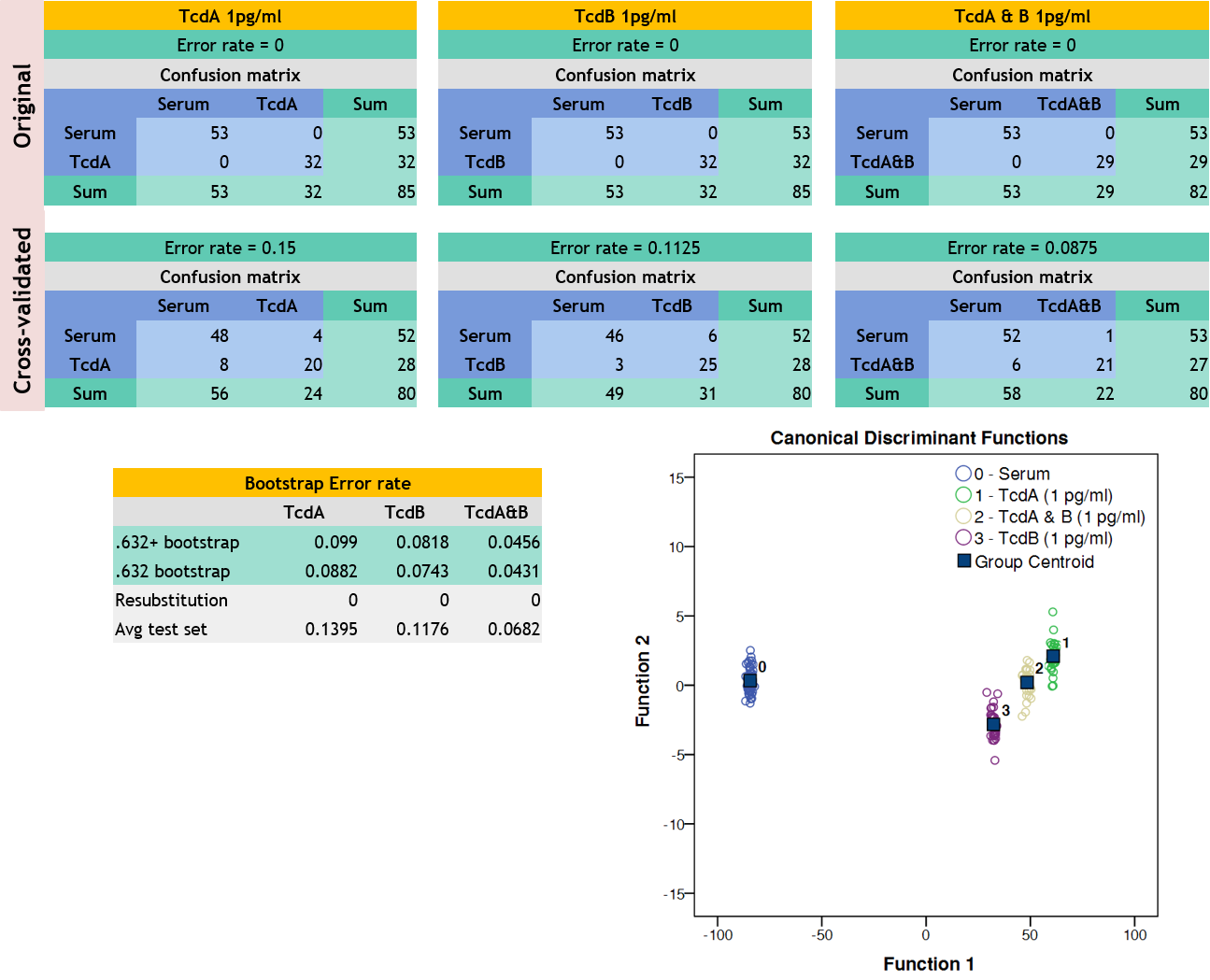
Serum samples were spiked with varying concentrations of TcdA, TcdB and both. RS was performed on air dried serum drop on a mirror polished stainless-steel slide. Raman spectra were obtained, background corrected, vector normalized and analyzed by Partial Least Square Linear Discriminant Analysis (PLS-LDA). PLS-LDA model accuracy was measured by cross-validation and bootstrap method.

Results:
At 1 pg/ml concentration, toxin spiked serum was distinguished from control serum 100%. PLS-LDA model performed well with cross-validated error rate ranging from 8 to 15% and bootstrap error rate ranging from 4 to 9%. Similar results were obtained for other concentrations of toxins (100 pg/ml, 1 ng/ml). 

Conclusion:
Raman spectroscopy has the potential to rapidly detect C. difficile toxins in serum at clinically relevant concentrations and may be useful as a diagnostic tool to modify therapy and and predict outcome in critically ill CDI patients.  

41.16 IN-VITRO MODEL OF MINOR AORTIC INJURY DEMONSTRATES SHEAR FORCES FACILITATING AORTIC CRACK PROPAGATION

J. Rabin1, A. Siddiqui2, J. Gipple2, Z. N. Kon3, B. Taylor3, T. M. Scalea1, H. W. Haslach2  1R Adams Cowley Shock Trauma Center,Surgery,Baltimore, MD, USA 2University Of Maryland,Mechanical Engineering,College Park, MD, USA 3University Of Maryland,Cardiac Surgery,Baltimore, MD, USA

Introduction:
Non-operative management is considered an appropriate treatment strategy for minor aortic injury, while blood pressure control and anti-impulse therapy are routinely utilized to help minimize injury progression. However a universal medical regimen for low grade intimal injuries has not been adopted and risks of injury progression not well described. This study investigates an in-vitro model of minor aortic injury to help identify risks of injury progression and factors associated with the mechanical failure of injured aortic tissue.   

Methods:
With IRB approval, ascending aortic tissue was obtained from the operating room after aortic aneurysm repair or heart transplant procurement, stored at 4 degrees Celsius in buffered solution and tested within 48 hours. Minimal aortic injury was modeled by creating a small partial thickness radial notch on the intimal surface of aortic tissue rings. These aortic rings were circumferentially expanded on a custom testing device under video acquisition until maximum diameter or rupture. The test video and aortic tissue were then analyzed to determine point of failure and the crack propagation length & angle. 

Results:
A total of 8 rings were obtained from 3 aneurysmal and 2 healthy aortas. Aneurysmal aortic tissue was obtained from a 71 year old female and 68 & 49 year old males. Healthy tissue was obtained from 2 males under the age of 30. All specimens demonstrated circumferential crack propagation from the root of the notch (fig 1) with an average angle 96.3±8.2 degrees between radius and crack. Length of propagation was longer in the aneurysmal tissues (6.60±4.19 mm vs 2.27±1.20 mm). There was no difference in initiation of crack propagation between healthy and aneurysmal tissue which occurred at an average of 1.8 times initial diameter.   

Conclusions:
Dilation of minimally damaged or injured aortic rings is associated with crack propagation and injury progression, which contributes to the aortic tissue's mechanical failure. This crack progression is consistent with an applied shear force and deformation within the layers of aortic tissue. Such shear forces are routinely generated through normal circumferential expansion that occurs with each pulsation, as the aortic wall stretches and relaxes, with the magnitude of these aortic shear forces affected by both pulse and blood pressure. The crack propagation demonstrated in this model illustrates the mechanical response to increased levels of applied shear which appears to compromise the structural integrity of the tissue and increases the risk of mechanical failure and aortic rupture. This suggests that strategies to reduce shear stress such as β- blockade, also be implemented in patients with minor aortic injury.  


41.17 Circulating DNA in Sepsis

D. Holden1, T. Murphy1, T. O. Baslanti1, Z. Wang1, G. Ghita1, S. Brakenridge1, A. Bihorac1, P. Efron1, F. Moore1, L. L. Moldawer1  1University Of Florida,College Of Medicine,Gainesville, FL, USA

Introduction: Nuclear DNA (ncDNA) and mitochondrial DNA (mtDNA) are damage associated molecular patterns (DAMPs) that are known to stimulate a pro-inflammatory response through pattern recognition receptors such as Toll-like receptors.  The association of circulating ncDNA and mtDNA with the persistence of organ dysfunction and the development of chronic critical illness (CCI) after sepsis are poorly understood. 

Methods: In this prospective observational cohort of critically ill surgical patients that developed sepsis, we measured circulating ncDNA and mtDNA levels on 0.5, 1, 4, 7, 14, and 21 days after sepsis by ddPCR. DAMP levels were then compared based on clinical outcomes, including the development of CCI (≥14 days ICU LOS with ongoing organ dysfunction) in comparison to those with rapid recovery (RAP), as well as by 1-year mortality. 

Results: We found that ncDNA and mtDNA were significantly higher in sepsis patients than in healthy controls. Additionally, ncDNA levels were significantly higher at 0.5, 1, 4 and 7 days after sepsis in patients that subsequently developed CCI as compared to RAP (Figure; p<0.05). ncDNA was correlated with serum lactate (Pearson: 0.85) and aspartate aminotransferase (Pearson: 0.97) consistent with shock severity and end organ tissue damage.  ncDNA levels peaked at 4 days after sepsis in both groups.  There were no significant differences in mtDNA levels between CCI and RAP at all time points. Elevated mtDNA levels at days 14 and 21 in hospitalized patients were significantly associated with 1 year survival (Figure 1). 

Conclusion: Elevated nuDNA levels early after sepsis are associated with persistent organ dysfunction and the development of CCI.  In the population of patients who experience prolonged hospital stays, the patients who have higher levels of circulating mtDNA do better, and more of these patients are alive at one year.  Since these same patients who have higher circulating mtDNA levels, do not have elevated circulating ncDNA levels, this means that the circulating mtDNA is not resulting from tissue death and cell necrosis.  Therefore for the patients who following sepsis, both experience prolonged hospital stays AND go on to survive greater than 1 year, there is something actively occurring in their bodies that is contributing to the release of circulating mtDNA.  This active release may be part of neutrophil NETosis, and it may be that patients with prolonged hospital stay who are capable of efficiently producing NETs and the associated circulating mtDNA have greater 1 year survival.


41.18 Single Dose Toxicity of Adenosine, Lidocaine, and Magnesium (ALM) Solution in a Murine Model

D. Rattigan1, J. Wang1, J. Marcotte1, R. Irons1, K. Cahill1, P. Zheng1, J. Gaughan1, S. Brown1, J. Carpenter1, G. Dobson2, J. P. Hazelton1  1Cooper University Hospital,Camden, NJ, USA 2James Cook University,College Of Medicine And Deintistry,Townsville, QUEENSLAND, Australia

Introduction:
Several common critical clinical pathologies both in military and civilian scenarios require rapid triage and prompt resuscitation. Traditional resuscitation for clinical scenarios such as sepsis and hemorrhagic shock include large volume infusions of isotonic crystalloid solution. more recent data has demonstrated that  low volume resuscitation techniques are a safe alternative with fewer subacute sequelae (1-3). Adenosine, lidocaine, and magnesium (ALM) in a high concentration solution is employed as a non-depoloraizing cardioplegic agent that reduces arrhythmias and reperfusion injury after cardiac resuscitation (4-7). In several animal studies, low concentration ALM solution has shown promising applications in various shock states (4-17), however, the baseline toxicity has never been characterized in healthy animals. The purpose of our study was to determine the baseline toxicity of this resuscitation fluid in a mouse model.

Methods:
In this study, 100 Healthy adult mice were given an intravenous bolus of increasing concentrations of ALM to establish the median lethal dose as well as any nonlethal toxicity. Mice that did not expire immediately were observed for several hours for any gross clinical toxicity and then for two weeks for any subacute toxicity. After two weeks, surviving animals were euthanized and the brain, heart, lungs, liver and kidneys were harvested for histology and apoptosis assay to determine if there was any occult toxicity.

Results:
Doses up to 75 times the previously studied therapeutic resuscitative dose of 1 cc/kg of 1/3/2.5mM ALM solution were well tolerated by all mice while higher doses demonstrated obvious clinical toxicity or mortality with the median lethal dose being a concentration 29.68-fold and a volume 1.9-fold that of the highest therapeutic volume ever studied in animals (4cc/kg) giving a therapeutic index of 55.65. TUNEL assay revealed no apoptotic cells in brain or heart tissue but did show some activity in kidney cells starting at doses 10 times the therapeutic concentration.

Conclusion:
The previously studied dosing of low concentration ALM is safe in healthy small animals with no obvious adverse clinical consequences and has a wide therapeutic index.  Studies in larger animals with more invasive hemodynamic and laboratory monitoring are required to ensure no subclinical toxicity prior to any human clinical trials.

41.13 Angiopoietin Dysregulation Following Severe Pediatric Trauma and Its Correlation with Endotheliopathy

R. P. Richter2, P. J. Hu1, R. M. Uhlich1, M. Shroyer3, J. D. Kerby1, J. F. Pittet4, R. T. Russell3, J. R. Richter1  1University Of Alabama at Birmingham,Acute Care Surgery,Birmingham, Alabama, USA 2University Of Alabama at Birmingham,Pediatric Critical Care Medicine,Birmingham, Alabama, USA 3University Of Alabama at Birmingham,Pediatric Surgery,Birmingham, Alabama, USA 4University Of Alabama at Birmingham,Critical Care Anesthesiology,Birmingham, Alabama, USA

Introduction:  The mechanisms and effects of vascular endotheliopathy in pediatric trauma are poorly understood. Angiopoietins are known mediators of endothelial cell (EC) integrity and are dysregulated during inflammatory states. Angiopoietin-1 (Agpt-1) helps maintain endothelial cell homeostasis. Agpt-2 is released after direct or indirect EC injury and promotes EC destabilization, thereby disrupting vascular endothelial integrity. The balance of these cytokines, quantified as Agpt-2-to-Agpt-1 ratio (Agpt-2:Agpt-1), reflects overall endothelial health and stability. We have recently established that plasma levels of syndecan-1 (Syn-1), an essential proteoglycan in the endothelial glycocalyx and a biomarker of endotheliopathy, correlate with mortality in pediatric trauma patients. However, at present, the behavior of the angiopoietins and their association with Syn-1 are not known in pediatric trauma. The objective of the current study was to (1) measure plasma levels of Agpt-1 and Agpt-2 following pediatric trauma and (2) correlate Agpt levels with plasma levels of Syn-1. As Agpt-2 levels positively correlate with illness severity in adult trauma, we hypothesize that Agpt-2 and the Agpt-2:Agpt-1 ratio significantly increase in injured patients compared to controls and have a positive correlation with Syn-1.

Methods:  We performed a prospective observational study comparing 45 pediatric trauma patients with 9 healthy pre-operative pediatric control patients at a level 1 pediatric trauma center from 2013 to 2016. Agpt-1 and Agpt-2 levels were measured in trauma patients upon hospital arrival and 24 hours after admission, from which Agpt-2:Agpt-1 ratios were derived. Syn-1 levels were measured at admission. The University of Alabama at Birmingham Institutional Review Board approved the study.

Results: Median age of trauma patients was 9 years (interquartile range, IQR, 6, 13), and the median injury severity score was 22 (IQR 16, 29). Compared to controls, Agpt-1 rose in trauma patients, though not significantly (p=0.335), and decreased to levels near control by 24 hours (p=0.112, Table). Agpt-2 rose significantly at admission (p=0.047) compared to controls and remained elevated after 24 hours (p=0.049), translating to a significantly elevated Agpt-2:Agpt-1 ratio at 24 hours (p=0.005). Syn-1 positively correlated with Agpt-2 levels at admission (r2=0.219, p=0.001) and 24 hours (r2=0.156, p=0.008) but not with Agpt-1 or the Agpt-2:Agpt-1 ratio.

Conclusion: Our findings reflect dysregulation of Agpt-1 and Agpt-2 after pediatric trauma. The correlation of Agpt-2 with Syn-1 suggests that Agpt-2 may be an important mediator of endotheliopathy after pediatric trauma and a potential therapeutic target.


41.14 Ceramide Contributes to Neutrophil Dysfunction in a PICS Murine Model

L. K. Winer1, A. M. Pugh1, E. Gulbins3, C. C. Caldwell1, V. Nomellini2  1University Of Cincinnati,Division Of Research, Department Of Surgery,Cincinnati, OH, USA 2University Of Cincinnati,Division Of Trauma, Critical Care, And Acute Care Surgery, Department Of Surgery,Cincinnati, OH, USA 3University Of Duisburg-Essen,Department Of Molecular Biology,Essen, GERMANY, Germany

Introduction:  Critically ill patients who survive the acute phase of sepsis can progress to persistent inflammation, immunosuppression and catabolism syndrome (PICS), and are at risk for secondary infection, organ failure, and death. Characteristics of PICS include increased circulating myeloid cells, decreased lymphocytes, and muscle loss. We recently reported that 8 days after cecal ligation and puncture (CLP), mice concurrently display the aforementioned features of PICS and are susceptible to lung infection. During lung infection, bronchial alveolar lavage analysis reveals impaired neutrophil accumulation and increased bacterial loads in PICS mice. However, the mechanisms by which PICS weakens the host response to infection remain unclear. Previous studies suggest that elevated levels of the membrane lipid ceramide induce cellular dysfunction. Here, we test the hypothesis that ceramide modulates neutrophil reactive oxygen species (ROS) production and chemotaxis during PICS. 

Methods:  Male CF-1 mice underwent 33% cecal ligation with a single 25-gauge needle puncture. Neutrophil dihydrorhodamine (DHR), a surrogate for ROS production, was measured by flow cytometry. Neutrophil chemotaxis was conducted on transwell plates with KC as the chemoattractant. Separate cohorts of healthy cells were treated with 10 μ M of C16-ceramide for 30 minutes and washed prior to DHR and chemotaxis assays. A student t-test was used for statistical comparison, and p-values ≤ 0.05 were considered statistically significant.

Results: We performed DHR and chemotaxis assays to (1) characterize neutrophils during PICS and (2) measure the effect of ceramide on neutrophil activity. We found that neutrophils isolated from PICS mice demonstrate a nearly 50% DHR increase (p<0.05, n=17) and a greater than 50% reduction in chemotaxis (p<0.05, n=17) compared with healthy controls. These effects were mimicked by ex vivo addition of C16-ceramide to healthy neutrophils, which increased DHR by nearly 10% (p<0.05, n = 6). Ceramide also decreased neutrophil chemotaxis by more than 30% (p<0.05, n = 7). Altogether, both PICS and ceramide increase neutrophil ROS and suppress chemotaxis. 

Conclusion: This study demonstrates that during PICS, there is a marked increase in neutrophil ROS production and impaired neutrophil chemotaxis. This combination of factors may underlie the decreased bacterial clearance and increased mortality previously observed in PICS mice. When applied to healthy mouse neutrophils, exogenous C16-ceramide produces a similar pattern of DHR expression and chemotaxis as that seen in vitro. We therefore speculate that C16-ceramide is associated with PICS neutrophil dysfunction, and may represent a therapeutic target in critical illness.
 

41.12 Effects of Propranolol and Clonidine on Bone Marrow Cytokines following Trauma and Chronic Stress

E. Miller1, T. J. Loftus1, J. Millar1, K. Kannan1, I. Alamo1, J. Plazas1, P. Efron1, A. Mohr1  1University Of Florida,Department Of Surgery,Gainesville, FL, USA

Introduction: Attenuating the neuroendocrine stress response with propranolol and clonidine has demonstrated efficacy in abrogating persistent injury-associated anemia. Several hematopoietic cytokines may contribute to this process. We hypothesized that propranolol and clonidine would decrease bone marrow expression of high mobility group box 1 (HMGB1) and increase bone marrow expression of interleukin 1 (IL-1), interleukin 10 (IL-10), stem cell factor (SCF), and B-cell lymphoma-extra large (Bcl-xL).

Methods: Male Sprague-Dawley rats were allocated to naïve control, lung contusion followed by hemorrhagic shock and daily chronic stress (LCHS/CS), LCHS/CS +propranolol, or LCHS/CS +clonidine (n=6-7/group). Chronic stress was performed by placement in a restraint cylinder for two hours daily until sacrifice and bone marrow collection on day seven. Bone marrow expression of HMGB1, IL-1a, IL-1b, IL-10, SCF, and Bcl-xL was assessed by polymerase chain reaction. Ratios of cDNA/B-actin were reported as mean ±standard deviation, ap<0.05 vs. naïve, bp<0.05 vs. untreated counterpart.

Results: Raw bone marrow cytokine expression values are listed in the table. Bone marrow HMGB1 expression was significantly increased following LCHS/CS compared to naïve animals, and was significantly decreased by propranolol (47% decrease, p=0.040) and clonidine (7% decrease, p=0.037). IL-1a was significantly decreased following LCHS/CS, and was significantly increased by propranolol (2,833% increase, p <0.001) and clonidine (1,842% increase, p<0.001). IL-1b was significantly decreased following LCHS/CS, and was significantly increased by propranolol (18,654% increase, p<0.001), and clonidine (10,073% increase, p<0.001). IL-10 was significantly decreased following LCHS/CS, and was not significantly affected by propranolol or clonidine. SCF was significantly decreased following LCHS/CS, and was significantly increased by propranolol (629% increase, p<0.001) and clonidine (468% increase, p<0.001). Bcl-xL was not significantly affected by LCHS/CS, and was significantly increased by propranolol (59% increase, p=0.006) and clonidine (77% increase, p<0.001).

Conclusion: Attenuating the neuroendocrine stress response with propranolol and clonidine modulates the bone marrow cytokine response to severe trauma and chronic stress, favoring effective erythropoiesis. Future research should assess the therapeutic value of propranolol and clonidine to prevent persistent injury-associated anemia among critically ill trauma patients.


41.10 Source Control Mitigates Hypothermia and Prevents Lymphopenia in Septic Mice

A. M. Pugh1, L. K. Winer1, V. Nomellini1,2, C. C. Caldwell1  1University Of Cincinnati,Division Of Research, Department Of Surgery,Cincinnati, OH, USA 2University Of Cincinnati,Division Of Trauma, Critical Care, And Acute Care Surgery,Cincinnati, OH, USA

Introduction: Hypothermia is shown to predict persistent lymphopenia in critically ill patients. Although less than 20% of septic patients present with hypothermia, this patient population demonstrates twice the mortality of febrile septic patients. The acute response to sepsis has been extensively investigated with the cecal ligation and puncture (CLP) murine model. However, this model lacks surgical source control and antibiotic administration. Our objective was to develop a sepsis model that included source control and could predict mortality. We hypothesize that a CLP-excision (CLP-E) murine model will mitigate hypothermia and lymphopenia after the acute phase of sepsis.


Methods:  Performed CLP on outbred mice with a 50% ligation of the cecum coupled with a through-and-through puncture using a 22-gauge needle. The acute phase of sepsis was determined by the onset of hypothermia, the nadir occurring between 3-4 hours post-CLP. Thus, 4 hours post-CLP, the necrotic cecum was excised, peritoneal wash was performed, and 2.5 mg/kg imipenem was administered. Monitored temperature using Anipills inserted during the CLP. Flow cytometry was used to characterize peritoneal neutrophils and enumerate splenic T cells.


Results: We first hypothesized that source control would improve survival. We demonstrated that CLP-E mortality was 12% compared to 67% in the CLP model. We observed that the persistent hypothermia in CLP mice normalized only after source control (mean = 33.9 vs 36.2?C, p<0.05). Additionally, peritoneal bacterial load was analyzed to verify source control. There was a 2-log decrease in bacterial load after source control in the CLP-E model. We next hypothesized that the activation of neutrophils taken from the infected foci could predict mortality. Peritoneal neutrophil CD11b intensity was significantly different between the mice that survived and those that did not (mean = 1.341×10⁵ vs. 0.7216×10⁵ MFI; p<0.05). A receiver operator characteristic (ROC) curve was then generated using neutrophil CD11b intensity as a predictor of mortality (AUC = 0.85). Lastly, we hypothesized that amelioration of hypothermia in CLP-E mice would prevent lymphopenia observed in the CLP model. Splenic CD4 T cell levels were depleted 32% in CLP mice compared to the untouched, while CLP-E CD4 counts were equivalent to the control group. Splenic CD8 T cell levels were not significantly different between CLP and untouched mice. However, there was a 42% increase in CLP-E mice.


Conclusion: In conclusion, we developed a murine sepsis model that includes surgical and antibiotic source control with the ability to predict mortality. Obtaining source control normalized core body temperature and prevented the lymphopenia seen in septic mice.


41.11 Pulsed Ultrasound of the Speen Reduces Inflammatory Response in Rats with Severe Sepsis

E. J. Charles1, B. Miao1, R. G. Sawyer1, Z. Yang1  1University Of Virginia,Surgery,Charlottesville, VA, USA

Introduction: The spleen is the largest secondary immune organ that mediates immunological processes and inflammatory responses. Our previous study using rats with severe septic shock demonstrates that the spleen plays a pivotal role in mediating inflammatory responses and contributes to shortened survival duration. Recent studies have elucidated a role for pulsed ultrasound (pUS) to modulate spleen-mediated inflammatory responses and attenuate renal ischemia-reperfusion injury.  We hypothesized that pUS treatment of the spleen improves survival in rats with severe septic shock by inhibiting inflammatory responses.

Methods: Male Sprague Dawley rats (n=3-4/group) underwent cecal ligation and incision (CLI) to induce severe sepsis or sham celiotomy.  After 2 hours, rats were treated with peritoneal lavage (300 mL normal saline) and intraperitoneal cefazolin (0.1 mg/g body weight). Three experimental groups were randomized: no further treatment (Control), splenectomy (Splx), or pUS treatment during washout, and compared to sham rats. pUS was administered with 7 MHz frequency and 1.2 bursting mechanical index for 1 second, and repeated every 6 seconds for 2 minutes (total exposure = 20 seconds). Plasma was obtained in all groups 4 hours after laparotomy.

Results: Control rats had significantly higher plasma levels of TNF-α (19.5±5 vs. 0±0 pg/mL, p=0.0007) and MCP-1 (6962.6±1088.2 vs. 127.7±59 pg/mL, p=0.0001) compared with Sham.  Both splenectomy and pUS treatment at 2 hours significantly reduced circulating levels of TNF-α (Splx: 1.8±0.7 pg/mL, p=0.002; pUS: 6.9±1.3 pg/mL, p=0.02) and MCP-1 (Splx: 2397.7±446.6 pg/mL, p=0.006; pUS: 3680.5±736.3 pg/mL, p=0.03) compared with Control (Figure).  Circulating levels of TNF-α and MCP-1 were not significantly different between Splx and pUS rats.  Phosphorylated Akt in the splenic tissue was significantly higher in pUS compared with Control (56.4±4.4 vs. 42.5±1.4 % of total Akt, p=0.048).  Additionally, pUS rats had significantly lower levels of splenic tissue IL-10 (p=0.048) and decreased leukocytosis (p=0.03) compared with Control rats.

Conclusion: Both pUS treatment of the spleen and splenectomy significantly reduce circulating levels of pro-inflammatory TNF-α and MCP-1 in rats with severe septic shock. pUS is a non-pharmacologic and noninvasive treatment that may improve survival in patients with severe sepsis by modulating the spleen and inhibiting the inflammatory response.