41.07 Pursuit of Biomarkers for Radiation Exposure Using a Two Model Approach

J. S. Vazquez1,2, J. L. Clifford3, M. C. Bravo4, T. Orfeo4, M. McLawhorn2, L. T. Moffatt2, K. E. Brummel-Ziedins4, J. W. Shupp1,2  1MedStar Washington Hospital Center,The Burn Center, Department Of Surgery,Washington, DC, USA 2Firefighters’ Burn And Surgical Research Laboratory,Washington, DC, USA 3U.S Army Center For Environmental Health Research,Fort Detricj, MD, USA 4University Of Vermont,Department Of Biochemistry,Colchester, VT, USA

Introduction:
Mass casualty incidents could potentially create polytrauma injuries with extensive ionizing radiation (IR) exposure. Studies have demonstrated that radiation-induced coagulopathy (RIC) and associated hemorrhage occurring as part of the acute radiation syndrome is one of the principle causes of death in humans from radiation exposure. This phenomenon can occur at doses near the LD50, where RIC can result in bleeding, microvascular thrombosis, and multi-organ failure. In order to gain detailed insight into the effects of IR on biological systems we conducted a two-model approach: 1) radiation dose response and time course experiments in mice involving a skin transcriptome analysis and 2) exposure of whole human blood ex-vivo to IR.

Methods:

Groups of C57 black mice (n=5/cohort) were exposed to whole body X-ray at total doses of 0, 1, 3, 6, or 20Gy. Skin biopsies were obtained at serial time points post irradiation (Day 0, 4, 7, 14, 21, 28) and RNA isolated for microarray analysis.  Ingenuity Pathways Analysis (IPA) software was used to identify gene networks with genes differentially expressed >2-fold between 20Gy and 0Gy groups at days 0, 4, and 7. Healthy human blood was exposed ex-vivo to IR at a dose of 5Gy and analyzed immediately post-exposure or at 40 min post-exposure by rotational thromboelastometry (ROTEM). Both groups were compared to non-irradiated blood as a control. Thrombin generation assays (TGA) using plasma isolated from exposed blood were performed.

Results:
Sammon plots showed that gene expression in mice clustered along both time and dose parameters. We observed a clear separation of the mice receiving the highest dose of radiation (20Gy), and within this group a separation with time following radiation exposure. An initial comparison was made for genes exhibiting a 2-fold or greater difference in expression between 0 and 20Gy exposure, with a P value < 0.01. IPA software revealed a number of significantly altered gene functional networks, including the hematological system as well as networks related to dermatological disease and conditions, molecular transport, cell death and survival, lipid metabolism, and others. A particular gene of interest is ADTRP (androgen-dependent tissue factor pathway inhibitor (TFPI) regulating protein) which was downregulated in the fourth ranked gene network at Day 0. The TGA and ROTEM analyses demonstrated that the ex vivo human blood samples were more procoagulant post irradiation.

Conclusion:
The mouse model reveals distinct dose and time dependent differences in gene expression. ADTRP appears to be downregulated which would lead to a decreased expression and activity of the natural anticoagulant TFPI. Decreased TFPI may account for the procoagulant profile seen in our ex-vivo human blood exposed to IR. ADTRP may be a formidable candidate marker of radiation exposure and RIC and is deserving of further studies.

41.08 Formalizing analysis of intentional trauma with a population-level iterated Prisoner’s Dilemma Model

O. Khanolkar2, G. An1  1University Of Chicago,Surgery,Chicago, IL, USA 2University Of Illinois At Chicago,College Of Medicine,Chicago, IL, USA

Introduction:  In 2016 Chicago experienced a near-historic level of intentional violence, and level that has unfortunately persisted; extensive data analysis of the various factors thought to affect violence was unable to explain why this occurred. This failure is due in part to the lack of a formal unifying framework that can integrate these multiple factors in a dynamic fashion. The fields of Game Theory and Behavioral Economics have provided frameworks to understand how population level phenomena arise from the behavior of individual actors. A classic Game Theoretic model is the Iterated Prisoner’s Dilemma (IPD), used to examine the evolution of cooperation and cheating (proxy for violence). A strategy termed “generous tit-for-tat” is the most evolutionarily successful approach to the IPD, which incorporates a parameter that essentially consists of forgiveness. We parameterize this model to incorporate socio-economic-racial factors into a conflict propensity/resolution matrix and implemented in a virtual population through agent-based modeling.

Methods:  Computational agents utilized the “generous tit-for-tat” IPD strategy where the “generosity” parameter” (GP) adapted based on prior encounters. The IPD reward matrix varied across an environmental scarcity metric corresponding to economic development, and GP varied on “racial” group identification. Two classes of agents represented community members and police. All simulations were initialized with the same GP, run to dynamic equilibrium, and the following metrics collected: GP population distributions, # of cheating/conflicts. An additional set of experiments simulated conflict resolution behavioral modification by boosting the GP among targeted agents (both random individuals and among police).

Results: There were 4 main findings: 1) Scarcity drove adaptation to decreased GP and increased cheating/conflict; 2) Scarcity drove convergence of decreased GP between the community and police; 3) “Racial” group identification exacerbated convergence of decreased GP between the community and police; and 4) Behavioral Modification had a temporary effect and required re-application to have a global effect, consistent with reported clinical findings.

Conclusion: This initial abstract Game Theoretic computational model demonstrated plausible behavior consistent with real world observations. The diverging GP based on scarcity is consistent with the concept that “rational” behavior in the real world needs to be appropriately contextualized; the formal demonstration of the baseline similarity of the agents offers potential that such a framework can be used to build empathy for disadvantaged communities, and between those communities and police. Formal integrative methods such as this prototype offer the possibility of developing evaluative frameworks that can better parse the generative factors leading to violence and aid in the design, development and optimization of potential interventions.


41.09 A CIRP-derived Peptide Attenuates Inflammation and Tissue Injury after Gut Ischemia-Reperfusion

J. T. McGinn1, W. Yang1,2, F. Zhang2, M. Aziz2, J. M. Nicastro1, G. Coppa1, P. Wang1,2  1Northwell Health,Surgery,MANHASSET, NEW YORK, USA 2The Feinstein Institute For Medical Research,Center For Immunology And Inflammation,MANHASSET, NEW YORK, USA

Introduction: Gut ischemia-reperfusion (I/R) can occur in shock and mesenteric occlusive diseases. I/R injury is the result of a maladaptive inflammatory response in addition to the ischemic insult. Gut I/R can cause local tissue damage, as well as remote organ damage, particularly lung injury. Extracellular cold-inducible RNA-binding protein (CIRP) functions as a damage-associated molecular pattern (DAMP) and has been demonstrated to be responsible for the damage occurring after I/R. A short peptide derived from CIRP, named C23, has demonstrated efficacy in blocking extracellular CIRP action by binding to CIRP’s binding site on Toll-like receptor 4 (TLR4)-myeloid differentiation factor 2 (MD2) complex. We hypothesize that C23 acts as a CIRP antagonist and reduces inflammation and tissue injury induced by gut I/R.

Methods: Male C57BL/6 mice (20-30 g) were subjected to 60-min of intestinal ischemia by clamping the superior mesenteric artery, after which the clamps were removed to allow reperfusion. Immediately after reperfusion, either normal saline (vehicle) or C23 peptide treatment (8 mg/kg body weight) was injected intraperitoneally into the animals. Four hours after reperfusion, blood, lungs and gut were collected for various analyses (Table 1). Animals without surgery served as the control group.

Results:After I/R, the serum levels of the organ injury markers LDH and AST were increased in the vehicle-treated animals, while C23 treated animals exhibited a significant reduction in LDH and AST by 48% and 53%, respectively. The serum levels of the proinflammatory cytokine TNF-a were elevated by 25-fold in the vehicle-treated mice, while this was decreased by 72% in C23-treated group. Similarly, TNF-a protein levels in the gut and IL-6 mRNA levels in the lungs were reduced by 69% and 86%, respectively, in the C23-treated group in comparison to the vehicle-treated group. Moreover, the expression of MIP-2 and the level of MPO in the lungs were dramatically increased after I/R and reduced by 91% and 25%, respectively, in the C23-treated group. Additionally, the expression of COX2 in the lungs after I/R was also decreased by 57% with C23 treatment as compared to vehicle group.

Conclusion:Treatment with the short peptide C23, an antagonist for extracellular CIRP activity, not only decreases inflammation at the local level in the gut but also systemically and remotely in the lungs after gut I/R. C23 treatment also shows a reduction of organ injury induced by I/R. Therefore, C23 peptide could be an effective therapeutic candidate in gut I/R injury.  


41.05 Coconut Water Reduces Hepatic Ischemia/Reperfusion Injury and Secondary Lung Injury

K. N. Wright1, A. Motameni1, J. Lakshmanan1, B. Zhang1, B. G. Harbrecht1  1University Of Louisville,Department Of Surgery,Louisville, KY, USA

Introduction:  Our laboratory previously demonstrated that coconut water decreased cytokine-induced iNOS mRNA and protein accumulation, nitrite production, and improved hepatocyte viability in our established model of in vitro hepatic inflammation. In the current study, we investigated the influence of coconut water on liver injury and inflammation and secondary lung injury after hepatic ischemia and reperfusion (IR). 

Methods:  Mice were randomized to drink either coconut water or standard tap water (n=6/group) for seven days prior to warm hepatic IR, where the portal vein was occluded for 60 minutes followed by reperfusion for 6 hours. Following reperfusion, liver, lungs, and serum were collected. Control sham animals were fed coconut water or standard tap water for seven days prior to laparotomy without hepatic IR. qRT-PCR was used to determine relative IL-6, IL-10, TNF-α, and iNOS mRNA levels. Alanine aminotransferase activity was determined by colorimetric assay (n=4/group). Necrosis and inflammation were assessed by whole mount hematoxylin and eosin (H&E) staining. Neutrophil infiltration was assessed by immunohistochemistry staining for myeloperoxidase.

Results: Liver injury after IR, as quantified by ALT, was attenuated in CW IR mice compared to control IR mice (783±140 U/mL vs 1892±108 U/mL, p<0.0005). CW also decreased expression of pro-inflammatory cytokines IL-6, TNF-α , and iNOS mRNA in mice liver tissue after hepatic IR as compared to control sham and CW IR mice (p<0.05). CW decreased expression of IL-6 in mice lung tissue after hepatic IR as compared to control sham and CW IR mice (p<0.05), but had no effect on expression of iNOS and TNF-α. Expression of IL-10 in lung tissue in CW sham, control IR, and CW IR mice were increased compared to control sham (p<0.05). Liver H&E staining showed decreased focal necrosis after IR in mice treated with CW. Lung H&E staining showed decreased cellular infiltrate after IR in mice treated with CW (Figure 1). Liver tissue from control IR mice showed a statistically significant increase in the number of neutrophils (55±8 per mm2) compared to control sham (21±4 per mm2) and CW IR mice (22±4 per mm2) (p<0.005). Lung tissue from control IR mice showed a statistically significant increase in the number of neutrophils (197±38 per mm2) compared to control sham (100±27 per mm2) and CW IR mice (101±18 per mm2) (p<0.05).

Conclusion: CW decreases inflammation and necrosis in liver and lung tissue of mice after hepatic ischemia and reperfusion by decreasing pro-inflammatory cytokines and neutrophil infiltration. CW could potentially be used in the clinical setting in the critically ill patient.

41.06 Heme oxygenase-1 promoter polymorphisms influences nosocomial infections following trauma

P. Waltz1, T. Cyr1, R. Namas1, Y. Vodovotz1, R. Shapiro1, B. Zuckerbraun1  1University Of Pittsburgh,Surgery,Pittsburgh, PA, USA

Introduction: HO-1 is an important protein to restore inflammatory and immune homeostasis following stress.  Promoter polymorphisms in the HO-1 gene influence the level of HO-1 expression in different organs and tissues to affect response to inflammation and disease.  Two specific polymorphisms have been suggested to exert functional importance, including a (GT)n dinucleotide length polymorphism and the T(-413)A single nucleotide polymorphism (SNP).  The length polymorphism is better studied with a clear understanding that longer (GT)n repeats exhibits lower HO-1 transcriptional activity.  The purpose of these studies was to test the hypothesis that HO-1 polymorphisms associated with higher production of HO-1 protein levels decreases the incidence of nosocomial infection in trauma patients.

Methods:  DNA collected prospectively and isolated from 403 trauma patients admitted to the intensive care unit was analyzed retrospectively for both polymorphisms.  Demographics, injury severity score, shock index, clinical outcomes including the development of nosocomial infection and multiple organ dysfunciton, as well as cytokines andchemokine analysis was performed.

Results:  Polymorphism distribution was similar to that previously described.  ~96.5 percent of patients that had a low expressing profile in one polymorphism type, had a high expressing polymorphism in the other type.  Nosocomial infection was seen in 22.3% of the population.  Individuals that had high expressing HO-1 polymorphisms in both types (AA/GTshort) demonstrated a 0% nosocmial infection rate (0/5 patients) while low expressing polymorphisms in both types (TT/GTlong) hd a 60% nosocmial infection rate (6/10) P<0.05.  In patients that presented in shock with higher shock indices, there was a higher rate of nosocmial infeciton.  The SNP polymorphisms (AA, AT, TT) were associated with a trend of increasing NI based upon the SNP.

Conclusion:  HO-1 polymorphisms associated with higher levels of HO-1 expression were associated with decreased nosomial infections following trauma.  Further study is warranted, but these polymorphism may help to understand risk and individualize care moving forward.
 

41.04 Effects of the Duration of Aortic Balloon Occlusion on Outcomes of Traumatic Cardiac Arrest in Swine

J. Xu1,2,3, P. Shen1,2,4, S. Xia1,2, S. Liu1,2, Z. Li3, M. Zhang1,2  1Department Of Emergency Medicine,Second Affiliated Hospital, Zhejiang University School Of Medicine,Hangzhou, ZHEJIANG, China 2Institute Of Emergency Medicine,Zhejiang University,Hangzhou, ZHEJIANG, China 3Department Of Emergency Medicine,Yuyao People’s Hospital, Medical School Of Ningbo University,Yuyao, ZHEJIANG, China 4Department Of Intensive Care Medicine,The First Hospital Of Jiaxing,Jiaxing, ZHEJIANG, China

Introduction: Early investigations demonstrated that aortic balloon occlusion (ABO) improved cardiac and cerebral perfusion during resuscitation and the success of resuscitation in non-traumatic cardiac arrest. Recent investigations demonstrated that ABO was effective in controlling traumatic hemorrhage, however, a prolonged occlusion could result in irreversible organ injury. Here we investigated the effects of different durations of ABO on outcomes of traumatic cardiac arrest. We hypothesized that ABO would improve resuscitation success, and a 30-min ABO would be better to produce post-resuscitation organ protection compared with 60-min ABO in a pig model of traumatic cardiac arrest.

Methods: In 27 male pigs weighing 33 ± 4 kg, 45% of estimated blood volume was removed within 20 mins. The animals were then subjected to 5 mins of untreated ventricular fibrillation and 5 mins of cardiopulmonary resuscitation (CPR). Coincident with the start of CPR, the animals were randomized to receive either 30-min ABO (n=7), 60-min ABO (n=8) or control (n=12). Meanwhile, fluid resuscitation was initiated by the infusion of normal saline with 1.5 times of hemorrhage volume in one hour, and finished by the reinfusion of 50% of the shed blood in another one hour. The resuscitated animals were observed for 24 hrs. Continuous and categorical variables were compared with one way analysis of variance and Fisher’s exact test, respectively.

Results:During CPR, coronary perfusion pressure was significantly greater in the 30-min and 60-min ABO groups than the control group (36.7±2.2 and 36.6±2.9 vs. 27.7±3.0 mmHg, both P<0.005). The success rate of resuscitation was higher in animals received ABO compared to the control group (7/7 and 8/8 vs. 9/12, P= 0.26 and 0.24). After resuscitation, ejection fraction was significantly increased and cardiac troponin I was significantly decreased in the 30-min ABO group than the 60-min ABO and control groups (6-hr ejection fraction, 66±4 vs. 58±6 and 59±5 %, both P<0.05; 6-hr cardiac troponin I, 343±34 vs. 421±73 and 433±46 pg/ml, both P<0.05). Neurologic dysfunction and cerebral injury were also significantly alleviated in the 30-min ABO group compared to the other two groups (24-hr neurological deficit score, 124±20 vs. 173±31 and 190±31, both P<0.005; 24-hr neuron specific enolase, 20.2±3.3 vs. 27.1±2.7 and 28.5±1.1 ng/ml, both P<0.005). Additionally, serum creatinine, blood urea nitrogen, intestinal fatty acid binding protein and diamine oxidase were lower in the 30-min ABO group than the other two groups although statistically insignificantly different.

Conclusion:In a pig model of traumatic cardiac arrest, ABO augmented the efficacy of resuscitation. A 30-min ABO was superior to 60-min ABO in improving post-resuscitation cardiac and neurologic outcomes without exacerbating the injuries of kidney and intestine.

41.03 Immune Genomic Expression Correlates with Discharge Location and Poor Outcomes in Trauma Patients

J. E. Vidosh1, D. E. Trimble1, M. P. Klueh1, E. Otles1, C. Wu1, L. M. Frydrych1, J. Cuschieri2, M. J. Delano1  1University Of Michigan,Department Of Surgery, Division Of Acute Care Surgery,Ann Arbor, MI, USA 2University Of Washington,Department Of Surgery,Seattle, WA, USA

Introduction: Patients who survive hospitalization following traumatic injury are discharged to either a skilled nursing facility (SNF), rehabilitation center (rehab), or home. Mortality is significantly higher in patients discharged to a SNF compared to rehab or home. The biological mechanisms associated with this increased mortality are unknown; however, the ability to identify patients with an increased mortality risk early during their course is crucial. We hypothesize that distinct genomic expression patterns exist in blood neutrophils, monocytes, and lymphocytes and that these patterns can predict discharge disposition early after injury.

Methods: Inflammation and the Host Response to Injury multicenter database was utilized to review data of 167 blunt trauma patients ≥18 years old. Patient discharge dispositions were identified as SNF, rehab, or home. Patient demographic and clinical data were compared using ANOVA. Affymetrix Glue Grant Human Transcriptome (GG-H) Arrays™ obtained at 7 standardized time points over 28 days were used to assess blood neutrophil, monocyte, and lymphocyte genomic expression. Microarray data was normalized across all subsets using Robust Multi-array Average™ (RMA) software. Pseudo-time ANOVA was incorporated by BRB Array Tools™ version 4.2.1 to compare the genomic changes using a FDR of 0.05%.

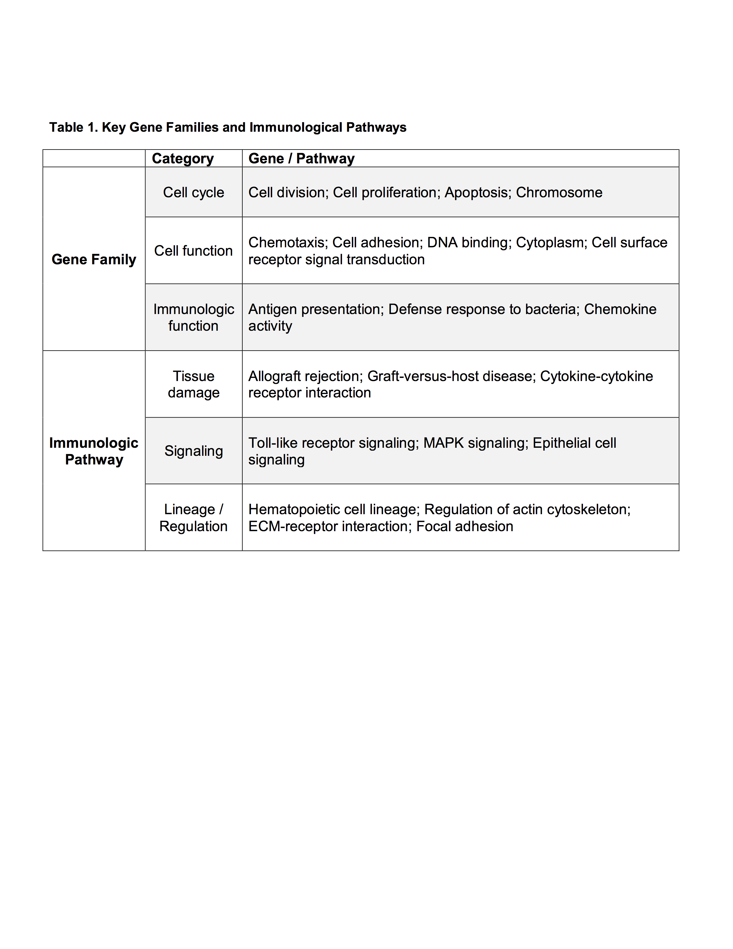
Results: When comparing discharge dispositions, SNF patients experienced greater rates of long-term organ failure (p<0.001). Analysis of genomic expression between discharge groups at 12 hours of admission demonstrated modest differences in lymphocytes and neutrophils, while significant differences were found in monocytes. Monocyte genomic differences persisted over 28 days. At 12 hours, 1187 monocyte genes significantly differed in expression. Over the 28 days, 1058 genes significantly differed. The most important gene families and immunological pathways represented are included in Table 1. The most represented ontological groups are those involved in wound healing and tissue regeneration, which may provoke SNF-associated organ failure.

Conclusion: Monocyte genomic expression differs significantly among severely injured trauma patients depending on discharge disposition, while only modest differences are seen in neutrophil and lymphocyte genomic expression. Monocyte genomic patterns can be detected within 12 hours of admission, persist over time, and are associated with an increased risk of organ failure. Although early genomic expression patterns can identify trauma patients with a poor clinical trajectory requiring SNF placement, more research is necessary to determine if early interventions can alter the clinical course, affect discharge disposition, and improve outcomes.

41.02 Paradoxical Suppression of a Subset of Circulating Mediators in the Most Severely Injured Patients

A. J. Lamparello1, R. A. Namas1, I. Billiar1, Y. Vodovotz1, T. R. Billiar1  1University Of Pittsburg,Pittsburgh, PA, USA

Introduction:
Trauma results in an increase in circulating inflammatory mediators referred to as a “cytokine storm.”  The magnitude of this increase correlates with complications and is thought to be dependent on the severity of injury.  However, a relatively limited number of inflammatory mediators have been characterized in human trauma.  Here, we measured the dynamic changes in 31 cytokines and chemokines in a large cohort of blunt trauma patients and analyzed the differences as a function of injury severity.

Methods:
We performed a retrospective study using a cohort of 472 blunt trauma survivors.  Plasma was sampled three times within the first 24 hours (including admission) and then daily from days 1 to 7 post-injury.  The samples were assayed using Luminex multiplex assay.  Three cohorts were analyzed as a function of injury severity.  Injury severity score (ISS) was calculated for each patient upon discharge and categorized as mild (ISS: 1-15, n=180), moderate (ISS: 15-24, n=170), or severe (ISS: ≥25, n=122).  Statistical difference (p < 0.05) between the groups was determined either by Student t test or χ2 test as appropriate.  Two-way analysis of variance was used to determine statistical significance of differing levels of inflammatory mediators.

Results:
Patients with severe injury had a significantly longer ICU length of stay (LOS), total LOS, days on mechanical ventilation, and rate of nosocomial infections compared to patients with mild or moderate injury based on ISS scores.  As expected, a number of inflammatory mediators exhibited elevations on admission and over time that positively correlated with injury severity (e.g., IL-6, IL-7, IL-17, sIL-2Rα, GM-CSF, IP-10/CXCL10, MIG/CXCL9, and MCP-1/CCL2).  In striking contrast, circulating levels of a subset of mediators, although significantly elevated in the mild and moderate injury groups (relative to healthy controls), were significantly suppressed in the most severely injured patients.  These mediators, which included IL-33, IL-9, IL-21, IL-22, IL-23, and IL-25, were lower during the first 24 hours post-admission and remained low throughout hospital stay.  Of note, several of these mediators have known tissue protective and reparative actions (e.g., IL-9, IL-22, and IL-33).

Conclusion:
Our novel findings identify a subset of mediators that are suppressed in the most severely injured patients.  These observations in injured humans provide potentially important insights into mechanisms associated with immune dysfunction following trauma.
 

40.20 The role of CDK5 in Prostate Cancer Transdifferentiation.

D. W. Howse1, A. Carter1, C. Morrissey2, J. Bibb1  1University Of Alabama at Birmingham,Surgery,Birmingham, Alabama, USA 2University Of Washington,Seattle, WA, USA

Introduction:

Prostate cancer is the most prevalent form of cancer and constitutes the 3rd leading cause of cancer related deaths among men. The standard initial treatment for prostate cancer is androgen deprivation therapy (ADT). Despite initial effectiveness, almost all prostate adenocarcinomas will become resistant to ADT over time. Once progressed to this stage, the cancer is then referred to as castration resistant prostate cancer (CRPC).  CRPC has a very poor prognosis with a median overall survival of approximately one-year with 90% of patients developing metastasis. CRPC may be divided into two categories: CRPC, and CRPC with a neuroendocrine phenotype (CRPC-NE). CPRPC-NE is considered the most highly aggressive form of prostate cancer.  Recently, we have shown that Cdk5, a non-traditional cyclin dependent kinase, contributes to the development and progression of many different types of neuroendocrine cancers. We hypothesize that Cdk5 and its downstream effector SUV39H1 contributes to the poor prognosis of CRPC. 

SUV39H1 is a H3K9m3 methyltransferase that, when disrupted, results in the relaxation of heterochromatin as well as chromosome instability. Phosphorylation of SUV39H1 disrupts its binding to heterochromatin, leading to early replication and replication stress. This project aims to assess whether Cdk5 is involved in the progression of prostate cancer through phosphorylation-mediated loss of function of SUV39H1. We hypothesize that aberrant activation of Cdk5 drives the conversion of CRPC to CRPC-NE through an SUV39H1-dependent pathway and that Cdk5 is an effective target for drug therapy.


Methods:


FFPE sections were obtained from Dr. Colm Morrissey from the University of Washington. The sections were 5 microns thick and were analyzed for Cdk5, p35/p25, chromogranin A, and synaptophysin. Phosphomietics of serine 391 of SUV39H1 have been made to elucidate the phosphorylation capacity of CDK5 and SUV39H1 as well as to transfect into cells to determine the effect on cell growth and migration.

Results:

Immunohistochemistry (IHC) illustrated that Cdk5 as well as its activators, p35/25, are present in patients that present with CRPC-NE. PC-3 cells (CRPC) exhibit expression of all of the required proteins for Cdk5 to phosphorylate SUV39H1.

Conclusion:

We have shown that the essential components in the Cdk5-SUV39H1 pathway are present in human tumors. Current studies with novel Cdk5 inhibitors, phosphorylation state-specific antibodies, and phosphomimetics will determine if SUV39H1 is required for the conversion of CRPC to CRPC-NE and the mechanisms responsible. Thus, we are providing new insights into the transdifferentiation from CRPC to CRPC-NE, toward the goal of finding treatment approaches for this most challenging form of cancer.
 

41.01 The Uniquely-Human CHRFAM7A Gene Alters Ligand Binding to the α -7 Nicotinic Acetylcholine Receptor

T. Chan1, E. Williams1, B. Eliceiri1, A. Baird1, T. Costantini1  1University Of California – San Diego,Division Of Trauma, Surgical Critical Care, Burns And Acute Care Surgery/Department Of Surgery,San Diego, CA, USA

Introduction: The α-7 nicotinic acetylcholine receptor (α7nAChR) is required for the anti-inflammatory activity of the vagus nerve and is thought to be essential to resolve inflammation after severe injury.  CHRFAM7A is a uniquely-human gene that encodes a human-specific subunit of the α7nAChR and a putative negative inhibitor of ligand binding. We previously demonstrated that CHRFAM7A expression is highly variable in human leukocytes suggesting a source of human variability in vagal anti-inflammatory responsiveness. We therefore hypothesized that expression of CHRFAM7A would alter ligand binding to the α7nAChR and give insight into human variability in the anti-inflammatory response to injury and infection.

Methods:  Transgenic mice were engineered to ubiquitously express the uniquely-human gene CHRFAM7A under control of the EF-1α promoter. CHRFAM7A gene expression was confirmed by PCR, quantitative RT-PCR and immunoblotting with an antibody raised to a peptide sequence unique to CHRFAM7A. Muscle tissue and peritoneal macrophages were harvested from these transgenic mice and ligand binding to α7nAChR compared to sibling-matched wild-type C57 mice.  Immunostaining of the neuromuscular junction was performed using α-bungarotoxin (α-BTX), a ligand specific for the α7nAChR. Macrophage α-BTX binding was measured using flow cytometry and immunohistochemistry.

Results: Human CHRFAM7A gene and protein expression was measured in transgenic mice but was undetectable in cells from wild-type animals. α-BTX co-stained with neurofilament at the neuromuscular junction in wild-type mice, however, α-BTX staining was absent in muscle from CHRFAM7A transgenic mice, demonstrating a loss of α7nAChR function. Similarly, CHRFAM7A expression in macrophages decreased α-BTX binding.

Conclusion: CHRFAM7A prevents binding of α-BTX to the α7nAChR. The variability of CHRFAM7A expression seen in humans may, therefore, contribute to human variability in the α7nAChR-dependent anti-inflammatory response to injury and infection. By extension, human CHRFAM7A expression may be responsible for the limited clinical effectiveness of vagal therapeutics that rely on a functional α7nAChR.


40.17 Human Biliary Tract Cancer Contains High Levels of S1P

Y. Hirose1, M. Nagahashi1, K. Yuza1, K. Miura1, J. Sakata1, T. Kobayashi1, H. Ichikawa1, Y. Shimada1, H. Kameyama1, K. Takabe2,3, T. Wakai1  1Niigata University Graduate School Of Medical And Dental Sciences,Division Of Digestive And General Surgery,Niigata, NIIGATA, Japan 2Roswell Park Cancer Institute,Breast Surgery, Department Of Surgical Oncology,Buffalo, NY, USA 3University At Buffalo Jacobs School Of Medicine And Biomedical Sciences, The State University Of New York,Department Of Surgery,Buffalo, NY, USA

Introduction: Biliary tract cancer, including gallbladder cancer, intra- and extrahepatic bile duct cancer, is one of the most lethal diseases among gastrointestinal cancer. Previous studies have suggested that chronic inflammation is involved in the disease process of biliary tract cancer. Sphingosine-1-phosphate (S1P) has emerged as a pleiotropic sphingolipid mediator that regulates many cellular functions associated with inflammation and cancer. Indeed, it has been previously suggested that S1P plays important roles in bile duct cancer progression in experimental model. However, the roles of S1P in human biliary tract cancer has yet to be clarified. The aim of this study is to determine the levels of sphingolipids including S1P and their metabolites in biliary tract cancer tissues and normal biliary tract tissues, and to clarify the difference in the levels of each sphingolipid between the two tissues.

Methods: We examined biliary tract cancer tissues (gallbladder cancer, N=5; intrahepatic bile duct cancer, N=2; and extrahepatic bile duct cancer, N=8) and normal biliary tract tissues (normal gallbladder mucosa, N=5; normal extrahepatic bile duct mucosa, N=12) in patients with biliary tract cancer. Sphingolipids including S1P and their metabolites of dihydro-S1P (DHS1P), sphingosine (Sph), and dihydro-Sph (DHSph) were quantified by liquid chromatography-electrospray ionization-tandem mass spectrometry (LC-ESI-MS/MS). The levels of each sphingolipid were compared between cancer tissues and normal tissues using the Mann-Whitney U test. All tests were two-sided and P<0.05 were considered statistically significant.

Results:In the comparison between all biliary tract cancer tissues (N=15) and all normal biliary tract tissues (N=17), the levels of S1P, DHS1P, Sph, and DHSph were significantly higher in cancer tissues than normal tissues (S1P, P=0.004; DHS1P, P=0.030; Sph, P=0.011; DHSph, P<0.001). In the comparison between intra- and extrahepatic bile duct cancer tissues (N=10) and normal bile duct mucosa (N=12), the levels of S1P, DHS1P, Sph, and DHSph were significantly higher in cancer tissues than normal tissues (S1P, P=0.004; DHS1P, P=0.030; Sph, P=0.011; DHSph, P<0.001). In the comparison between gallbladder cancer tissues (N=5) and normal gallbladder mucosa (N=5), the levels of DHSph were significantly higher in cancer tissues than normal tissues (P=0.016), but there were no significant difference in the levels of S1P, DHS1P and Sph.

Conclusion:To our knowledge, this is the first study to reveal the levels of sphingolipids including S1P in human biliary tract cancer patients by mass spectrometry. The high levels of sphingolipids in the cancer tissue may indicate the important roles of S1P in disease process of biliary tract cancer in human patients.


40.18 Dysregulation of Sphingolipids in Human Hepatocellular Carcinoma

K. Miura1, M. Nagahashi1, Y. Hirose1, T. Kobayashi1, J. Sakata1, H. Kameyama1, Y. Shimada1, H. Ichikawa1, K. Takabe2,3, T. Wakai1  1Niigata University Graduate School Of Medical And Dental Sciences,Division Of Digestive And General Surgery,Niigata, NIIGATA, Japan 2Roswell Park Cancer Institute,Breast Surgery, Department Of Surgical Oncology,Buffalo, NY, USA 3University At Buffalo Jacobs School Of Medicine And Biomedical Sciences, The State University Of New York,Department Of Surgery,Buffalo, NY, USA

Introduction:  Hepatocellular carcinoma (HCC) is now the third leading cause of cancer deaths worldwide, with over 500,000 people affected. Sphingolipids including sphingosine-1-phosphate (S1P) and ceramide have emerged as key regulatory molecules, controlling various aspects of cancer biology and implicated in the mechanism of action of cancer chemotherapeutics. S1P is known to play important roles in cancer cell survival and progression. We previously demonstrated that S1P is a crucial mediator of cancer-induced angiogenesis and lymphangiogenesis and promote metastasis (Cancer Research 2012). On the other hand, ceramide has been considered to mediate anti-proliferative responses, such as cell growth inhibition, apoptosis induction, senescence modulation and autophagy. Ceramide can be de-acylated to give sphingosine, and sphingosine in turn can be phosphorylated to produce S1P. The dynamic balance between S1P and sphingosine/ceramide is referred to as the “sphingolipid rheostat” and influences cancer cell fate. Although important roles of sphingolipids in cancer progression has been revealed in experimental models, its roles in human HCC patients are yet to be determined. In this study, we measure the levels of sphingolipids including S1P and ceramides in tumor and normal liver tissue by state-of-the-art mass spectrometry.

Methods:  Tumor and normal liver tissue were obtained from 20 patients with HCC. Sphingolipids were measured by liquid chromatography-electrospray ionization-tandem mass spectrometry (LC-ESI-MS/MS). Wilcoxon matched-pairs signed rank test was performed to compare the levels of each sphingolipid between tumor and normal tissue, and P<0.05 was considered as statistically significant.

Results: The levels of sphingolipids including sphingosine, dihydro-sphingosine, S1P, dihydro-S1P, and ceramides were detected successfully in the tumor and normal liver tissue from 20 HCC patients. The levels of S1P and dihydro-S1P in tumor tissue were significantly higher than those in normal liver tissue with almost four-fold increase (P<0.0001 and P<0.0001). Further, sphingosine and dihydro-sphingosine in tumor tissue were also significantly higher than those in normal tissue (P<0.001 and P<0.01). Finally, the levels of each ceramide species (C14:0, C16:0, C18:1, C18:0, C20:0, C22:0, C24:1, C24:0, C26:0) in tumor tissue were significantly higher than those in normal tissue (P<0.0001 for each species).

Conclusion: Our results indicate important role of sphingolipids in HCC. Further study will be required to investigate the distinct roles of each sphingolipid in human patients. 


40.19 Effect of HO-3867, a novel curcumin analog, on cholangiocarcinoma

H. Kazik1, S. Kunnimalaiyaan1, T. Gamblin1, M. Kunnimalaiyaan1  1Medical College Of Wisconsin,Surgical Oncology/Surgery,Milwaukee, WI, USA

Introduction: Cholangiocarcinoma (CCA) is rare, but lethal bile duct cancer which remains difficult to diagnose and treat. Currently, systemic therapies and surgery remain the viable solutions. However, CCA responds poorly to these therapies and only a few patients are candidates for potentially curative surgical resection. Therefore, an urgent need for the identification of molecularly targeted compound(s) remains. HO-3867, a curcumin analog, has shown efficacy as an inhibitor of signal transducer and activator of transcription 3 (STAT3) in ovarian cancer cell proliferation but the effects of HO-3867 in other cancer types including CCA is unknown. Therefore, the objective of this study is to test the hypothesis that HO-3867 will effectively inhibit CCA cellular proliferation.

Methods: Effect of HO-3867 (0-10 µM) on two human CCA cell lines, CCLP-1 and CC-SW-1 cellular proliferation was measured through MTT assay, colony forming ability, and real time confluency assay. Cell lysates were analyzed via western blot to determine the effect of HO-3867 on STAT3 phosphorylation as well as the levels of pro and anti-apoptotic proteins.

Results:Treatment with HO-3867 significantly reduced cellular proliferation, colony formation, and cell confluency in a dose- and time-dependent fashion in both cell lines. Following treatment with HO-3867, induction of apoptosis was evident in CCLP-1 through an increase in pro-apoptotic marker (cleaved poly ADP ribose polymerase) and a decrease in anti-apoptotic marker (cyclin D1 and survivin). HO-3867 treatment reduced the levels of phosphorylation of STAT3.

Conclusion:HO-3867 significantly reduced the growth of both CCLP-1 and CC-SW-1. To our knowledge, this is the first study on the effect of HO-3867 in CCA cell lines in vitro and provides rationale for further preclinical analysis.


40.15 γH2AX Expression Discriminates Between Benign and Malignant Biliary Tract Biopsy Specimens

Y. Hirose1, M. Nagahashi1, H. Ishikawa1, K. Yuza1, K. Miura1, H. Ichikawa1, Y. Shimada1, H. Kameyama1, J. Sakata1, T. Kobayashi1, T. Wakai1  1Niigata University Graduate School Of Medical And Dental Sciences,Division Of Digestive And General Surgery,Niigata, NIIGATA, Japan

Introduction: Preoperative discrimination between benign and malignant biliary structures based on small biopsy specimens is difficult. 5.2 to 24.5 percent of biliary structures resected as malignant proved to be benign after histological examination of the surgical specimens. Recent studies have demonstrated that γH2AX, a sensitive marker for DNA double-strand breaks (DSBs), are detected at higher levels in cancer than in precancerous or normal lesions. However, γH2AX expression in biopsy specimens of biliary structures have not been examined. The aim of this study was to determine the usefulness of γH2AX expression in biopsy specimens to discriminate between benign and malignant tissues.

Methods: We examined biopsy specimens of six extrahepatic bile duct cancer tissues, seven inflammatory tissues of the bile duct, and five normal bile duct tissues. The diagnoses of these samples were confirmed by postoperative histological examination or clinical follow-up. Immunohistochemical examination of γH2AX was performed for each biopsy specimen. The nuclear staining pattern was classified into two patterns: focal staining pattern, characterized by focus formation in the nucleus; and diffuse staining pattern, characterized by peripheral– or pan-nuclear staining. The labeling index (LI) of γH2AX-positive cells was calculated for each staining pattern. Continuous variables were compared using the Mann-Whitney U test or the Kruskal-Wallis test. All tests were two-tailed, and P-values less than 0.05 were considered statistically significant.

Results:Kruskal-Wallis test revealed that the LI of γH2AX-positive cells with focal staining was significantly different between cancerous, inflamed, and normal biopsy tissues . Moreover, Dunn's multiple comparison test revealed that there was a significant difference of LI of γH2AX-positive cells with focal staining between cancer tissues and normal tissues (P < 0.01). On the other hand, there was no significant difference in the LI of γH2AX-positive cells with diffuse staining among the three different types of tissues (Kruskal-Wallis test; P > 0.05).

Conclusion:To our knowledge, this is the first study to examine the LI of γH2AX-positive cells in human biliary biopsy samples. The LI of γH2AX-positive cells with focal staining might be a useful biomarker to discriminate between benign and malignant biliary tissues.


40.16 Possible Role of HOX genes in Pancreatic Ductal Adenocarcinoma Survival

K. Takabe1, E. Katsuta1, L. Yan2  1Roswell Park Cancer Institute,Breast Surgery, Department Of Surgical Oncology,Buffalo, NY, USA 2Roswell Park Cancer Institute,Department Of Biostatistics And Bioinformatics,Buffalo, NY, USA

Introduction:  Pancreatic adenocarcinoma (PDAC) is one of the most aggressive cancers with severe prognosis in general; however, we sometimes encounter exceptional long term survivors.  Although there have been many study to explore the genes that are responsible for these differences in prognosis of PDAC, it remains unknown. The HOX genes are a family of homeodomain-containing transcription factors that determine cellular identity during development. This study aimed to investigate the clinical relevance of HOX genes on the PDAC patient survival. 

Methods:  RNA-sequencing and clinical data were obtained from the Cancer Genome Atlas (TCGA) through cBioportal. Gene expression was compared between patients who survived more than 5 years (good prognosis group) and patients who died within 3 years of diagnosis (poor prognosis group). Patients whose cause of death was not pancreatic cancer in bad prognosis group were excluded for the analysis. The cut-off value of gene expression for survival analysis was determined by automated scanning and selecting the threshold yielding the lowest p-value for the each gene.

Results: Among pancreatic cancer TCGA cohort, 94 PDAC patients were confirmed of their date of death or lived longer than 5 years. 4 and 82 patients were classified as good and poor prognosis group, respectively. 1 and 3 patients were diagnosed as Stage IIA and IIB in good prognosis group, 1, 5, 9, 62, 2 and 3 patients were diagnosed as Stage IA, IB, IIA, IIB, III and IV in poor prognosis group, respectively. 120 genes were extracted as differentially expressed gene with adjusted p<0.05. Among 120 genes, 8 genes were HOX genes, including HOXA9 (logFC=7.18, adj. p<0.001), HOXA7 (logFC=3.26, adj. p<0.001), HOXA4 (logFC=2.18, adj. p<0.001), HOXA6 (logFC=5.38, adj. p<0.001), HOXA5 (logFC=3.85, p<0.001), HOXA10 (logFC=4.19, adj. p=0.001), HOXA2 (logFC=1.75, adj. p=0.036) and HOXA13 (logFC=2.87, adj. p=0.039). All of them were upregulated in good prognosis group. Then we compared the survival based upon HOX expression in whole 154 PDAC in TCGA cohort. High expression of HOXA2 (median OS: 19.8 month vs 16.8 month, p=0.005, median DFS: 16.9 month and 9.6 month, p=0.001), HOXA4 (median OS: 20.6 month vs 12.5 month, p<0.001, median DFS: 17.1 month and 9.5 month, p<0.001), and HOXA5 (median OS: 19.9 month vs 12.5 month, p<0.001, median DFS: 17.1 month and 9.5 month, p=0.001) showed significantly better both OS and DFS. 

Conclusion: This is the first report that high expression of HOX genes associate with exceptional long term survivor in PDAC. Further studies are warranted to investigate the mechanism how these gene expressions contribute to patient survival.

40.13 Changes in MiR-200 Family and RASSF2 Expression in Colorectal Cancer and Normal Adjacent Epithelium

V. Stephen1, J. Carter1, J. Hallion1, M. R. Eichenberger1, E. M. Hattab2, S. Galandiuk1  1University Of Louisville,Department Of Surgery,Louisville, KY, USA 2University Of Louisville,Department Of Pathology,Louisville, KY, USA

Introduction:

Colorectal cancer (CRC) is the second leading cause of cancer-related death in the U.S. MicroRNAs (miRNAs) are small, non-coding RNA molecules that downregulate protein expression and have a role in carcinogenesis by dysregulating oncogenes and tumor suppressor genes. MiRNAs from the miR-200 family (miR-200a, -200b, -200c, -141, -429) are associated with many cancers, playing a role in blocking epithelial to mesenchymal transition (EMT). RASSF2, another target of the miR-200 family, is a negative regulator for the known proto-oncogene K-Ras. We have previously observed that miR-200 is upregulated and RASSF2 is downregulated in CRC cell lines and that modulating miR-200 expression can alter cell proliferation in CRC cell lines. We expanded upon this work by examining miR-200 and RASSF2 expression in fresh frozen CRC tissue and histologically normal adjacent epithelial tissue obtained from CRC patient resection specimens.

Methods:

CRC and normal adjacent tissue samples were obtained from 5 patients diagnosed with stage III CRC from our institution's biorepository. Specimens were cut onto slides and stained. Cells of interest (cancer or normal epithelium) were captured using the ArcturusXT Laser Capture Microdissection System, using H&E slides as reference (Fig. 1A). RNA was then extracted and expression of miR-200 and RASSF2 mRNA was measured via qRT-PCR. Protein was extracted and measured via Western Blot.

Results:

We observed a downregulation of miR-200a, miR-200b, miR-141, and miR-429 (p=0.047, 0.119, 0.007, and 0.027 respectively), and no change in miR-200c (p=0.589) in CRC tissue samples compared to normal adjacent epithelium (Fig. 1B). We also observed a 2.7-fold downregulation of RASSF2 mRNA (p=0.021) (Fig. 1C) and a 6-fold downregulation of RASSF2 protein in CRC tissue relative to normal adjacent epithelium (p=0.092) (Fig. 1D).

Conclusion:

Although our data concerning RASSF2 were consistent with those obtained from CRC cell lines, that for miR-200 was not. RASSF2 is a negative regulator of K-Ras, an oncogenic signaling protein. It is advantageous for cancer cells to downregulate RASSF2 expression in any environment. MiR-200, on the other hand, has two dichotomous roles. Upregulation may be able to block RASSF2, but downregulation helps induce EMT. In cell culture, there is no advantage in promoting EMT and metastasis, but this advantage exists in vivo, particularly in later or more aggressive cancers. Further investigations considering earlier stages of CRC may shed light on this discrepancy. Although we were able to alter RASSF2 expression and cell proliferation by modulating miR-200 expression in cell culture, these data from human tissue identify limitations of the miR-200 family as a therapeutic target in CRC.

40.14 High Levels of Sphingolipids in Human Pancreatic Cancer

K. Yuza1, M. Nagahashi1, Y. Hirose1, M. Nakajima1, K. Miura1, H. Ichikawa1, Y. Shimada1, J. Sakata1, H. Kameyama1, T. Kobayashi1, K. Takabe2,3, T. Wakai1  1Niigata University Graduate School Of Medical And Dental Sciences,Division Of Digestive And General Surgery,Niigata City, NIIGATA, Japan 2Roswell Park Cancer Institute,Breast Surgery, Department Of Surgical Oncology,Buffalo, NY, USA 3University At Buffalo Jacobs School Of Medicine And Biomedical Sciences, The State University Of New York,Department Of Surgery,Buffalo, NY, USA

Introduction:
Pancreatic cancer is one of the most lethal diseases and it often spreads quickly before it causes any symptoms. Elucidation of the underlying mechanisms how pancreatic cancer progresses and metastasizes is the key to improve outcome. Sphingosine-1-phosphate (S1P), a bioactive lipid mediator plays critical roles in cancer progression. S1P is involved in numerous cellular functions such as cell proliferation, migration, survival, angiogenesis and lymph angiogenesis, all of which are related to cancer progression and metastasis. Although it is expected that S1P plays an important role in pancreatic cancer progression based on the previous findings of experimental models, its role in human pancreatic cancer has never been revealed. We hypothesized that sphingolipids including S1P are produced higher in pancreatic cancer compared with normal pancreas tissue.

Methods:
Tumor and non-cancerous pancreas tissue were obtained from 10 patients with pancreatic cancer. Both tumor and non-cancerous pancreas tissue were collected from the same resected specimen, and non-cancerous tissue was collected from normal pancreas as far away as possible from the cancer site. Sphingolipids including S1P and their metabolites were measured by liquid chromatography-electrospray ionization-tandem mass spectrometry (LC-ESI-MS/MS). The levels of each sphingolipid were compared between tumor and non-cancerous pancreas tissue by using the Wilcoxon matched-pairs signed rank test. All of the tests were two-sided and P values < 0.05 were considered to be statistically significant.

Results:
Levels of sphingosine, dihydro-sphingosine, S1P, and dihydro-S1P in the pancreatic cancer and normal pancreas tissue were all successfully determined. The levels of all these sphingolipids were universally higher in the cancer tissue than in the normal pancreas tissue (P<0.001 for sphingosine, dihydro-sphingosine, and S1P; P<0.05 for dihydro-S1P). We also determined the levels of each species of ceramide (C14:0, C16:0, C18:1, C18:0, C20:0, C22:0, C24:1, C24:0, C26:1 and C26:0) in the pancreatic cancer and normal pancreas tissue. We found that C14:0 alone was significantly higher in the cancer tissue than in the normal pancreas tissue.

Conclusion:
Levels of sphingolipids in cancer tissue are generally higher than normal pancreas tissue in patients with pancreatic cancer. The high levels of S1P and its metabolites in cancerous tissues implicate the important role of S1P in pancreatic cancer.
 

40.10 Stemness, EZH2, and Notch in Melanoma

P. Elliott1, G. Kaushik1, B. Roy1, S. Umar1, S. Anant1, J. M. Mammen1  1University Of Kansas,Surgery,Kansas City, KS, USA

Introduction: Epigenetic mechanisms and chromatin modifications emerged as key players in cancer development. Enhancer of Zeste Homolog 2 (EZH2), a histone methyltransferase, is the main component of the polycomb-repressive complex 2 (PRC2) involved in epigenetic modification. Increased activity of EZH2 has been reported to be associated with different cancer types including melanoma (mel). Evidence for functional role of EZH2 in melanomagenesis, in particular to stem signaling, is poorly understood. We focused on the interplay between epigenetic modifier Enhancer of Zeste Homolog 2 (EZH2) and stem cells regulation via notch stem cell signaling pathway. 

Methods: We used various mel lines and normal melanocytes (melcytes). The hexoseaminidase assay, colony formation assay (CFA), migration assay and spheroid assay (SA) were used to determine cell growth, proliferation and stemness. Protein expression studies and mRNA levels in cells were done by using standard Immunoblotting technique and qPCR. A lentiviral expression system was used to knockdown as well as to ectopic overexpress the gene of interest in various mel lines and melcytes respectively. 

Results:From TCGA database, we observed higher expression levels (> 60%) EZH2 mRNA in cutaneous as compared to uveal mel.   EZH2 protein is highly expressed in most mel cell types as compared to normal melcytes with very low or undetected EZH2 levels (Figure 1) (n=3, p < 0.01). This higher expression level of EZH2 was further confirmed by IHC staining of tissue slides. We studied the effects of EZH2 knockdown by using shRNA virus on growth/proliferation (approximately 40%), CFA, SA and migration of SKMEL-28 mel cells. We observed reduction in cell growth, spheroid size and cell migration in shRNA expressed cells. EHZ2 knockdown showed reduction in colony size in shRNA expressed mel. Notch receptor proteins play an important role in mel stemness and signaling. We studied the correlation of EZH2 with Notch signaling pathway. We further studied mRNA levels of various Notch receptors in UACC275 cells with Ezh2 knockdown using siRNAs. Knockdown significantly reduced the levels of Notch-1 mRNA . Other Notch mRNAs (Notch-2, Notch-3 and Notch-4) were not significantly affected. Higher expression pattern of Notch-1 protein in mel lines was similar to Ezh2 expression patterns. EZH2  was overexpressed ectopically in normal melcytes to study physiological effect on proliferation. Over expression of EZH2 in melcytes increased the proliferation of melcytes. EZH2 overexpression also affected the protein levels of Notch-1 in melcytes. 

Conclusion:Ezh2 and notch signaling showed cross talk in mel cells. More studies required to support association of EZH2 with notch signaling in mel stem cells. 


40.11 High Expression of DMT1 Indicated Better Prognosis in Non-B Non-C Hepatocellular Carcinoma

T. Hoki1, E. Katsuta2, L. Yan3, K. Takabe2,4, F. Ito1,4,5  1Roswell Park Cancer Institute,Center For Immunotherapy,Buffalo, NY, USA 2Roswell Park Cancer Institute,Breast Surgery, Department Of Surgical Oncology,Buffalo, NY, USA 3Roswell Park Cancer Institute,Department Of Biostatistics And Bioinformatics,Buffalo, NY, USA 4State University Of New York At Buffalo,Department Of Surgery, University At Buffalo Jacobs School Of Medicine And Biomedical Sciences,Buffalo, NY, USA 5Roswell Park Cancer Institute,Department Of Surgical Oncology,Buffalo, NY, USA

Introduction:

Hepatocellular carcinoma (HCC) is the sixth most common malignancy with poor prognosis worldwide. HCC commonly develops in patients with underlying chronic liver disease. Higher iron accumulation is present in chronic liver disease and is known to be a risk factor in the development of HCC. The divalent metal-ion transporter-1 (DMT1) is a primary importer of non-heme iron, and is ubiquitously expressed throughout the body, with highest expression in the proximal duodenum, which is the main site of iron uptake. We previously reported that mal-regulation of iron metabolism caused by increased DMT1 expression in the duodenum induced hepatocarcinogenesis. Increased expression of DMT1 in tumorous tissue has also been shown to be associated with carcinogenesis and progression in colorectal and esophageal adenocarcinoma. However, the role of DMT1 in liver of HCC patients remains unknown.

Methods:
Clinical and RNA-seq data were obtained from the Cancer Genome Atlas (TCGA). Gene expression level was compared among each AJCC Stage, and each viral infectious status. Patients were divided into two groups based on DMT1 expression level for survival analysis using Kaplan-Meier method followed by Log-rank test. The cut-off value was determined by automated scanning and selecting the threshold yielding the lowest p-value.

Results:
Of 442 HCC patients in the TCGA cohort, tumor RNA-seq data were obtained from 342 patients with overall survival (OS) and etiology data. The prevalence of HBV, HCV, dual HBV-HCV, and non-B non-C were 97(28.4%), 47(13.7%), 6(1.8%), and 192(56.1%), respectively. Clinical stages were available for 327 patients, and the patients number of stage I, II, III, and IV were 165(50.5%), 75(22.9%), 82(25.1%), and 5(1.5%), respectively. The median observation period was 19.3 months (range, 0–120.73m). There was no significant difference in the expression level of DMT1 among each clinical stage (stage I/II/III/IV) and each viral infection status (HBV/HCV/dual HBV-HCV/non-B non-C). To investigate the impact of DMT1 expression on patients’ prognosis, OS was compared between high and low expression groups in the whole cohort as well as in different viral infectious status. Interestingly, high expression of DMT1 showed better survival in non-B non-C patients (5-year OS rate: 65.2% vs 32.7%, p=0.038), HBV patients (5-year OS rate: 78.3% vs 61.9%, p=0.006), and HBV and/or HCV-infected patients (5-year OS rate: 65.7% vs 36.5%, p=0.001) compared to low expression group. Whereas there was no significant difference in OS between high and low expression groups in HCV patients (p=0.193) and the whole cohort (p=0.069).

Conclusion:
HCC with increased expression of DMT1 has better OS for HBV, HBV and/or HCV, and non-B non-C patients. These findings may imply that DMT1 in HCC tumors plays different role among different viral infectious status patients, and play roles to suppress tumor progression other than iron transportation.

40.12 Targeting Synaptic Vesicle 2 Proteins for Treating Neuroendocrine Tumors

J. Whitt1, J. Ou2, X. Liu2, H. Chen1, R. Jaskula-Sztul1  1University Of Alabama at Birmingham,Department Of Surgery,Birmingham, Alabama, USA 2University Of Alabama at Birmingham,Biomedical Engineering,Birmingham, Alabama, USA

Introduction: Neuroendocrine (NE) malignancies may arise from hormone producing cells located throughout the body. Although advances in diagnostics have led to an increased ability to detect localized and well-differentiated tumors, many neuroendocrine tumors (NETs) don’t produce symptoms until after metastasis. Moreover, patients with liver metastases have a 5-year overall survival rate of less than 30 percent. The identification of a new theranostic agent is necessary to improve the clinical outcome for patients. We propose the use of synaptic vesicle membrane protein 2(SV2A) as an alternative therapeutic target for advanced NETs.

Methods:

Recombinant botulinum heavy chain (rHCR) was produced using an IPTG-inducible expression vector in E. coli BL21. The rHCR was His-Tag purified and stored in PBS buffer before usage.

Cytotoxicity:  H727, TT, and MZ cells were plated at a density of 5000 cells/well in 96-well plates and incubated under standard conditions overnight. The next day, cells were treated with 10, 100, or 500 nmol/L of rHCR and incubated for 72 hr. Following incubation, cell viability was assessed by ATP quantification using the CellTiter-Glo(Promega) assay. Fresh NE tumors were dissociated and injected into polydimethylsiloxane bioreactors in a matrigel and collagen suspension for 3D culture experiments. The viability of 3D cultures incubated with various doses of rHCR was assessed by measuring the uptake of the near-infrared dye IR-783 using an IVIS imaging system.

Western blot: H727, TT, and MZ cells were seeded in 6-well plates at a density of 3×10⁵ cells/well for 24 h followed by treatment with 100nmol/L for 72 h. Total cellular proteins were isolated and analyzed to assess the level of SV2A expression and the effect of rHCR on the expression levels of NET marker proteins.

Immunohistochemistry: Deparaffinized tissue culture slides were incubated with SV2A primary antibody in 1% BSA and incubated overnight at 4?C. Slides were rinsed twice with TBS containing 0.025% Triton, followed by 0.3% H2O2 for 15 min. Slides were then incubated with HRP-conjugated secondary antibody for 1 h at room temperature.

Detection of protein-protein interaction: Precleared cell lysate was incubated with glutathione-agarose beads in the presence of 10 ug of GST-tagged rHCR for 2 h at 4?C with end-over end mixing. Bound proteins were eluted with 20 mM reduced glutathione and analyzed by SDS-PAGE.

Results:All NET cell lines and tumor samples showed SV2A expression. In vitro and 3D bioreactor studies showed no significant change in cell viability at 100 nmol/L. SV2A was detected by Western blot after incubation with GST-tagged rHCR and glutathione-agarose beads.

Conclusion:The detection of SV2A in neuroendocrine cell lines and patient derived samples indicates that SV2A is a potential new therapeutic target for neuroendocrine tumors. The lack of cytotoxicity of rHCR supports its use for targeted delivery of drugs to NETs.