44.10 Duration of Doxycycline Treatment Impacts Hernia Repair Strength

C. Totten1, J. Tharappel1, J. S. Roth1  1University Of Kentucky,General Surgery/Surgery/Medicine,Lexington, KY, USA

Introduction: Incisional hernia formation compliates up to 30% of abdominal operations. Doxycycline, an inhibitor of MMPs, can enhance the hernia repair strength, increase collagen 1 depositon and reduce collagen 3 deposition. The optimal timing and duration of treatment is not well understood.  The present study evaluates the effect of duration of doxycycline treatment on hernia repair strength and collagen deposition in an animal model.

Methods: 42 male Sprague Dawley rats underwent hernia creation and repair with a polypropylene mesh. Animals were randomly assigned into 6 groups: (1) 16 weeks of doxycycline treatment with 8 week additional survival  (24 weeks total) (2) 16 weeks of saline treatment and 8 weeks additional survival (3) 8 weeks of doxycycline treatment and 8 weeks additional survival (4) 8 weeks of saline treatment and 8 weeks additional survival, (5) 16 weeks of doxycycline treatment with no additional survival, (6)16 weeks of saline treatment and no additional survival. After the assigned treatment and interval times,  abdominal walls with implanted mesh were harvested and tensiometric testing and biochemical analyses were performed.

Results:42 animals underwent hernia repair with survival. Animals administered doxycycline for 8 weeks demonstrated no improvement in hernia repair strength relative to controls ( Group 1- 20.46 N vs. Group 2 -18.85 N, NS) . Animals receiving 16 weeks of doxycycline with 24-week total post hernia repair survival time demonstrated enhanced abdominal wall strength relative to control (Group 3 -18.55 N vs Group 4 – 15.95 N, p=.06). Animals treated continuously with doxycycline for 16 weeks up until the time of necropsy demonstrated improved abdominal wall strength (Group 5 – 23.77 N vs. Group 6 – 20.63 N, p=.008). Collagen 1 deposition was increased and MMP-2 was decreased in the animals treated with doxycycline for 16 weeks.

Conclusion:In an experimental model of hernia repair, doxycycline administration for 16 weeks was associated with enhanced tensiometric strength and greater collagen 1 deposition than placebo whereas 8 weeks of doxcycyline therapy was not effective. In light of this finding, future studies evaluating the impact of doxycycline upon hernia repair outcomes should utilize longer durations of therapy.  Future studies are needed to ascertain the benefits of doxycycline in hernia repair beyond 16 weeks duration. Translational studies utilizng doxycycline are required to evaluate the impact upon clinical outcomes. 


44.09 Mechanisms mediating stress-induced vulnerability to gastrointestinal inflammation

B. Vickers1, C. Graham2, A. Chakraborti2, A. Moon2, J. Bibb2, G. Kennedy2  1Rhodes College,Memphis, TN, USA 2University Of Alabama at Birmingham,Gastrointestinal Surgery,Birmingham, Alabama, USA

Introduction:  Inflammatory bowel disease (IBD), including ulcerative colitis and Crohn's disease, is a chronic and relapsing inflammatory disorder of the intestine. Approximately 1 million Americans are currently diagnosed with IBD and as many as 70,000 new cases are diagnosed each year. One of the main factors contributing to IBD is stress, which has become a rampant problem in America and is considered a high-ranking health concern. Unfortunately, the relationship between IBD and stress has not been thoroughly investigated and the contributions of stress to IBD pathology are not well understood. We hypothesize that stress decreases susceptibility to intestinal inflammation.

Methods:  To better understand the pathways that connect stress and IBD, C57Bl/6 mice were administered a Chronic Unpredictable Stress (CUS) regimen followed by 7 consecutive days of  a subthreshold dose of  Dextran Sodium Sulfate (DSS), a chemical inducer of colitis. After completion of CUS +/- DSS, animals were sacrificed, fecal samples were collected, and RNA was isolated from the proximal colon.  

Results: To assess the effects of CUS and/or DSS on colon inflammation, an Enzyme-Linked Immunosorbent Assay (ELISA) was performed to quantify Lipocalin-2 (LCN-2) protein levels in fecal samples. We found that the LCN-2 levels in the CUS + DSS mice were significantly higher than control. Additionally, Quantitative Real Time Polymerase Chain Reaction (qRT-PCR) was used to quantify mRNA transcript levels of three pro-inflammatory cytokines (IL-1β, IL-6, TNFα) in the proximal colon. Interestingly, CUS + DSS mice showed a significant increase in TNFα over control or either treatment alone.  

Conclusion: We found that CUS and DSS alone failed to induce inflammation, but the combination of CUS and DSS showed a significant increase in colon inflammatory mediators suggesting that chronic stress may lower gut inflammatory threshold thereby increasing susceptibility to colitis.  In the future, we plan to assess CUS in an IL10-/- colitis model as well as in the context of high fat/high carbohydrate diet. 

44.08 Increased expression of long Non-coding RNA H19 is Associated with Colon Adenocarcinoma Recurrence

S. J. O’Brien1, C. Fiechter1, M. Paas1, A. Rochet1, S. Galandiuk1  1University Of Louisville,Price Institute Of Surgical Research, The Hiram C. Polk Jr. M.D. Department Of Surgery,Louisville, KY, USA

Introduction:

Colorectal adenocarcinoma is the fourth leading cause of cancer-related death in the United States. Non-coding RNAs have recently been identified as critical mediators of tumor biology. Long non-coding RNAs (lncRNAs) are diverse in their mechanisms but are known mediators of tumor progression; H19 is a well characterized lncRNA involved in the regulation of P53 and in cancer progression. The aim of this study was to identify the association of tumor H19 expression with recurrence-free and overall survival in a colon cancer data set.

Methods:

The clinical dataset from The Cancer Genome Atlas colon adenocarcinoma data set was downloaded using FirebrowseR. The normalized expression of H19 from the associated RNA-seq data set was downloaded using cBioportal. Univariable and multivariable cox proportional regression analysis were used to identify an association between H19 expression in cancer tissue at the time of resection and recurrence-free, and overall survival. 

Results:

Four hundred and eighteen patients were included in this study. Median age was 69 years (IQR 58-75) Two hundred and nineteen patients (52%) were men. Most tumors were located in the sigmoid colon (135/418- 32%).  Two hundred and thirty-nine patients (57%) had stage 1 or 2 cancers. Lymphatic invasion was present in 36% patients (152/418). The median expression of H19 in the data set defined high or low expression groups. The only difference between groups was that high H19 expression was associated with stage 3 and 4 disease (p=0.048). There was no difference in the overall survival between the low and high H19 groups (Log Rank=0.481). High H19 expression was associated with reduced recurrence-free survival (Log-Rank=0.007) (Figure 1). On univariable regression analysis, high H19 expression, stage 3 or 4 disease, and lymphatic invasion were associated with disease recurrence (Hazard ratio=1.861, 95%CI:1.181-2.932, p=0.007, HR=2.498, 95%CI: 1.622-3.847, P<0.001, and HR= 1.928, 95%CI: 1.233-3.014, p=0.004 respectively). On multivariable regression analysis, only high H19 expression (HR=1.686, 95%CI: 1.046-2.720, p=0.032) and stage 3 or 4 disease (HR=2.888, 95%CI: 1.317-3.976, P=0.003) remained statistically significant predictors of disease recurrence.

Conclusion

H19 was associated with advanced stage of tumor disease and is a significant predictor of recurrent cancer on multivariable analysis. The results of this study are in keeping with in vitro studies in that H19 has a number of oncogenic mechanisms of action, including increased proliferation and mediation of epithelial-mesenchymal transition. As it is a critical molecule in cell regulation, it may have both prognostic and therapeutic uses in the future.


44.07 CNP-miR146a Alters Macrophage Phenotype in Diabetic Wounds

L. Dewberry1, C. Zgheib1, M. M. Hodges1, S. A. Hilton1, J. Hu1, J. Xu1, K. W. Liechty1  1University Of Colorado Denver,Department Of Surgery,Aurora, CO, USA

Introduction:

Diabetic wounds are characterized by chronic inflammation. Persistent, proinflammatory macrophage polarization (M1) has been implicated in this pathophysiology. Previous work in our lab has demonstrated that miR-146a downregulates inflammation in the wound, and that the delivery of miR-146a conjugated to cerium oxide nanoparticles (CNP-miR146a) improves diabetic wound healing. We hypothesize one of the mechanisms of this improved wound healing is through the promotion of macrophage polarization from M1 to M2, which leads to resolution of inflammation.

Methods:
Using a murine model, two 8mm dorsal wounds were created using eleven-week-old male mice homozygous for the Leprdb mutation (db/db) and age-matched male nondiabetic heterozygous littermates (db/+). At the time of wounding, one group (n=5) was injected with 10micromolar CNP-146a in each wound. This was compared to the db/db group injected with PBS (n=5) and the db/+ group (n=5). Wounds were harvested at day 10, and macrophages were isolated from with wound homogenate using magnetic separation. Genetic expression was evaluated with real-time quantitative PCR.

Results:
At day 10, diabetic wounds treated with CNP-146a demonstrated decreased expression of the markers associated with the M1 phenotype, TNF, STAT-1, and IL-1B, compared to diabetic wounds treated with PBS, with the level of expression similar to those seen in the non-diabetic wounds.

Conclusion:
Administration of CNP-146a to a diabetic wound returns the level of inflammation in macrophages to that of a non-diabetic wound and supports the hypothesis that administration of CNP-146a may restore the transition of macrophages from M1 to M2 to facilitate normal wound healing.

44.06 Activation of the Vacuolar H+-ATPase via CaSR Activation in the Rat

M. J. Barahona1, J. Ollodart1, T. M. Gisinger1,3, V. M. Baratta1, Y. W. Stroehl1,2, D. Mulligan1, J. P. Geibel1,4  1Yale University, School of Medicine,Department Surgery,New Haven, CT, USA 2Charité University Medicine Berlin,Faculty Of Medicine,Berlin, BERLIN, Germany 3Paracelsus Medical University,Department Of Medicine,Salzburg, SALZBURG, Austria 4Yale University School Of Medicine,Department Of Cellular And Molecular Physiology,New Haven, CT, USA

Introduction:  Gastroesophageal reflux disease is a prevalent chronic disorder, yet symptom management is often difficult to achieve or maintain. Traditional treatments involve proton pump inhibitors and H2-receptor antagonists, though many patients have persistent symptomatology. Recently, an unaccounted apical vacuolar H+-ATPase was identified and found to be activated in the absence of H,KATPase activity. This H+-ATPase is activated by the calcium-sensing receptor (CaSR), which is in turn modulated by the calcium-activated chloride channel (CaCC). Production of gastric acid via this mechanism can be inhibited by blocking gastric CaCC. Tannic acid has been shown to block the CaCC in other organ systems. Here, we demonstrate that modulation of CaSR via the calcimimetic R568 and CaCC, via tannic acid can indirectly influence acid secretion through the vacuolar H+-ATPase.

Methods:  Gastric glands from rats were isolated via a hand dissection technique. The individual glands were then perfused in vitro with a K+-free HEPES (4-(2-hydroxyethyl)-1-piperazineethanesulfonic acid) to suppress H,KATPase activity. The control group was perfused with 400 nM R568 hydrochloride, a positive allosteric modulator and agonist of CaSR, while the experimental group was perfused with 400 nM R568 hydrochloride and 200 µM tannic acid. Next, the extracellular calcium concentration was elevated to 5.0 mM in the presence and absence of R568 and tannic acid. The H+-ATPase activity was selectively monitored with the rate of proton extrusion (ΔpH/min) on individual parietal cells by observing a change and recovery rate of intracellular pH after acid loading the cells with various solutions, leaving only the H+-ATPase as an active proton secretory pathway.

Results: After exposure to R568 in glands from fasted animals there was increased vacuolar H+-ATPase activity with normal extracellular (1.0 mM) Ca2+ with a rate of proton extrusion increase of 0.01255 ± 0.00120 ΔpH/min. Elevations in Ca2+ concentration (1.0 mM to 5.0 mM) caused a further increase in K+-independent H+ secretion 0.01621 ± 0.00087 ΔpH/min. When tannic acid, an inhibitor of CaCC was added to the perfusates with 1.0 mM Ca2+ and 5.0 mM Ca2+there was a significant inhibition of proton secretion under all conditions with a lower proton extrusion relative to the control 0.00296 ± 0.00027 ΔpH/min (p<0.0001) and  0.00409 ± 0.00053 ΔpH/min (p<0.0001), respectively.

Conclusion: An increase in extracellular calcium concentration leads to CaSR stimulation and elevated H+-ATPase activity. In this study, we demonstrate tannic acid-induced inhibition of the H+-ATPase through inactivation of the CaCC. The inhibition of H+-ATPase activity along with H,KATPase activity theoretically may lead to complete blockade of gastric acid production under resting and stimulated conditions. The CaSR along with CaCC can be important new pharmacologic targets to suppress acid secretion.
 

44.05 A Thermogelling Hyaluronic Acid Vaginal Stent to Reduce Postoperative Vaginal Fibrosis

J. Hakim1, O. Wyman2, S. Keswani1  1Texas Children’s Hospital,Pediatric Adolescent Gynecology,Houston, TX, USA 2Baylor College Of Medicine,Houston, TX, USA

Introduction:  There is a need for new therapies to prevent vaginal fibrosis. Up to 50,000 girls and 213,000 adult women yearly in the United States require vaginal reconstructive surgery. There is a high rate (up to 73%) of vaginal fibrosis after these surgeries. Sequelae from vaginal fibrosis can be life-long and lead to significant reductions in quality of life. Currently, treatments are limited to conjugated estrogen (CEE) creams in combination with vaginal stents. Unfortunately, current CEE creams cannot assess delivered estrogen due to cream egress from the vagina and the cumbersome nature of vaginal stents leads to early discontinuation. We have created a novel hydrogel that utilizes “click” chemistry to make a thermogelling hyaluronic acid (HA) drug eluting hydrogel. We sought to compare local estrogen delivery with Premarin cream to sustained estrogen delivery with a novel CEE-eluding NorbHA hydrogel on vaginal wound healing.

Methods:  Norbornene-functionalized hyaluronic acid (NorbHA) and tetrazine-functionalized hyaluronic acid (TetHA) was synthesized. Hydrogel was composed of a 1:1 ratio of NorbHA:TetHA with various concentrations of estradiol (E2), 1.5 MDa HA, or a combination of both. The vaginal stents were placed into a full-thickness 1 mm murine vaginal excisional injury model. Animals were sacrificed at days 0, 2, 3, 7, 10. Macrophage inhibiting factor 1 (MIF1), transforming growth factor-β3 (TGFβ3) expression and vascular endothelial growth factor (VEGF) expression were analysed as markers of inflammation and angiogenesis respectively. Histology and immunohistochemistry were used to assess wound resolution and estrogen receptor (ER) density.

Results: Dissolution parameters demonstrated that drug release was still possible until 72h in the thermogelling hydrogel compared to CEE cream (12h). There was a burst release of E2 over the first 12h period followed by a steady-state release while HA had a burst release at 24 h. A statistically significant decrease in MIF1 expression was found in the group containing both HA and E2 compared to the CEE cream between the day 0 and day 3 timepoints. Greater expression of TGFβ3 and VEGF were found in the groups containing E2, or E2/HA compared to the estrogen cream by day 2. ER density increases with E2 delivery but appears to be dependent on circulating estrogen levels. 

Conclusion: Vaginal tissue healing is enhanced through a novel thermogelling vaginal hydrogel with sustained estrogen and HA release compared to local exogenous estrogen cream. Improvements in both the anti-inflammatory and pro-angiogenic effects of sustained estrogen delivery included reduction in neutrophil and macrophage infiltration, modulating ECM degradation and stabilizing collagen. Further development of this platform may provide a substantial increase in efficacy of E2 delivery to the vaginal tissue and reduce post-operative vaginal fibrosis.


44.04 NOD2 polymorphism is associated with stricutring phentotype in Crohn's disease

J. Hallion1, S. O’Brien1, C. Fiechter1, M. B. Ekman1, J. Burton1, M. Eichenberger1, S. Galandiuk1  1Price Institute of Surgical Research,University Of Louisville,Louisville, KENTUCKY, USA

Introduction: Crohn’s disease is a common inflammatory disorder of the gastrointestinal tract. It has a multifactorial etiology with immunological, environmental, and genetic factors. Phenotypically, Crohn’s disease was classified (Montreal Classification) using age of onset, disease location, and disease behavior. Single nucleotide polymorphisms (SNPs) are common single base pair genetic variants between individuals and are associated with disease phenotype in a number of diseases. Recent genome wide exploratory studies have identified SNPs that are associated with a stricturing phenotype in Crohn’s disease. The aim of this study was to determine if SNP allele frequencies truly correlate with stricturing Crohn’s disease phenotype in a mid-western Caucasian United States population.


Methods:  Patients with Crohn’s disease were selected from a prospectively maintained university surgical digestive practice database. Blood samples were drawn during clinic appointments. Patients with stricturing (Montreal B2) and non-stricturing (Montreal B1) phenotypes were eligible for inclusion. Clinical data were extracted from an electronic database and patients were matched for gender and race. Genomic DNA was extracted from the blood sample, and 4 SNPs (NOD2 rs2066844 R702W, FUT2 rs601338, IL23-R rs1004819, ATG16L1 rs2241880 T300A) were assessed using a TaqMan genotyping assay.

Results: One hundred and sixty-three patients with Crohn’s disease were included in this study; 65% (106/163) were female. Age of onset (Montreal A) was equally distributed between the stricturing and non-stricturing groups (p=0.408). There was no difference in disease location (p=0.814), or the presence of upper GI disease (p=0.274) between the groups. There was no difference in allele frequency or genotype frequency between the stricturing and non-stricturing groups for FUT2, ATG16L1, or IL23-R (Table 1). The T allele (p=0.004) and the TT genotype (p=0.035) in the NOD2 R702W SNP were significantly associated with a non-stricturing phenotype.

Conclusion: We confirmed a significant association between a non-stricturing phenotype and the TT genotype and the T allele in the NOD2 R702W SNP. NOD2 is a well characterized gene that is involved in Crohn’s disease pathophysiology and the results of this study validate the results of large genome wide association studies in a United States population. Further study is required to determine if the NOD2 R702W T allele is a protective factor against stricturing disease.
 

44.03 The Other Side of the Sword: The Role of Autophagy in Survival and Tumor Cell Growth

B. L. Rademacher1, T. Steeno1, K. A. Matkowskyj2, A. Auyeung1, E. H. Carchman1  1University Of Wisconsin,Surgery,Madison, WI, USA 2University Of Wisconsin,Pathology And Laboratory Medicine,Madison, WI, USA

Introduction:  Autophagy is thought to have a dual role in cancer; protective against carcinogenesis, while an important pro-survival mechanism for tumor cells, especially in the setting of treatment (chemotherapy or radiation). We have previously been shown, both pharmacologically and genetically, that autophagy is important in preventing anal carcinogenesis. However, the role of autophagy in already established cancer, in terms of tumor growth and “patient” survival, has yet to be examined. We hypothesized that autophagy is a protective mechanism used by tumor cells to address their increased metabolic needs and that by knocking out autophagy we would decrease tumor growth rates and improve survival. 

Methods:  We generated a conditional, genetic knockout mouse model for the essential autophagic protein Atg7. The generated mice carry Atg7f/f gene globally. They also carry an inducible CreER transgene is expressed from an epithelial-specific K14 transcriptional promotor, and its activity is induced by topical exposure to 4-OH tamoxifen (4-OH TAM), resulting in the knockout of the essential autophagic protein Atg7. For this study, the anuses of the mice were treated with the carcinogen, 7,12 dimethylbenz[a]anthracene (DMBA), until anal cancer developed (2-5 mm in diameter upon entry into the study). Fourteen of the mice were then treated with 4-OH TAM to knockout autophagy locally while twelve were not. Tumor sizes were then measured weekly until euthanasia requirements were met or the mouse died. Tumor volumes were then calculated and normalized to day 0 (date of treatment with 4-OH TAM or not). These tumor volumes were then log transformed, then regressed linearly against time in days, resulting in a slope that equates to the tumor growth rate. Differences in tumor growth rates and mouse survival between the two treatment groups were then examined via independent samples t-test and Kaplan-Meier statistics with Log-Rank comparison, respectively. 

Results: Mice treated with 4-OH TAM had a statistically significant longer mean survival compared to control mice not treated with tamoxifen (98 days vs 49 days, p-value 0.018). In terms of tumor growth rates, there was a trend to decreased mean tumor growth rates in the 4-OH Tam treated group compared to the control group (0.00167 mm/day +/- 0.003 vs 0.00129 mm/day+/-, p-value 0.725).

Conclusion: The localized knockout of autophagy in anal tumors resulted in a trend in decreased growth rates with a significant increase in mouse survival. Increased sample sizes are needed to reduce the variance in tumor growth rates to further elucidate the role of autophagy in tumor growth. 


44.02 Farnesoid-X Receptor Inhibition Protects the Intestinal Barrier by Upregulating Cell Junction Genes

M. Nguyen1, F. Li2, L. Hung2, O. Escobar1, C. Gayer1  1Children’s Hospital Los Angeles,Pediatric Surgery,Los Angeles, CA, USA 2Children’s Hospital Los Angeles,Single Cell, Sequencing, CyTOF Core Laboratory,Los Angeles, CA, USA

Introduction: Farnesoid-X receptor (FXR) is a nuclear receptor involved in bile acid homeostasis. Our lab has shown that FXR activation interferes with the epidermal growth factor receptor (EGFR) pathway, decreasing cell proliferation. Furthermore, we have shown that the intestinal epithelial barrier of FXR knockout (FXRKO) mice is protected from lipopolysaccharide (LPS)-induced injury compared to that of wild-type (WT) mice, as measured by serum fluorescence after oral gavage with FIT-C dextran. We hypothesized that gene expression of molecules in the EGFR pathway and tight junctional proteins would be increased in FXRKO compared to WT mice.

Methods: WT and FXRKO mice were treated with intraperitoneal injections of either normal saline or LPS (n=4-6 in all treatment groups). After sixteen hours, mice were sacrificed, and RNA was extracted from mucosal scrapings of terminal ileal samples. Gene expression profiling was perfomed by RNA-seq, and differentially expressed genes were identified by DESeq2. Genes demonstrating statistically significant (adjusted p<0.05, Benjamini-Hochberg FDR) differential gene expression were cross-referenced with the NIH DAVID Bioinformatics Resources 6.8 database for molecular function and pathway analysis.

Results: Principal component analysis revealed distinct clustering by genotype, indicating a baseline difference between FXRKO and WT mice. We also found distinct clustering by treatment group. A unique LPS response was observed in FXRKO versus WT mice (Figure 1, each row is one of 494 genes with adjusted p=<0.05). Of note, expression of EGFR was upregulated by LPS treatment in FXRKO mice compared to WT mice, as was expression of Pik3ca and Pic3c2a, members of the PI3K family, and Map3k1, a member of the MAPK family. Other genes demonstrating unique LPS response in FXRKO mice included Ddx3x, Yes1, and Arhgap5, which are involved in pathways related to cadherin binding, adherens junctions, and focal adhesions, respectively.

Conclusion: Acute intestinal injury as modelled by LPS injection significantly alters the gene expression profile of FXRKO mice in comparison to WT mice, specifically affecting pathways involved in intestinal barrier integrity and restitution. These data provide avenues for investigation into specific genes and molecular pathways to understand the mechanism through which the intestinal barrier is protected from acute injury in FXRKO mice. Understanding the role of FXR in acute injury will help identify therapeutic targets for the treatment of diseases such as necrotizing enterocolitis and inflammatory bowel disease.


44.01 Upregulation of miR-146a Suppresses Inflammatory Responses in Bleomycin-induced Acute Lung Injury

C. Zgheib1, S. A. Hilton1, L. C. Dewberry1, J. Hu1, J. Xu1, S. Seal3, L. Hernandez-Lagunas2, E. Nozik-Grayck2, K. W. Liechty1  1Laboratory for Fetal and Regenerative Biology, Department of Surgery, University of Colorado Denver School of Medicine and Children’s Hospital Colorado,Aurora, CO, USA 2Developmental Lung Biology and Cardiovascular Pulmonary Research Laboratories, University of Colorado,Aurora, CO, USA 3Department of Material Science Engineering, AMPAC, Nanoscience Technology Center, College of Medicine, University of Central Florida,Orlando, FL, USA

Introduction:

Acute lung injury is characterized by an initial inflammatory response accompanied by acute respiratory distress and surfactant deficiency, which can progress to chronic pulmonary fibrosis. Following lung injury, the respiratory epithelium releases pro-inflammatory mediators which promote the recruitment of neutrophils and subsequently macrophages into the injury sites, further increasing cytokine production and modulation of the extracellular matrix, including collagen. We have previously shown that following injury, delivery of miR146a – an anti-inflammatory microRNA – via conjugation with cerium oxide nanoparticles (CNP), significantly reduced inflammation and promoted healing of impaired diabetic wounds. Thus, we hypothesized that intra-tracheal delivery of CNP-miR146a at the time of bleomycin injury may reduce pulmonary inflammation and subsequent fibrosis.


Methods:

Using an established murine model, juvenile (10 week) male C57BL/6 mice, underwent intra-tracheal instillation of bleomycin (3 units/kg), PBS (n = 5), or bleomycin (n = 6) and a single dose of CNP-miR146a (100 ng, n = 6). Animals were euthanized 14 days post-injury for lung inflation and tissue harvest. Tissue was homogenized and total RNA was extracted. QPCR analysis was used to measure the gene expression of the following inflammatory markers IL-6, IL-1b, and TNFα.


Results:

Our data revealed that bleomycin-induced lung injury in mice resulted in significant upregulation of pro-inflammatory cytokine tumor necrosis factor-alpha (TNF-α), IL-6, and IL-1β, compared to PBS treated controls. Interestingly, intra-tracheal instillation of CNP-miR146a at the time of bleomycin injury showed decreased expression of these pro-inflammatory markers. This data is in agreement with our previous findings that demonstrated less mucosal sloughing/hemorrhage, decreased CD45+inflammatory cells, and markedly decreased lung fibrosis in the CNP-miR146a-treated bleomycin group compared to the bleomycin treatment alone.


Conclusions:

This study results’ indicate that CNP-miR146a decreases the inflammation associated with the bleomycin induced lung injury. CNP-miR146a is a promising therapeutic that could limit the consequences of acute lung injury and warrants further evaluation.

23.10 Inhibition of ADAMTS-4 Prevents the Development of Aortic Aneurysms and Dissections in Mouse

P. Ren1,2, M. Hughes1,2, L. Zhang1,2, Y. Zheng1,2, S. Krishnamoorthy1,2, J. Coselli1,2,3, Y. Shen1,2,3, S. LeMaire1,2,3  1Baylor College of Medicine,Cardiothoracic Surgery,Houston, TEXAS, USA 2Texas Heart Institute,Cardiovascular Surgery,Houston, TEXAS, USA 3Baylor College of Medicine,Cardiovascular Research Institute,Houston, TEXAS, USA

Introduction: We have previously shown that ADAMTS-4, a disintegrin and metalloproteinase with thrombospondin motifs 4, was significantly elevated in aortic tissue from patients with aortic aneurysms and dissections (AAD), and that ADAMTS-4 contributed to aortic destruction and sporadic AAD development in a mouse model. The current study aimed to determine whether pharmacologic inhibition of ADAMTS-4 can prevent AAD development in mice.

Methods: Four-week old male wild type C57BL/6J mice (n=83) were challenged with high-fat diet for 8 weeks and received angiotensin II infusion during the last 4 weeks. The challenged mice were randomly assigned to receive water (control group, n=50) or 75mg/kg/day of ADAMTS inhibitor pentosan polysulfate sodium (NaPPS) (treatment group, n=33). Incidence of AAD was evaluated by two independent observers. Aortic inflammatory cell infiltrate, ECM destruction, SMC apoptosis, and ADAMTS-4 levels were measured.

Results: NaPPS treatment reduced the development of AAD. The overall incidence of AAD decreased from 78% in the control group to 58% in the NaPPS treatment group (P=0.05). AAD reduction was particularly significant in the descending aorta (36% in control group vs 15% in NaPPS treatment group [P=0.05]) and in the suprarenal aorta (60% in control group vs 33% in NaPPS treatment group [P=0.03]). Furthermore, aortas from the NaPPS treatment group showed greater reductions in ADAMTS-4 expression, macrophage infiltration, elastic fiber destruction and SMC apoptosis as compared to controls. 

Conclusion: ADAMTS inhibitor NaPPS partially reduced the challenge-induced aortic destruction, inflammation, and the development of AAD in mice. Thus, pharmacologically targeting ADAMTS-4 may be a promising therapeutic strategy for preventing AAD formation and progression.
 

23.09 Endothelial CEPT1 Promotes Aortic Atherogenesis

C. Yang1,2, L. Belaygorod1, C. Feng3, C. F. Semenkovich3, M. Zayed1  1Washington University,Section Of Vascular Surgery,Department Of Surgery, Washington University School Of Medicine,St. Louis, MO, USA 2Huazhong University Of Science And Technology,Department Of Vascular Surgery,Union Hospital, Tongji Medical College,Wuhan, Hubei, China 3Washington University,Division Of Endocrinology, Metabolism, And Lipid Research, Department Of Medicine,,St. Louis, MO, USA

Introduction: Intracellular lipid metabolism is essential for endothelial cell activation and function. However, it is unknown to what extent this affects atheroprogression. We recently observed that choline ethanolamine phosphotransferase (CEPT1), a central enzyme in phospholipogenesis, is upregulated in peripheral arterial plaque of patients with clinical risk factors such as diabetes. In these patients, plaque phospholipid profiles were also reflective of increased CEPT1 activity. We hypothesize that CEPT1 may be an important EC regulator of plaque progression. To test this, we evaluated aortic atheroprogression in an adult apoe-/- mice following selective conditional endothelial knockdown of cept1.

Methods: Tamoxifen-induced VE-Cadherin driven cre expression and recombination of Loxp (Lp) flanked exon3 of the cept1 was used to induce selective gene knockdown in the endothelium of adult apoe-/- mice (cept1Lp/Lp apoe-/-). Six week old apoe-/- and cept1Lp/Lp apoe-/- mice were maintained on a western diet (42% fat content) for 12 weeks. Aortic atheroprogression was then evaluated and quantified following Oil red O staining. 

Results:Whole mount aortic staining revealed that cept1Lp/Lp apoe-/- mice had markedly reduced aortic plaque volumes, particularly in the areas of high sheer stress such as the aortic arch (p < 0.05; Fig1A) and infrarenal aorta (p < 0.05; Fig1B). Histologic analysis of the aortic root intima also revealed significantly reduced Oil red O staining in cept1Lp/Lp apoe-/- mice (p < 0.05; Fig1C).

Conclusion:Conditional knockdown of cept1 in the endothelium of adult apoe-/- mice is atheroprotective. This novel observation demonstrates the critical role of CEPT1 in plaque progression, and invites further investigation regarding its potential therapeutic targeting to reduce atheroprogression.
 

23.08 Remote Ischemic Conditioning to Decrease Postoperative Complications After Major Abdominal Surgery

J. Rosado1, A. Alvarez1, K. P. Oberoi1, G. Dikdan1, S. R. Pentakota1, S. Husain2, B. Koneru1  1Rutgers New Jersey Medical School,Surgery,Newark, NEW JERSEY, USA 2Rutgers New Jersey Medical School,Genomics Center,Newark, NJ, USA

Introduction:  Remote ischemic conditioning (RIC) modulates inflammation after ischemia reperfusion, and may decrease postsurgical inflammation and complications. Here we present preliminary data regarding peripheral blood leukocyte gene expression in subjects from a prospective randomized a phase II clinical trial of RIC in progress (NCT03234543).

Methods: Adults undergoing abdominal surgery (duration > 2 hours, hospital stay > 2 days) are randomized (1:1) to receive either RIC or sham intervention immediately before surgery and on postoperative days 1 and 2. Each RIC intervention comprises 3 cycles of 5/5 minutes of inflation/deflation of mid-thigh cuff in one lower extremity. Inflation pressures were 250 mmHg for the first and systolic pressures plus 50 mmHg for the others. Sham interventions comprise cuff inflation pressure of 20 mmHg.  Primary outcome is 30-day complications measured as comprehensive complications index. Peripheral blood was collected for plasma and RNA before (R0) and 1hr post surgery (R1), and 1 hour after 2nd and 3rd interventions (R2 and R3). Leukocyte mRNA transcripts were examined in R0 and R3 samples of both groups using RNA sequencing (30 million reads) for global expression profiles (n=6 each; fold change >1.5 and false discovery rate p<0.05) and RT-PCR for cytokine transcripts (IL-1β, IL-6, IL-8, IL-10, and TNF-α; n=8 each; Wilcoxon p< 0.05).

Results: 45 of planned 100 subjects (23/22 RIC/No RIC groups) were enrolled. At baseline (R0), mRNA profiles significantly differed between two groups in only 14 transcripts, and none were related to inflammation. 122 transcripts had significant differential expression between two groups at R3. 39/122 were transcripts of immunoglobulins, and expression levels of all were increased in No RIC. Importantly, TMED7, LGALS2, GZMH, and IL32 transcripts encoding proteins, which promote inflammation via IL-1 signaling, macrophage M1 phenotype, granzyme, and p38 kinase and NF-kb pathways, respectively were increased in No RIC. Transcripts of RELL1, IL1RAP, FKBP5, which encode member of TNF-a receptor family, IL-1 receptor accessory protein, and a cellular glucocorticoid responsiveness protein, respectively were decreased in No RIC.  RT-RCR data showed a trend towards increased expression of IL-10 in No RIC that did not reach significance (p=0.10).

Conclusion: Preliminary results from this novel trial suggest that remote ischemic conditioning in the perioperative period down regulates expression of leukocyte proinflammatory genes. Further studies of gene expression in additional subjects and other time points and studies of plasma acute phase proteins and complement components are in progress. Also, the associations between the molecular data and the primary outcome will be examined. 


23.07 Impact Of Traumatically Brain-Injured Donors On Outcomes After Heart Transplantation

A. Suarez-Pierre1, T. C. Crawford1, X. Zhou1, C. Lui1, C. D. Fraser1, E. Etchill1, K. Sharma2, R. Higgins1, G. J. Whitman1, A. Kilic1, C. W. Choi1  1Johns Hopkins University School Of Medicine,Cardiac Surgery,Baltimore, MD, USA 2Johns Hopkins University School of Medicine,Cardiology,Baltimore, MARYLAND, USA

Introduction: Heart transplant recipients of traumatically brain-injured (TBI) donors have been reported to have inferior survival and increased rates of coronary artery vasculopathy in single-center studies with limited sample sizes. This study sought to examine the impact of TBI donors on outcomes after heart transplantation. 

Methods:  We identified all adult heart transplants performed between January 2007 and December 2016 (inclusive) in the OPTN database. Patients undergoing repeat or heterotopic heart transplantation were excluded from the study. Recipients were dichotomized based on donor cause of death (TBI versus non-TBI), propensity-scored across 22 variables with known associations with mortality, and matched 1:1 without replacement. The primary endpoint was all-cause mortality at 1-, 3-, and 5-years after transplantation. Secondary endpoints were 3- and 5-year survival conditional on 1-year survival and rates of coronary artery vasculopathy at 1-, 3-, and 5- years after transplant. 

Results: In the study period, 20,244 patients underwent heart transplantation. TBI was the primary cause of death of all donors (53.4%; 10,816/20,244) and among TBI donors, blunt injury (59.6%; 6,443/10,816) and gunshot wound (35%; 3,781/10,816) were the most common mechanisms of traumatic brain injury. Propensity matching generated 6,919 pairs with adequate covariate balance (all standardized mean differences < 0.07). Risk-adjusted survival was similar between recipients of TBI donors and non-TBI donors at 1-year (90.5% vs 90.0%, log-rank p=0.32), 3-years (84.1% vs 83.3%, log-rank p=0.25), and 5-years (78.1% vs 77.5%, log-rank p=0.34). Risk-adjusted survival conditional on 1-year survival, was also similar at 3-years (92.8% vs 92.6%, log-rank p=0.57) and 5-years (86.2% vs 86.1%, log-rank p=0.74). The risk-adjusted rates of coronary artery vasculopathy did not differ either at 1-year (8.0% vs 7.7%, log-rank p=0.60), 3-years (20.6% vs 20.4%, log-rank p=0.77), or 5-years (30.6% vs 30.4%; long-rank p=0.78).

Conclusion: In the largest analysis of TBI donors in heart transplantation, we found similar survival and rates of coronary artery vasculopathy to those who received hearts from non-TBI donors out to 5 years. These findings should allay concerns over continued transplantation with this unique donor population.

23.06 Persistent Gender Disparities in Access to Kidney Transplantation

C. Holscher1, C. Haugen1, K. Jackson1, A. Kernodle1, S. Bae1, J. Garonzik Wang1, D. Segev1  1Johns Hopkins University School Of Medicine,Baltimore, MD, USA

Introduction: While national policies direct organ allocation for waitlisted candidates, the decision to list a candidate for transplantation is made at the center- and patient-level. Historically, women have had decreased access to kidney transplantation (KT). We sought to investigate if gender disparities in access to KT have improved over time. 

Methods: To explore temporal trends in access to KT, we studied 1,511,863 adults (age 18-99) with incident end-stage renal disease (ESRD) using the United States Renal Data System (USRDS) from 2000 to 2015. We divided the study period into four eras and compared characteristics of patients who were and were not listed for transplantation (Chi-square and Student’s t tests), and tested if waitlisting changed over time (Cuzick test of trend).  We used Cox regression to determine the association between era and access to transplantation while controlling for candidate factors.  As a sensitivity analysis to determine whether a differential risk of death before waitlisting impacted our inferences, we used a competing risk regression using the Fine and Gray method with a 5% random sample.

Results: The proportion of ESRD patients who were subsequently waitlisted decreased over time (13.2% in 2000-2003 to 8.7% in 2012-2015, p<0.001). Compared to those who were never waitlisted, waitlist registrants were less likely to be female (37% vs 45%, p<0.001), were younger (mean 50 vs. 66 years, p<0.001), were more likely to be African American (32% vs 28%, p<0.001), were more likely to be Hispanic (20% vs. 13%, p<0.001), and were more likely to have private insurance (38% vs. 17%) or be uninsured (13% vs 6%, p<0.001). After controlling for age, race, ethnicity, and prior insurance, men had similar access to KT over time (per 4-year era, aHR 1.00, 95% CI 1.00-1.01, p=0.6), while women had less access (aHR 0.80, 95% CI 0.20-0.81, p<0.001) that worsened with time (interaction p<0.001) (Figure). For context, in 2000-2003, women were 20% less likely to be waitlisted for kidney transplant (aHR 0.80, 95% CI 0.78-0.82, p<0.001), while in 2012-2015 this worsened to 22% less likely (aHR 0.78, 95% CI 0.76-0.80, p<0.001). Our sensitivity analysis using a competing risk regression also showed persistent gender disparities in waitlisting.

Conclusion: Despite decades of studies showing that women have less access to kidney transplantation, gender disparities in access to KT have not improved over time, rather they have worsened. Further focus and novel interventions are needed to improve access for female KT candidates. 


23.05 Tissue Factor-Targeted Peptide Amphiphile as an Injectable Therapy for Hemorrhage

M. Klein1, H. Kassam1, M. Karver2, M. Struble2, L. Palmer2, N. Tsihlis1, S. Stupp2, B. Gavitt4, T. Pritts3, M. Kibbe1  1University Of North Carolina At Chapel Hill,General Surgery,Chapel Hill, NC, USA 2Northwestern University,Chicago, IL, USA 3University Of Cincinnati,Cincinnati, OH, USA 4United States Air Force School of Aerospace Medicine,Cincinnati, OH, USA

Introduction: Non-compressible torso hemorrhage is a leading cause of preventable death in civilian and battlefield trauma. We sought to develop a tissue factor (TF)-targeted nanofiber that can be given intravenously to slow hemorrhage until definitive bleeding control can be obtained.  TF was chosen because it is only exposed to the intravascular space upon vessel disruption. Peptide amphiphile (PA) monomers that self-assemble into nanofibers were chosen as the delivery vehicle.  Here, we systematically analyzed the binding interface of TF to factor VII to generate a TF-binding sequence.  We hypothesize that TF-targeted nanofibers will localize to the site of bleeding in a liver hemorrhage model.

Methods:  PA monomers were synthesized by solid-phase peptide synthesis, purified by high pressure liquid chromatography (HPLC), and characterized by HPLC-mass spectrometry (HPLC-MS).  A TF-binding sequence SFEEARE (SFE) was added to the PA backbone.  SFE-PAs (75% by weight) were co-assembled with PA backbone (20%) and a fluorescently labeled TAMRA PA (5%) to create TF-targeted nanofibers.  Non-targeted PA nanofibers served as controls.  Nanofiber formation was assessed via conventional transmission electron microscopy (TEM) and structure by circular dichroism (CD) spectroscopy.  Male Sprague Dawley rats (250-290g) underwent a liver punch hemorrhage model in which the liver was exposed via a midline laparotomy, followed by tail vein injection of the nanofiber (2.5mg).  A 12mm biopsy punch was used to injure the left lateral lobe of the liver.  Shed blood was collected with pre-weighed gauze for 30 minutes and expressed as percent of total blood volume.  Data are presented as mean±SEM and analyzed by ANOVA.

Results: PAs were >95% pure, as shown by HPLC-MS.  Co-assemblies of PAs formed nanofibers, as shown by TEM, and displayed the expected β-sheet signal characteristic of PA nanofibers when analyzed by CD spectroscopy.  Injection of the SFE-PA nanofiber demonstrated binding to the site of liver injury using fluorescent microscopy, with uninjured liver showing minimal fluorescence and control nanofibers showing minimal fluorescence.  Somewhat surprisingly, given that there was no therapeutic molecule on the PA nanofiber, total blood loss for rats that received the SFE-PA nanofiber was 36% lower than sham rats (14.5±0.95% vs. 22.8±1.1%, n=6, P<0.05).  Blood loss for rats injected with the non-targeted PA nanofiber was similar to sham rats (21.59±2.8%, n=6).

Conclusion: We have successfully synthesized, purified, and characterized a novel PA nanofiber that targets sites of active bleeding in a rat model of hemorrhage.  Not only did our injectable TF-targeted PA nanofiber localize to the site of injury, it appears to decrease blood loss in the setting of abdominal hemorrhage. Incorporation of a therapeutic agent will make this promising technology even more effective in treatment of non-compressible torso hemorrhage.


23.04 Sex Differences in Murine Myocutaneous Flap Revascularization

J. S. Brandenburg1, R. M. Clark1, B. B. Coffman4, G. Sharma2, H. J. Hathaway3, E. R. Prossnitz2, T. R. Howdieshell1,3  1University Of New Mexico HSC,Surgery,Albuquerque, NM, USA 2University Of New Mexico HSC,Molecular Medicine,Albuquerque, NM, USA 3University Of New Mexico HSC,Cell Biology,Albuquerque, NM, USA 4University Of New Mexico HSC,Pathology,Albuquerque, NM, USA


Introduction:   Sex differences in susceptibility to ischemia/reperfusion injury have been documented in humans.  Premenopausal women have a lower risk of ischemic heart disease than age-matched men, whereas after menopause, the risk is similar or even higher in women.  However, little is known about the effects of sex on cutaneous wound revascularization.

Methods:   A cranial-based, peninsular-shaped myocutaneous flap was surgically created on the dorsum of 10-12 week old male (n=10) and female (n=10) C57BL6 mice.  Planimetric analysis of digital photographic images was utilized to determine flap viability.  Real-time flap perfusion, ischemia, and functional revascularization was determined by laser speckle contrast imaging (LSCI).  Image analysis of CD31-immunostained sections confirmed flap microvascular anatomy, density, and surface area.  Values are expressed as means ± SEM.  Student’s t test was performed when comparing two groups, and two-way ANOVA used when comparing multiple groups.  Statistical significance was accepted at a p value < 0.05.

Results:  Flaps created on female mice were engrafted to the recipient site resulting in nearly complete viability at post-operative day 10.  In contrast, distal full thickness myocutaneous necrosis was evident at 10 days post-surgery in male mice (percent flap necrosis: female 5.8 ± 0.9% versus male 30.8 ± 4.7%, p < 0.05).  Over the 10 day study interval, LSCI documented functional flap revascularization in all regions of interest (ROI) in female mice, but minimal distal flap reperfusion in male mice (day 10 distal ROI perfusion: female 191.5 ± 9.2 PU versus male 138.9 ± 11.7 PU, p < 0.05).  Day 10 histologic sections immunostained to detect CD31 confirmed significant increases in distal flap vessel count and vascular surface area in female compared to male mice (vessel count: female 127 ± 12 vessels/mm2 versus male 38 ± 6 vessels/mm2, p < 0.05; vascular surface area: female 7,899 ± 85 µ2/mm2 versus male 2,335 ± 40 µ2/mm2, p < 0.05).

Conclusions:  In a graded-ischemia wound healing model, flap revascularization was more effective in female mice.  The recognition and identification of sex-specific wound healing differences may lead to a better understanding of the underlying mechanisms of myocutaneous revascularization and drive novel discovery to improve soft tissue wound healing.


23.03 Gut Microbiota and Pancreatic Cancer Crosstalk via Immune System: Evidence of a Unique Symbiosis

V. Sethi1, S. Kurtom1, I. Fernandez2, J. Pignac-Kobinger2, A. Ferrantella1, C. Jacob1, P. Roy1, P. Sharma1, M. T. Abreu2, S. Roy1, S. Ramakrishnan1, A. Saluja1, V. Dudeja1  1University Of Miami,Surgical Oncology,Miami, FL, USA 2University Of Miami,Gastroenterology,Miami, FL, USA

Introduction:  Pancreatic ductal adenocarcinoma (PDAC) is characterized by a unique immunosuppressive milieu which makes it particularly resistant to modern checkpoint blocking drugs like anti-PD1 and anti-CTLA4.  Like many other cancers, PDAC is associated with a changed ‘dysbiotic’ gut microbiota. Whether this dysbiotic state promotes cancer or is a neutral-bystander is unclear. We aimed to study as well as investigate the immunotherapeutic implications of this relationship in mice. 

Methods: Wildtype mice-pups (guests) were co-housed with two different types of ‘host’ mice: cancer-bearing KPC (KrasG12D/+;Trp53R172H/+;Pdx-1-Cre) mice or with cancer-naïve mice. Due to coprophagy, the gut microbiota transferred horizontally from hosts to guests. After 2 months of co-caging, the guest mice were given subcutaneous KPC-PDAC and tumor progression was followed. In subsequent experiments, wildtype mice were depleted of their entire gut microbiome by oral antibiotics and growth of orthotopic, subcutaneous and metastatic PDAC was compared between them and controls. Experiments were repeated in various genetic backgrounds. Specific antibiotics were also combined with immune checkpoint-inhibitors in a therapeutic fashion. Pancreatic tumors and metastases were immunophenotyped and were probed for bacteria.

Results: Mice co-housed with KPC cagemates had more aggressive tumor-kinetics than mice co-caged with cancer-naïve mice. Eradicating the gut microbiota by oral antibiotics shrunk tumors in all models of PDAC. This effect disappeared in immunodeficient Rag1-/- mice but not in Tlr4-/- knockout mice. Antibiotics significantly enhanced the efficacy of anti-PD1 and anti-CTLA4 in decreasing tumor burden. Very interestingly, pancreatic tumors grew viable bacteria on culture medium and this was prevented by poorly-absorbable oral antibiotics. Similarly, 16S rRNA amplicon sequencing of PDAC metastases revealed  the presence of a metastatic microbiome similar to the gut, thereby suggesting a gut-to-tumor translocation of bacteria. Flowcytometry results indicated that gut bacteria induced a myeloid cell-mediated immunosupression inside the tumor and antibiotics induced a Th1/Tc1 cell mediated anti-tumor immunity. Neutralizing various nodes of the gut bacteria-induced immunosuppressive axis by monoclonal antibodies abrogated the tumor-suppressing effects of antibiotics thereby, revealing a possible mechanism. 

Conclusion: Our preclinical data presents evidence of the tumor-enhancing potential of gut dysbiosis in cancer-bearing hosts and highlights a potential role of oral antibiotics as novel immunotherapeutic agents against primary and metastatic pancreatic cancer.

23.02 A National Survey of Sexual Harassment Among Surgeons

A. Nayyar1, S. Scarlet1, P. D. Strassle1, D. W. Ollila1, L. M. Erdahl3, K. P. McGuire2, K. K. Gallagher1  1University Of North Carolina At Chapel Hill,Department Of Surgery,Chapel Hill, NC, USA 2Virginia Commonwealth University,Department Of Surgery,Richmond, VA, USA 3University Of Iowa,Department Of Surgery,Iowa City, IA, USA

Introduction:

Emerging data suggests that experiencing sexual harassment in the workplace is a common occurrence for women across all professions, which can be harmful personally and professionally.  While recent studies have reported the incidence of sexual harassment in medicine, no study has examined the nature or scope of sexual harassment experienced by surgeons.


Methods:

An anonymous, electronic survey was distributed via a web-based platform to members of American College of Surgeons (ACS), Association of Women Surgeons (AWS), and through targeted social media platforms from April-July 2018. Questions pertained to workplace experiences and frequency of sexual harassment in the past 12 months. Sexual harassment was defined according to the U.S. Equal Employment Opportunity Commission. Fisher’s exact and Mantel-Haenszel tests were used to assess differences in respondent characteristics.

Results:

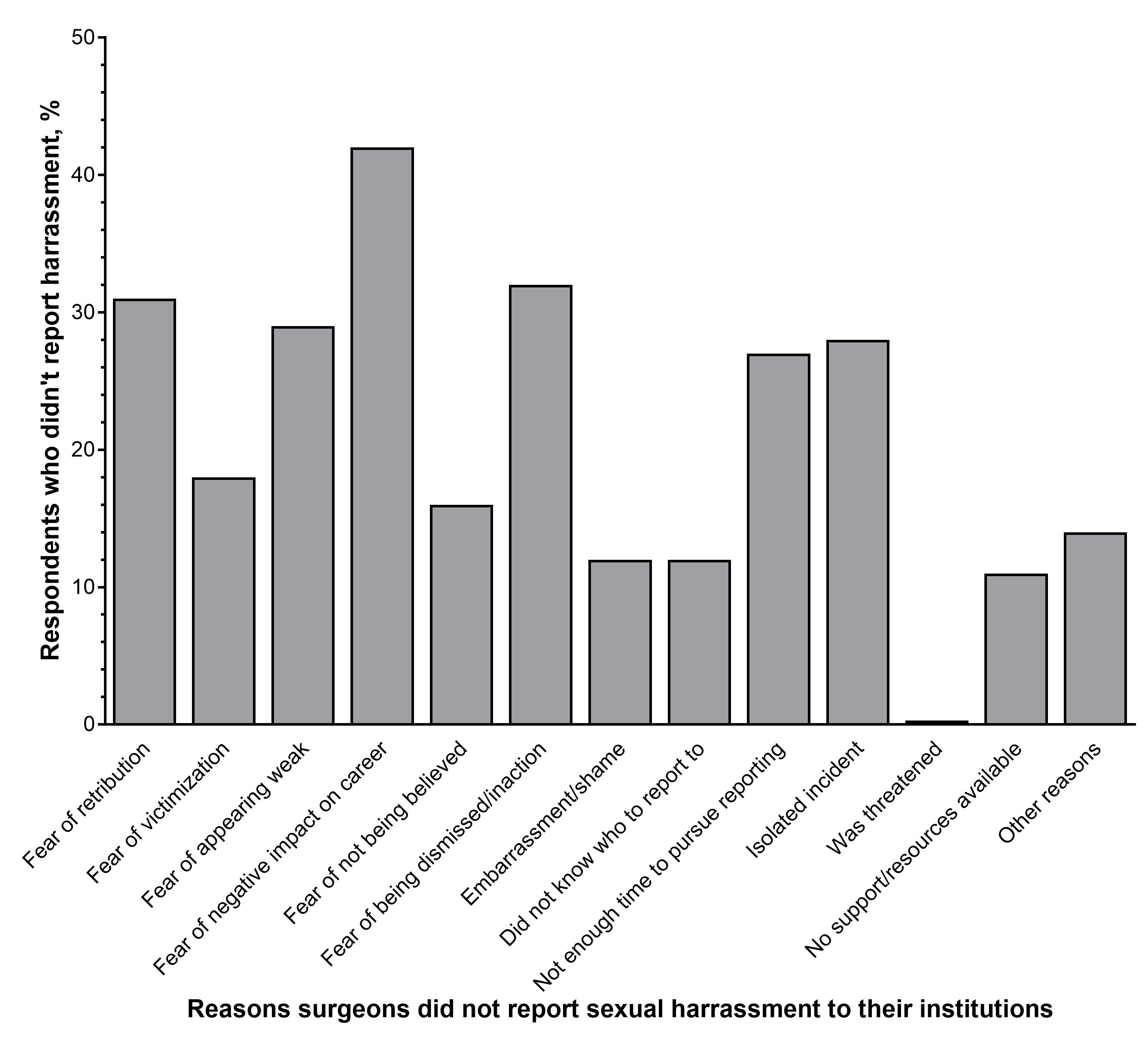
1,005 individuals completed the survey. 74% (n=744) identified as women, a response rate of 18% among US women surgeons. 25% (n=249) of respondents identified as men which represents 1% of male surgeons in the US – these responses were analyzed separately. Respondents reported employment by an academic institution (51%), community medical center (13%), private practice (15%), or other (19%). For both genders, the most common specialties were general surgery (34%), trauma surgery (10%), and surgical oncology (8%). Overall, 58% (n=432) of women surgeons experienced sexual harassment within the 12-month period preceding the survey, compared to 25% (n=61) of men, p<0.0001. Among women, the most common forms of harassment reported were “verbal or physical conduct (e.g. body language)” (53%), “unwanted sexual advances or physical contact” (23%), and “comments about sexual orientation” (10%). Women trainees were more than twice as likely to experience harassment as compared to attending surgeons (OR 2.52, 95% CI 1.78, 3.56, p<0.0001). The majority (84%) of incidents of harassment reported by women as part of the survey were not reported to any institutional authority. The most common reasons for not reporting was “fear of a negative impact on my career” (43%), “fear of retribution” (32%), and “fear of being dismissed and/or inaction towards perpetrator” (31%) (Figure).  

Conclusion:

Our study indicates that there is an alarming prevalence of unreported sexual harassment experienced by women surgeons in the US.   Combined with the documented sexual harassment of women physicians in other medical specialties, we believe this finding demonstrates an urgent need to improve the safety of the healthcare workplace, not just for women surgeons, but for all.
 

23.01 Fetal Enzyme Therapy in Sly Syndrome Crosses Blood-Brain Barrier and Induces Immunologic Tolerance

Q. Nguyen1, R. G. Witt1, C. Eikani1, B. Wang1, T. C. Mackenzie1  1University Of California – San Francisco,San Francisco, CA, USA

Introduction:
Sly Syndrome, or Mucopolysaccharidosis Type 7 (MPS7), is an autosomal recessive disorder in which missing β-glucuronidase (GUSB) results in systemic cellular dysfunction and in utero demise. Postnatal enzyme replacement therapy (ERT) is unable to treat neurologic phenotype because the blood brain barrier is closed after birth. Furthermore, patients can develop an immune response to the missing protein after repeated doses, which limits the treatment efficacy. In this study, we investigate whether administration of the enzyme in utero, prior to formation of the blood brain barrier and to maturation of the immune system will result in brain penetration and induce tolerance.

Methods:
We bred heterozygous MPS7 mice and injected the resultant fetuses at E14.5 with recombinant human GUSB or control phosphate-buffered saline (PBS) through maternal midline laparotomy. To assess for tissue enzyme levels, we harvested brain tissue at E18-P1 and performed chromogenic GUSB enzyme activity assays. To compare against postnatal treatment, we also injected adult MPS7 mice with enzyme and harvested brain tissue 4 days after injection. To assess for long-term immune response to enzyme, we performed serial post-natal enzyme injections, then challenged mice with GUSB plus Complete Freund’s Adjuvant (CFA) at 6-8 weeks of age and then measured antibody production using ELISA for IgG1, IgG2b, IgG2c, and IgG3.  Mice challenged with CFA continued to receive serial enzyme booster injections.

Results:
For the brain penetration studies, we administered enzyme or PBS in utero (n=9), or postnatally (n=6). Enzyme replacement resulted in detectable brain enzyme activity after in utero but not after postnatal treatment (Fig. 1A). For the tolerance studies, we detected a significant decrease in IgG1 antibody production with IUERT compared to both the untreated and post-natal ERT groups (n=91) (Fig. 1B). During the course of the ELISA studies, we found that some mice expired spontaneously after receiving booster injections following the CFA challenge, likely secondary to anaphylaxis, but that in utero exposure to the enzyme was protective against treatment-related death (Fig. 1C).

Conclusion:
We have found that IUERT delivers enzyme to brain tissue, and also successfully induces long-term tolerance to the missing enzyme. Future studies will examine the subtype of brain cells that take up the enzyme as well as quantify levels of IgE which likely contribute to the anaphylaxis. These findings have broad applicability for using the fetal environment to induce tolerance and treat neurologic disorders, thus opening the field of fetal surgery to include emerging molecular therapies.