40.16 Possible Role of HOX genes in Pancreatic Ductal Adenocarcinoma Survival

K. Takabe1, E. Katsuta1, L. Yan2  1Roswell Park Cancer Institute,Breast Surgery, Department Of Surgical Oncology,Buffalo, NY, USA 2Roswell Park Cancer Institute,Department Of Biostatistics And Bioinformatics,Buffalo, NY, USA

Introduction:  Pancreatic adenocarcinoma (PDAC) is one of the most aggressive cancers with severe prognosis in general; however, we sometimes encounter exceptional long term survivors.  Although there have been many study to explore the genes that are responsible for these differences in prognosis of PDAC, it remains unknown. The HOX genes are a family of homeodomain-containing transcription factors that determine cellular identity during development. This study aimed to investigate the clinical relevance of HOX genes on the PDAC patient survival. 

Methods:  RNA-sequencing and clinical data were obtained from the Cancer Genome Atlas (TCGA) through cBioportal. Gene expression was compared between patients who survived more than 5 years (good prognosis group) and patients who died within 3 years of diagnosis (poor prognosis group). Patients whose cause of death was not pancreatic cancer in bad prognosis group were excluded for the analysis. The cut-off value of gene expression for survival analysis was determined by automated scanning and selecting the threshold yielding the lowest p-value for the each gene.

Results: Among pancreatic cancer TCGA cohort, 94 PDAC patients were confirmed of their date of death or lived longer than 5 years. 4 and 82 patients were classified as good and poor prognosis group, respectively. 1 and 3 patients were diagnosed as Stage IIA and IIB in good prognosis group, 1, 5, 9, 62, 2 and 3 patients were diagnosed as Stage IA, IB, IIA, IIB, III and IV in poor prognosis group, respectively. 120 genes were extracted as differentially expressed gene with adjusted p<0.05. Among 120 genes, 8 genes were HOX genes, including HOXA9 (logFC=7.18, adj. p<0.001), HOXA7 (logFC=3.26, adj. p<0.001), HOXA4 (logFC=2.18, adj. p<0.001), HOXA6 (logFC=5.38, adj. p<0.001), HOXA5 (logFC=3.85, p<0.001), HOXA10 (logFC=4.19, adj. p=0.001), HOXA2 (logFC=1.75, adj. p=0.036) and HOXA13 (logFC=2.87, adj. p=0.039). All of them were upregulated in good prognosis group. Then we compared the survival based upon HOX expression in whole 154 PDAC in TCGA cohort. High expression of HOXA2 (median OS: 19.8 month vs 16.8 month, p=0.005, median DFS: 16.9 month and 9.6 month, p=0.001), HOXA4 (median OS: 20.6 month vs 12.5 month, p<0.001, median DFS: 17.1 month and 9.5 month, p<0.001), and HOXA5 (median OS: 19.9 month vs 12.5 month, p<0.001, median DFS: 17.1 month and 9.5 month, p=0.001) showed significantly better both OS and DFS. 

Conclusion: This is the first report that high expression of HOX genes associate with exceptional long term survivor in PDAC. Further studies are warranted to investigate the mechanism how these gene expressions contribute to patient survival.

40.14 High Levels of Sphingolipids in Human Pancreatic Cancer

K. Yuza1, M. Nagahashi1, Y. Hirose1, M. Nakajima1, K. Miura1, H. Ichikawa1, Y. Shimada1, J. Sakata1, H. Kameyama1, T. Kobayashi1, K. Takabe2,3, T. Wakai1  1Niigata University Graduate School Of Medical And Dental Sciences,Division Of Digestive And General Surgery,Niigata City, NIIGATA, Japan 2Roswell Park Cancer Institute,Breast Surgery, Department Of Surgical Oncology,Buffalo, NY, USA 3University At Buffalo Jacobs School Of Medicine And Biomedical Sciences, The State University Of New York,Department Of Surgery,Buffalo, NY, USA

Introduction:
Pancreatic cancer is one of the most lethal diseases and it often spreads quickly before it causes any symptoms. Elucidation of the underlying mechanisms how pancreatic cancer progresses and metastasizes is the key to improve outcome. Sphingosine-1-phosphate (S1P), a bioactive lipid mediator plays critical roles in cancer progression. S1P is involved in numerous cellular functions such as cell proliferation, migration, survival, angiogenesis and lymph angiogenesis, all of which are related to cancer progression and metastasis. Although it is expected that S1P plays an important role in pancreatic cancer progression based on the previous findings of experimental models, its role in human pancreatic cancer has never been revealed. We hypothesized that sphingolipids including S1P are produced higher in pancreatic cancer compared with normal pancreas tissue.

Methods:
Tumor and non-cancerous pancreas tissue were obtained from 10 patients with pancreatic cancer. Both tumor and non-cancerous pancreas tissue were collected from the same resected specimen, and non-cancerous tissue was collected from normal pancreas as far away as possible from the cancer site. Sphingolipids including S1P and their metabolites were measured by liquid chromatography-electrospray ionization-tandem mass spectrometry (LC-ESI-MS/MS). The levels of each sphingolipid were compared between tumor and non-cancerous pancreas tissue by using the Wilcoxon matched-pairs signed rank test. All of the tests were two-sided and P values < 0.05 were considered to be statistically significant.

Results:
Levels of sphingosine, dihydro-sphingosine, S1P, and dihydro-S1P in the pancreatic cancer and normal pancreas tissue were all successfully determined. The levels of all these sphingolipids were universally higher in the cancer tissue than in the normal pancreas tissue (P<0.001 for sphingosine, dihydro-sphingosine, and S1P; P<0.05 for dihydro-S1P). We also determined the levels of each species of ceramide (C14:0, C16:0, C18:1, C18:0, C20:0, C22:0, C24:1, C24:0, C26:1 and C26:0) in the pancreatic cancer and normal pancreas tissue. We found that C14:0 alone was significantly higher in the cancer tissue than in the normal pancreas tissue.

Conclusion:
Levels of sphingolipids in cancer tissue are generally higher than normal pancreas tissue in patients with pancreatic cancer. The high levels of S1P and its metabolites in cancerous tissues implicate the important role of S1P in pancreatic cancer.
 

40.11 High Expression of DMT1 Indicated Better Prognosis in Non-B Non-C Hepatocellular Carcinoma

T. Hoki1, E. Katsuta2, L. Yan3, K. Takabe2,4, F. Ito1,4,5  1Roswell Park Cancer Institute,Center For Immunotherapy,Buffalo, NY, USA 2Roswell Park Cancer Institute,Breast Surgery, Department Of Surgical Oncology,Buffalo, NY, USA 3Roswell Park Cancer Institute,Department Of Biostatistics And Bioinformatics,Buffalo, NY, USA 4State University Of New York At Buffalo,Department Of Surgery, University At Buffalo Jacobs School Of Medicine And Biomedical Sciences,Buffalo, NY, USA 5Roswell Park Cancer Institute,Department Of Surgical Oncology,Buffalo, NY, USA

Introduction:

Hepatocellular carcinoma (HCC) is the sixth most common malignancy with poor prognosis worldwide. HCC commonly develops in patients with underlying chronic liver disease. Higher iron accumulation is present in chronic liver disease and is known to be a risk factor in the development of HCC. The divalent metal-ion transporter-1 (DMT1) is a primary importer of non-heme iron, and is ubiquitously expressed throughout the body, with highest expression in the proximal duodenum, which is the main site of iron uptake. We previously reported that mal-regulation of iron metabolism caused by increased DMT1 expression in the duodenum induced hepatocarcinogenesis. Increased expression of DMT1 in tumorous tissue has also been shown to be associated with carcinogenesis and progression in colorectal and esophageal adenocarcinoma. However, the role of DMT1 in liver of HCC patients remains unknown.

Methods:
Clinical and RNA-seq data were obtained from the Cancer Genome Atlas (TCGA). Gene expression level was compared among each AJCC Stage, and each viral infectious status. Patients were divided into two groups based on DMT1 expression level for survival analysis using Kaplan-Meier method followed by Log-rank test. The cut-off value was determined by automated scanning and selecting the threshold yielding the lowest p-value.

Results:
Of 442 HCC patients in the TCGA cohort, tumor RNA-seq data were obtained from 342 patients with overall survival (OS) and etiology data. The prevalence of HBV, HCV, dual HBV-HCV, and non-B non-C were 97(28.4%), 47(13.7%), 6(1.8%), and 192(56.1%), respectively. Clinical stages were available for 327 patients, and the patients number of stage I, II, III, and IV were 165(50.5%), 75(22.9%), 82(25.1%), and 5(1.5%), respectively. The median observation period was 19.3 months (range, 0–120.73m). There was no significant difference in the expression level of DMT1 among each clinical stage (stage I/II/III/IV) and each viral infection status (HBV/HCV/dual HBV-HCV/non-B non-C). To investigate the impact of DMT1 expression on patients’ prognosis, OS was compared between high and low expression groups in the whole cohort as well as in different viral infectious status. Interestingly, high expression of DMT1 showed better survival in non-B non-C patients (5-year OS rate: 65.2% vs 32.7%, p=0.038), HBV patients (5-year OS rate: 78.3% vs 61.9%, p=0.006), and HBV and/or HCV-infected patients (5-year OS rate: 65.7% vs 36.5%, p=0.001) compared to low expression group. Whereas there was no significant difference in OS between high and low expression groups in HCV patients (p=0.193) and the whole cohort (p=0.069).

Conclusion:
HCC with increased expression of DMT1 has better OS for HBV, HBV and/or HCV, and non-B non-C patients. These findings may imply that DMT1 in HCC tumors plays different role among different viral infectious status patients, and play roles to suppress tumor progression other than iron transportation.

35.08 Clinical Impact of Genetic Alterations According to Primary Tumor Sidedness in Colorectal Cancer

Y. Shimada1, Y. Tajima1, M. Nagahashi1, H. Ichikawa1, M. Nakano1, H. Kameyama1, J. Sakata1, T. Kobayashi1, Y. Takii2, S. Okuda3, K. Takabe4,5, T. Wakai1  1Niigata University Graduate School Of Medical And Dental Sciences,Division Of Digestive And General Surgery,Niigata, , Japan 2Niigata Cancer Center Hospital,Department Of Surgery,Niigata, , Japan 3Niigata University Graduate School Of Medical And Dental Sciences,Division Of Bioinformatics,Niigata, , Japan 4Roswell Park Cancer Institute,Breast Surgery,Buffalo, NY, USA 5University At Buffalo Jacobs School Of Medicine And Biomedical Sciences,Department Of Surgery,Buffalo, NY, USA

Introduction: Right-sided colorectal cancer (RCRC), which is derived from midgut, has different molecular and biological characteristics compared with left-sided colorectal cancer (LCRC) which is derived from hindgut. Recently, several unplanned retrospective analyses revealed the differences between RCRC and LCRC in prognosis and response to targeted therapy. We hypothesized that primary tumor sidedness is a surrogate for non-random distribution of genetic alterations, and is a simple and useful biomarker in patients with Stage IV CRC. To teste this hypothesis, we investigated the genetic alterations using comprehensive genomic sequencing (CGS), and analyzed the clinical impact of primary tumor sidedness in patients with Stage IV CRC.

Methods:  One-hundred-eleven Stage IV CRC patients with either RCRC or LCRC were analyzed. We investigated genetic alterations using 415-gene panel, which includes the genetic alterations associated with resistance to anti-EGFR therapy. The differences of clinicopathological characteristics and genetic alterations were analyzed between RCRC and LCRC using Fisher’s exact test. The differences in response to targeted therapies, and clinical significance of residual tumor status were analyzed between RCRC and LCRC using log-rank test. 

Results: Thirty-four patients (31%) and 77 patients (69%) had RCRC and LCRC, respectively. Histopathological grade 3 was significantly associated with RCRC (P = 0.042). Pulmonary metastasis was significantly associated with LCRC (P = 0.012), and peritoneal metastasis was significantly associated with RCRC (P = 0.002). Regarding residual tumor status, R0 resection of both primary and metastatic lesions showed significantly better overall survival compared with R2 resection in both RCRC and LCRC (P = 0.026 and 0.002, respectively). Regarding genetic alterations, RCRC has more genetic alterations associated with resistance to anti-EGFR therapy (BRAF, ERBB2, FGFR1, KRAS, PIK3CA, PTEN) compared with LCRC (P = 0.040). In 73 patients with anti-VEGF therapy, there was no significant difference on progression-free survival (PFS) between RCRC and LCRC (P = 0.866). Conversely, in 47 patients with anti-EGFR therapy, RCRC showed significantly worse PFS than LCRC (P = 0.019).

Conclusion: RCRC is more likely to have the genetic alterations associated with resistance to anti-EGFR therapy compared with LCRC, and shows resistance to anti-EGFR therapy. Primary tumor sidedness is a surrogate for non-random distribution of molecular subtypes in CRC.
 

32.04 Impact of Frailty on Failure to Rescue After Low Risk and High Risk Inpatient Surgery

R. Shah1, K. Attwood6, S. Arya2, D. E. Hall3, J. M. Johanning5, N. N. Massarweh4  1Henry Ford Health System,General Surgery,Detroid, MI, USA 2Emory University School Of Medicine,Division Of Vascular And Endovascular Therapy/ Department Of Surgery,Atlanta, GA, USA 3University Of Pittsburg,Center For Health Equity Research And Promotion, Veterans Affairs Pittsburgh Healthcare System,Pittsburgh, PA, USA 4Baylor College Of Medicine,VA HSR&D Center For Innovations In Quality, Effectiveness And Safety, Michael E DeBakey VA Medical Center,Houston, TX, USA 5University Of Nebraska College Of Medicine,Department Of Surgery,Omaha, NE, USA 6Roswell Park Cancer Institute,Surgical Oncology,Buffalo, NY, USA

Introduction:  Failure to rescue (FTR), or death after a potentially preventable complication, is a nationally endorsed, publically reported quality measure. However, little is known about the impact of frailty on FTR—in particular, after lower risk surgical procedures.

Methods:  Retrospective cohort study of 984,550 patients from the National Surgical Quality Improvement Program (2005-2012) who underwent inpatient general, vascular, thoracic, cardiac and orthopedic operations. Frailty was assessed using the clinically applicable Risk Analysis Index (RAI) and patients were stratified into five groups based on RAI score (<=10, 11-20, 21-30, 31-40 and >40). Procedures were categorized as low (≤1%) or high mortality risk (>1%). The association between RAI, the number of post-operative complications (0, 1, 2, 3+), and FTR was evaluated using hierarchical modeling. 

Results: Among the most frail (RAI >30) patients in the cohort, ~20% were aged 55 years or younger. Regardless of procedural risk, increasing RAI score was associated with both an increased occurrence of post-operative complications and the number of complications. For those who underwent low risk surgery, major complication rates were 3.2%, 8.6%, 13.5%, 23.8% and 36.4% for RAI scores of <=10, 11-20, 21-30, 31-40 and > 40, respectively and for patients undergoing high risk surgery, the corresponding rates of major complications were 13.5%, 23.7%, 31.1%, 42.5% and 54.4%, respectively. Stratifying by the number of complications, significant increases in FTR rates were observed across RAI categories after both low and high risk procedures (Figure 1; trend test, p<0.001 for all). Increasing RAI was associated with an increased risk of FTR that was most pronounced after low risk procedures. For instance, the odds ratios (ORs) for FTR after 1 major complication for patients undergoing a low risk procedure were 4.8 (3.7, 6.2), 8.1 (5.9, 11.2), 19.3(12.6, 29.6) and 48.8 (22.7, 104.9) for RAI scores of 11-20, 21-30, 31-40 and > 40, respectively and for patients undergoing a high risk procedure, the corresponding ORs were 2.6 (2.4, 2.8), 5.2 (4.8, 5.6), 9.3 (8.5, 10.3) and 19.5 (16.8, 22.6) respectively. 

Conclusion: Frailty has a dose-response relationship with complications and FTR that is similarly apparent after low and high risk inpatient surgical procedures.  Tools facilitating rapid assessment of frailty during preoperative assessment, may help provide patients with more accurate estimates of surgical risk and could improve patient engagement in peri-operative interventions that enhance physiologic reserve and can potentially mitigate aspects of procedural risk.


23.08 Priming with IL-7/15 to Generate Metabolically Fit CD8+ T Cells in the Tumor Microenvironment

S. Patel1, T. Hoki1, T. Yamauchi1, K. A. Collins1, C. A. Eppolito1, A. J. Francois1, J. V. Welch1, J. A. DiTursi1, K. Odunsi1,2,3, F. Ito1,4,5  1Roswell Park Cancer Institute,Center For Immunotherapy,Buffalo, NY, USA 2Roswell Park Cancer Institute,Department Of Gynecologic Oncology,Buffalo, NY, USA 3Roswell Park Cancer Institute,Department Of Immunology,Buffalo, NY, USA 4State University Of New York At Buffalo,Department Of Surgery, University At Buffalo Jacobs School Of Medicine And Biomedical Sciences,Buffalo, NY, USA 5Roswell Park Cancer Institute,Department Of Surgical Oncology,Buffalo, NY, USA

Introduction:
Current approaches to adoptive cell therapy (ACT) with antigen-specific T cells are limited by the difficulty of obtaining sufficient numbers of T cells against targeted antigens with effective in vivo characteristics. Whereas interleukin (IL)-2 has been widely used for generation of antitumor T cells in vitro clinically, dose-dependent effects of IL-2 on differentiation of T cells are associated with decreased proliferative and self-renewal capacity in vivo. IL-7 and IL-15 are also common γ  chain cytokines that play pivotal roles in homeostasis, proliferation, and maintenance of memory CD8+ T cells. Accumulating evidence largely from examining hematological malignancies indicates that the combined use of IL-7 and IL-15 (IL-7/15) can produce T cells that confer superior antitumor immunity in vivo. However, antitumor efficacy of IL-7/15-primed T cells in an orthotopic tumor model has not been rigorously evaluated.

Methods:
Pmel-1 T-cell receptor transgenic CD8+ T cells were activated with the cognate antigen gp100 expressed on B16 melanoma in IL-7/15 for 6 days. IL-2 was used for a control. Phenotype and function as well as metabolic profile of IL-2- and IL-7/15-primed T cells were evaluated. To determine in vivo antitumor efficacy, C57BL/6 mice bearing subcutaneous B16F10 melanoma were treated with adoptive transfer of IL-2- or IL-7/15-primed Pmel-1 T cells, followed by systemic administration of IL-2, and vaccination with gp100, anti-CD40 antibody, and toll-like receptor (TLR) agonist to augment antitumor efficacy of transferred T cells.

Results:
Cell expansion was significantly higher when T cells were activated in IL-7/15 at day 6 compared with ones in IL-2. IL-7/15-primed T cells consisted of a higher proportion of less-differentiated CD44+CD62L+ T cells, and secreted significantly more IL-2 against the target antigen compared to IL-2-primed T cells while both had comparable effector function such as specific lysis of targets and IFNγ  production in vitro. Furthermore, IL-7/15-primed T cells had higher mitochondrial spare respiratory capacity than IL-2-primed T cells, suggesting that IL-7/15-primed T cells have capacity to produce more ATP in case of a sudden increase in energy demand. In line with this, adoptively-transferred IL-7/15-primed T cells expressed significantly higher Ki67 than IL-2-primed T cells in the tumor microenvironment (TME). Significantly delayed tumor growth and improved survival were observed in mice treated with IL-7/15-primed T cells compared to IL-2-primed T cells.

Conclusion:
Taken together, our studies suggest that IL-7/15 modulates the metabolic programming of T cells to promote more robust and efficient CD8+ T cells that can proliferate in the TME. In particular, IL-7/15-primed T cells have higher self-renewal and spare respiratory capacity with potent effector function that correspond to significantly improved survival in an orthotopic tumor model.
 

23.05 CRISPR/Cas9 Genome Editing of Tumor-specific CD8+ T cell-derived Induced Pluripotent Stem Cells

T. Yamauchi1, H. Saito2,3, T. Hoki1, F. Ito1,2,4,5  2University Of Michigan,Surgery,Ann Arbor, MI, USA 3Kanazawa Medical University,Biochemistry,Kanazawa, ISHIKAWA, Japan 4State University Of New York At Buffalo,Surgery,Buffalo, NY, USA 5Roswell Park Cancer Institute,Surgical Oncology,Buffalo, NY, USA 1Roswell Park Cancer Institute,Center For Immunotherapy,Buffalo, NY, USA

Introduction: Current approaches to adoptive T cell therapy are limited by the difficulty of obtaining sufficient numbers of T cells against targeted antigens with effective in vivo characteristics.  This limitation can be overcome by using induced pluripotent stem cells (iPSCs) that could provide an unlimited source of autologous T cells. We and others have shown that iPSC-derived regenerated T cells have potent antitumor efficacy in vitro and in vivo. Recently, the type II bacterial CRISPR (clustered regularly interspaced short palindromic repeats)/Cas (CRISPR-associated) system was developed as an efficient and versatile technology for genome editing in eukaryotic cells and whole organisms. The potential of iPSCs can be further enhanced by genome engineering and then used to study individual gene function, track cells or endogenous proteins with a knock-in reporter, and correct genetic defects for gene therapy. 

Methods: We reprogrammed T-cell receptor (TCR) transgenic CD8+ T cells into pluripotency, and established a syngeneic mouse model for evaluating in vitro and in vivo antitumor reactivity of regenerated T cells from iPSCs bearing a rearranged TCR of known antigen specificity. Plasmids encoding rtTA and tet-O-Cas9-2A-mCherry were obtained and co-transduced using the lentiviral system. The Cas9 expression was analyzed after the doxycycline treatment, and clonally-derived Cas9-iPSCs were obtained.

Results: Pluripotency of TCR transgenic T cell-derived iPSCs (TiPSCs) were confirmed with immunostaining of embryonic stem cell (ESC) markers, RT-PCR (reverse transcription-polymerase chain reaction) analysis of pluripotency-associated transcription factors, and whole genome expression profiling by microarray analysis that demonstrated a high degree of similarity in their gene expression patterns and close correlation values with ESCs, but distinct from parental CD8+ T cells. Cytogenetic analysis revealed derived TiPSCs maintained normal karyotype. The TiPSCs differentiated to embryoid bodies in vitro, and upregulation of marker genes for all three germ layers was detected by immunostaining. Their differentiation capacity was further confirmed by teratoma formation in immune-deficient mice in vivo. Moreover, we confirmed that the TiPSCs retained the same rearranged configuration of TCR chain genes as the original TCR transgenic T cells. TiPSCs were, then, subjected to the lentivirus-mediated transduction of tetracycline-inducible Cas9 vectors. The transduction efficacy was confirmed by the mCherry fluorescence and the RT-PCR against the Cas9 sequence.

Conclusion: Our results indicate successful reprogramming of antigen-specific T cells and lentivirus-mediated transduction of tetracycline-inducible Cas9 vectors into TiPSCs, which will allow us to generate an unlimited number of phenotypically defined, functional and expandable genome-edited autologous antigen-specific T cells.

23.04 CD40 signaling is required for expansion of terminally-differentiated CX3CR1+ CD8+ T cells

F. Ito1,2,3, T. Hoki2, C. A. Eppolito2, A. J. Francois2, K. Odunsi2,4,5, T. Yamauchi2  3State University Of New York At Buffalo,Surgery,Buffalo, NY, USA 1Roswell Park Cancer Institute,Surgical Oncology,Buffalo, NY, USA 2Roswell Park Cancer Institute,Center For Immunotherapy,Buffalo, NY, USA 4Roswell Park Cancer Institute,Gynecologic Oncology,Buffalo, NY, USA 5Roswell Park Cancer Institute,Immunology,Buffalo, NY, USA

Introduction: Successful immunotherapeutic treatment of chronic infectious diseases and cancer requires the generation of a strong cellular immune response. Combined CD40 antibody and toll-like receptor (TLR) (CD40/TLR) stimulation has been found to mediate potent cellular immunity in the context of tumor immunology and cancer immunotherapy. However, the mechanisms of enhanced antitumor efficacy by CD40/TLR stimulation remain elusive.

Methods: To investigate phenotype and function of CD40/TLR vaccine-stimulated antigen-specific T cells, we used MC38 colon adenocarcinoma and B16 melanoma models. To evaluate endogenous T cell response, MC38 tumor-bearing C57BL/6 mice were treated with mutated-neoantigen peptide, agonistic CD40 antibody, and poly I:C (TLR3). To assess phenotype and function of adoptively-transferred T cells, we used pmel-1 T cell receptor (TCR) transgenic CD8+ T cells specific for the gp100 melanocyte differentiation antigen expressed on B16 melanoma. In vitro–activated Pmel-1 CD8+ T cells were adoptively transferred into C57BL/6 mice bearing subcutaneous B16 melanomas. Systemic administration of IL-2 and vaccination with the gp100, anti-CD40 antibody, and Imiquimod (TLR7) were used to enhance antitumor immunity of transferred T cells. Tissues including blood, spleen, and tumors were harvested for further analysis. In both tumor models, control mice were treated with no vaccination, CD40 antibody alone or TLR agonist alone.

Results: In both tumor models, mice treated with combined CD40/TLR vaccination had significantly decreased tumor growth and improved survival compared to no vaccination, CD40 antibody alone or TLR agonist alone. Interestingly, vaccination with the cognate antigen and CD40/TLR not only expanded antigen-specific CD8 T cells in both tumor models, but also facilitated them to express the chemokine receptor, CX3CR1. CX3CR1+ CD8+ T cells were found to express higher levels of killer-cell lectin like receptor G1 (KLRG1), TNF-related apoptosis-inducing ligand (TRAIL), perforin, and granzyme, suggesting terminally-differentiated subset. The generation of CX3CR1+ CD8+ T cells was greatly facilitated by CD40 antibody while TLR agonist increases the expansion of total number of antigen-specific CD8 T cells. Importantly, total number and frequency of CX3CR1+ CD8+ T cells were significantly decreased in tumor-bearing CD40 knockout (KO) mice, indicating that host expression of CD40 is required for generation and expansion of CX3CR1+ CD8+ T cells.

Conclusion: Effective vaccination with the cognate antigen and CD40/TLR accompanies generation of tumor-specific terminally-differentiated CX3CR1+ CD8+ T cells dependent on CD40 signaling.

23.02 Defining CD8+ T-cell Subsets that are Rescued by PD-1/PD-L1 Blockade in the Tumor Microenvironment

T. Yamauchi1, T. Hoki1, C. A. Eppolito1, A. Francois1, K. Odunsi1,2,3, F. Ito1,4,5  1Roswell Park Cancer Institute,Center For Immunotherapy,Buffalo, NY, USA 2Roswell Park Cancer Institute,Gynecologic Oncology,Buffalo, NY, USA 3Roswell Park Cancer Institute,Immunology,Buffalo, NY, USA 4State University Of New York At Buffalo,Surgery,Buffalo, NY, USA 5Roswell Park Cancer Institute,Surgical Oncology,Buffalo, NY, USA

Introduction: Cancer immunotherapies that target the T-cell immune checkpoints, such as programmed cell death-1 (PD-1) and its ligand (PD-L1) have shown unprecedented success for the treatment of a variety of malignancies including melanoma. Although a significant number of cancer patients benefit from immune checkpoint inhibitors (CPIs), many fail to have clinical responses.   A better understanding of the mechanisms that regulate CD8+ T-cell responses in the tumor microenvironment is required to improve immunotherapies that restore function in exhausted CD8+ T cells.  Although heterogeneity of effector CD8+ T cells in the tumor microenvironment (TME) has been recognized, their functions and roles are ill-defined.

Methods: We have evaluated phenotypical and functional heterogeneity of tumor-infiltrating lymphocytes (TILs) after adoptive transfer of ex vivo primed pmel-1 T cell receptor (TCR) transgenic CD8+ T cells specific for the gp100 melanocyte differentiation antigen expressed on B16 melanoma. In vitro–activated Pmel-1 CD8+ T cells were adoptively transferred into C57BL/6 mice bearing subcutaneous B16 melanomas that had been established for 11-14 days. Systemic administration of IL-2 and vaccination with anti-CD40 antibody and toll-like receptor (TLR) agonist were used to enhance antitumor immunity of transferred T cells. Tumor and spleen were harvested for functional analysis of adoptively transferred T cells.

Results: We found that the chemokine receptor, CX3CR1 identified three distinct effector CD8+ T-cell subsets, CX3CR1 negative (-), intermediate (int), and high (hi) in blood, spleen and the TME.  A CX3CR1hi subset contained terminally-differentiated CD8+ T cells that expressed higher levels of killer-cell lectin like receptor G1 (KLRG1), TNF-related apoptosis-inducing ligand (TRAIL), perforin, and granzyme. Significantly more CX3CR1int CD8+ T cells expressed CD25 compared to other subsets, suggesting this is the subset that is rapidly proliferate and preferentially generate terminally-differentiated T cells. Unexpectedly, despite their terminally differentiated status, a CX3CR1hi CD8+ T-cell subset expressed significantly lower levels of co-inhibitory receptors, PD-1, LAG3, and TIGIT compared to CX3CR1 and CX3CR1int CD8+ T-cell subsets in the TME. In line with this, proliferation and cytokine production of CX3CR1 and CX3CR1int CD8+ T-cell subsets were significantly decreased in the TME compared to CX3CR1hi CD8+ T-cell subset. Importantly, PD-1/PD-L1 blockade significantly improved effector functions of CX3CR1 and CX3CR1int CD8+ T-cell subsets in the TME.

Conclusion: The chemokine receptor, CX3CR1 defines distinct effector CD8+ T-cell subsets in periphery and in the TME. Tumor-infiltrating CX3CR1 and CX3CR1int CD8+ T-cell subsets express high levels of co-inhibitory receptors, PD-1, LAG3, and TIGIT, and their effector functions are improved by PD-1/PD-L1 blockade. 

11.01 Outcomes after CRS-HIPEC by Facility: Do Higher Volumes Matter?

K. N. Partain1, E. Gabriel1, K. Attwood2, C. Powers3, M. Kim3, S. P. Bagaria1, S. N. Hochwald3  1Mayo Clinic – Florida,Department Of Surgery, Section Of Surgical Oncology,Jacksonville, FL, USA 2Roswell Park Cancer Institute,Department Of Biostatistics,Buffalo, NY, USA 3Roswell Park Cancer Institute,Department Of Surgical Oncology,Buffalo, NY, USA

Introduction:  Cytoreductive surgery with hyperthermic intraperitoneal chemotherapy (CRS-HIPEC) offers favorable outcomes for select patients with appendiceal and colorectal cancer (CRC). Studies have suggested that this procedure should be performed at high-volume centers, which can limit access to treatment. The purpose of this study was to determine the association between treatment volume and outcomes for CRS-HIPEC.

Methods: This was a retrospective analysis using the National Cancer Data Base, 2004-2013. CRS-HIPEC treatment centers were stratified by low-volume (<10 cases/decade), middle-volume (11-20), and high-volume (>20). Patients who received any systemic chemotherapy were excluded. The primary, long-term outcome was overall survival (OS). Secondary, short-term outcomes included the number of lymph nodes examined in the surgical specimen, post-operative hospital length of stay (LOS), unplanned readmission rate, and 30- and 90-day mortality. 

Results: A total of 749 cases were identified: 303 at low-volume, 138 at middle-volume, and 308 at high-volume centers. Carcinomatosis of appendiceal origin was present in 84.5% of cases, with the remainder of CRC origin. Table 1 summarizes the baseline demographic and clinical characteristics among the three types of centers. Overall, the cases treated among different centers were similar with respect to age, race, insurance status, and comorbid status (as reported by the Charlson-Deyo comorbidity score). The average distance traveled was highly variable (low: 54.5 miles, middle: 238.3 miles, high: 364.1 miles; p<0.001). There was no difference in the average number of lymph nodes examined (low: 13.7, middle: 14.0, high: 12.4; p=0.33), readmission rates (low: 8.7%, middle: 8.9%, high 6.7%; p=0.87), 30-day morality (low: 0.9%, middle: 0.8%, high:1.8%; p=0.59), or 90-day mortality (low: 4.1%, middle: 3.4%, high:4.7%; p=0.83). There was a difference in the average hospital LOS (low: 13.9 days, middle: 17.3 days, high: 19.2 days; p=0.008). The median follow-up for OS was 48.3 months (range 0.5 – 101.8 months). There was no significant association between case volume and median OS (low: 45.8 months, middle: 58.4 months, high: 59.4 months; p=0.43).

Conclusion: Contrary to the push for centralization of CRS-HIPEC, this data suggests that CRS-HIPEC can be completed at lower volume performing centers to achieve similar short- and long-term outcomes compared to higher performing centers. Development of CRS-HIPEC programs in geographic areas of need may be beneficial for patients located far from centralized facilities.

1.11 Common Driver Mutation and Smoking History Affect Tumor Mutation Burden in Lung Adenocarcinoma

M. Nagahashi1, S. Sato2, K. Yuza1, Y. Shimada1, H. Ichikawa1, S. Watanabe3, S. Okuda4, K. Takabe5,6, M. Tsuchida2, T. Wakai1  1Niigata University Graduate School Of Medical And Dental Sciences,Division Of Digestive And General Surgeyr,Niigata City, NIIGATA, Japan 2Niigata University Graduate School Of Medical And Dental Sciences,Division Of Thoracic And Cardiovascular Surgery,Niigata City, NIIGATA, Japan 3Niigata University Graduate School Of Medical And Dental Sciences,Department Of Respiratory Medicine And Infectious Disease,Niigata City, NIIGATA, Japan 4Niigata University Graduate School Of Medical And Dental Sciences,Division Of Bioinformatics,Niigata City, NIIGATA, Japan 5Roswell Park Cancer Institute,Breast Surgery, Department Of Surgical Oncology,Buffalo, NY, USA 6University At Buffalo Jacobs School Of Medicine And Biosciences, The State University Of New York,Department Of Surgery,Buffalo, NY, USA

Introduction:
Although the expectation of immune checkpoint inhibitors, such as anti-PD-1 therapy, is tremendously high based on its dramatic efficacy, only a limited number of patients respond. Even worse, there is no definite biomarker to identify which tumor responds to the therapy. Recent progress in the genomic analysis using next-generation sequencing (NGS) technology has enabled comprehensive detection of mutations and tumor mutation burden (TMB) in cancer patients. The high TMB (TMB-H) tumor is defined as one with high somatic mutational rates, which correlates with the generation of neo-antigens and potential clinical response to immunotherapies. In this study, we analyzed the TMB in patients with lung adenocarcinoma utilizing NGS, and clarified the characteristics of patients with TMB-H in relation to common driver mutations of lung adenocarcinoma and smoking history.

Methods:
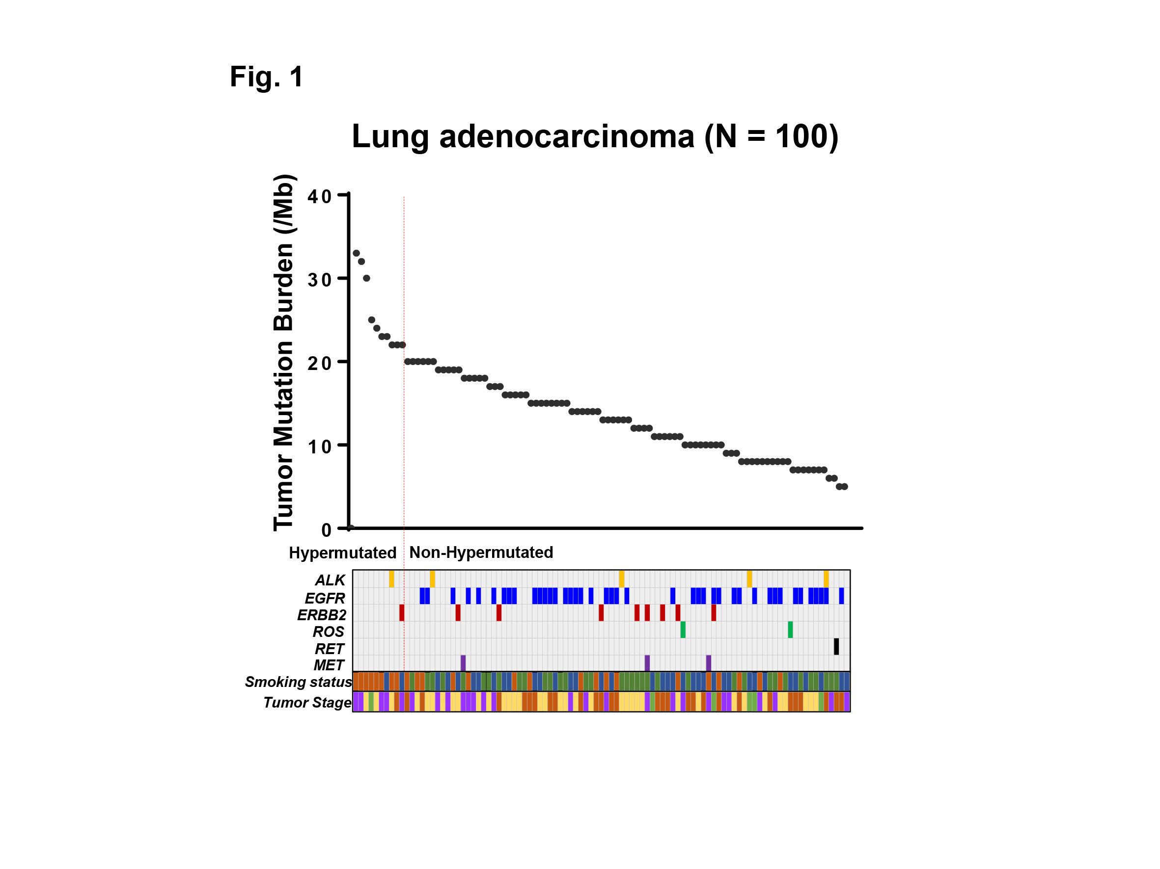
Genomic aberrations were determined in Japanese patients with lung adenocarcinoma (N=100) using NGS of 415 known cancer genes, and TMB was determined in each patient. High TMB was defined as 20 or more mutations per megabase of sequenced DNA. Common driver mutations, including ALK, EGFR, ERBB2, ROS, RET, MET, and smoking status were compared with TMB to examine their association.

Results:
The median TMB was 13.5 (range 5 – 33) per megabase among 100 patients with lung adenocarcinoma examined. Ten out of 100 (10%) patients showed TMB-H, and 90 (90%) patients showed low TMB (TMB-L) (Fig. 1). Only 2 out of 10 (20%) patients with TMB-H had one of common driver mutations (one had ALK, and the other had ERBB2 mutation), while 57 out of 90 (63%) patients with TMB-L had one of the driver mutations including ALK, EGFR, ERBB2, ROS, RET, MET (P<0.05) (Fig. 1). Of note, no EGFR mutation was observed in patients with TMB-H. Eight out of 10 (80%) patients with TMB-H had current smoking history, while 17 out of 90 (19%) patients with TMB-L had the history, respectively (P<0.001). Moreover, a group of current smokers without driver mutations had the highest TMB in our cohort.

Conclusion:
We analyzed the TMB in patients with lung adenocarcinoma utilizing NGS-based analysis. Our data indicates that the common driver mutations and smoking history are associated with TMB-H in lung adenocarcinoma, which may impact treatment strategies for these patients.
 

1.10 Combined TLR/CD40 Stimulation Potentiates an Immunogenic Neoantigen Vaccine

T. Hoki1, T. Yamauchi1, C. A. Eppolito1, A. J. Francois1, K. Odunsi1,2,3, F. Ito1,4,5  1Roswell Park Cancer Institute,Center For Immunotherapy,Buffalo, NY, USA 2Roswell Park Cancer Institute,Department Of Gynecologic Oncology,Buffalo, NY, USA 3Roswell Park Cancer Institute,Department Of Immunology,Buffalo, NY, USA 4State University Of New York At Buffalo,Department Of Surgery, University At Buffalo Jacobs School Of Medicine And Biomedical Sciences,Buffalo, NY, USA 5Roswell Park Cancer Institute,Department Of Surgical Oncology,Buffalo, NY, USA

Introduction:
Cancer neoantigens are derived from nonsynonymous, tumor-specific mutations that create de novo epitopes for T cells, and bypass central thymic tolerance. Although they are highly immunogenic and induce immune responses in humans, the overall success of vaccination studies that target cancer neoantigens has so far been limited. To boost cell-mediated immunity against epithelial tumors, signaling through CD40 has been used with promising results. Toll-like receptor (TLR) agonists have also been implemented as adjuvants. Furthermore, combinatorial stimulation of TLRs and CD40 generates expansion of CD8+ T cells targeting nonmutated self-antigens compared with either agonist alone. However, therapeutic efficacy of combined TLR/CD40 stimulation in the setting of neoantigen vaccine remains elusive. 

Methods:
To this end, we used murine MC38 colon adenocarcinoma cells that harbor a single-epitope mutation within the Adpgk protein with the neo-epitope presented in MHC-I H-2Db molecules. C57BL/6 mice were inoculated subcutaneously with MC38 cells. MC38 tumor-bearing mice were treated with soluble Adpgk mutant epitope in combination with TLR agonist, anti-CD40 antibody or both. 

Results:
Therapeutic vaccination with the Adpgk mutant peptide combined with TLR agonist and an anti-CD40 antibody (TLR/CD40) significantly slowed tumor growth and improved survival. This was associated with expansion and terminal differentiation of CD8+ T cells in the periphery as well as in the tumor microenvironment. Significantly increased terminally differentiated neoantigen-specific CD8+ T cells in blood and spleen were identified using the tetramer staining assay.

Conclusion:
These studies provide the rational basis for the use of TLR and CD40 agonists together as essential adjuvants to optimize vaccines designed to elicit therapeutic immunity against cancer neoantigens.

1.09 The Significance of Phospho-Sphingosine Kinase 1 in the Lymphatic Spread of Esophageal Carcinoma

H. Ichikawa1, M. Nagahashi1, M. Nemoto1, T. Hanyu1, T. Ishikawa1, Y. Kano1, Y. Muneoka1, Y. Hirose1, Y. Shimada1, J. Sakata1, T. Kobayashi1, H. Kameyama1, T. Kazuaki2,3, T. Wakai1  1Niigata University Graduate School Of Medical And Dental Sciences,Division Of Digestive And General Surgery,Niigata, NIIGATA, Japan 2Roswell Park Cancer Institute,Breast Surgery, Department Of Surgical Oncology,Buffalo, NY, USA 3University At Buffalo Jacobs School Of Medicine And Biomedical Sciences,The State University Of New York, Department Of Surgery,Buffalo, NY, USA

Introduction: Lymphatic invasion and lymph node metastasis are main mode of spread in esophageal squamous cell carcinoma (ESCC). Sphingosine-1-phosphate (S1P), a pleiotropic bioactive lipid mediator, is one of the important molecule in cancer progression. Sphingosine kinase 1 (SphK1) is activated by phosphorylation and produce S1P. Phospho-SphK1 (pSphK1) were also elucidated to confer to the lymphatic spread of cancer in previous studies. However, the significance of pSphK1 in the progression of ESCC have not been fully investigated to date.

Methods: We retrospectively analyzed the cases of 96 patients who underwent esophagectomy without preoperative therapy for ESCC from 2000 to 2008. Immunohistochemistry of the surgically resected specimens was conducted using the primary polyclonal antibody against pSphK1. Sixty-one of the 96 patients (63.5%) had tumors with high pSphK1 expression (pSphK1-high group), and the others had ones with low pSphK1 expression (pSphK1-low group). We compared clinicopathological factors and overall survival after esophagectomy between the two groups.

Results: Pathological N category according to TNM classification of UICC 7th edition was significantly higher in pSphK1-high group (P < 0.01). Median number of lymph node metastasis (pSphK1-high: 2 vs. pSphK1-low: 0; P < 0.01), the proportion of tumor with lymphatic invasion (67.2% vs. 17.1%; P < 0.01) and that with intramural metastasis (26.2% vs. 2.9%; P < 0.01) were significantly higher in pSphK1-high group than in pSphK1-low group. Multivariate analysis revealed that the presence of lymphatic invasion was independently associated with high expression of pSphK1 (Odds ratio = 8.45; P < 0.01). The 5-year overall survival rate after esophagectomy in the pSphK1-high group was significantly lower compared to the pSphK1-low group (50.8% vs. 67.3%; P = 0.01).

Conclusions: We provide the first evidence of the association between high expression of pSphK1 and lymphatic spread in ESCC. Our study demonstrated that S1P signaling pathway is worth investigating to further understand the molecular mechanism of lymphatic spread in ESCC.

1.02 High Expression of SLCO2B1 is Associated with Prostate Cancer Recurrence after Prostatectomy

T. TERAKAWA1,2, E. Katsuta3, M. Fujisawa2, K. Guru1, K. Takabe3  1Roswell Park Cancer Institute,Urology,Buffalo, NY, USA 2Kobe University,Urology,Kobe, , Japan 3Roswell Park Cancer Institute,Surgical Oncology,Buffalo, NY, USA

Introduction:
Solute carrier organic anion (SLCO) genes encode organic anion transport proteins, which is a family of transport proteins that influx number of substrates into the cells including androgens. Among them, high expression of SLCO2B1 has been shown to be associated with the resistance to androgen deprivation therapy and with the prognosis of the patients with hormone sensitive prostate cancer. Here, we hypothesized that the high expression of SLCO2B1 in human prostate cancer may increase influx of androgens to remaining undetectable dormant cancer cells thus is associated with worse disease-free survival after radical prostatectomy.

Methods:
Clinical and RNA-seq data were all obtained from the Cancer Genome Atlas (TCGA). Patients were classified as either high or low expression of SLCO2B1 by the mean value. Overall survival (OS) and disease-free survival (DFS) were compared between high and low expression group in whole cohort, as well as subgroups based upon Gleason score (GS≤6, =7 or ≥8). Gene set enrichment analysis (GSEA) were conducted between high and low expression group.

Results:
Of all patients, 193 and 305 patients were classified as SLCO2B1 high and low expression group, respectively. The patients with high expression of SLCO2B1 were found to have more aggressive cancer characteristics, including high Gleason score (p<0.001), higher T stage (p<0.001), and positive surgical margin (p=0.011). Among all patients, 5-year OS and DFS were 97.8% and 71.2%, respectively. High expression group showed significantly worse DFS after radical prostatectomy (5-year DFS rate: 60.3% vs 78.7%, p=0.026), whereas there was no significant difference in overall survival between these two groups (5-year OS rate: 99.3% vs 96.8%, p=0.923). The patients with higher Gleason score had significantly higher levels of SLCO2B1 expression (GS≤6, vs GS=7; p=0.045, GS=7 vs GS≥8; p=0.002). Significant difference in DFS between high and low expression groups were only observed in the patients with GS≥8 (5-year DFS rate: 38.6% vs 70%, p=0.005), and not in the patients with GS≤7 (GS≤6; p=0.640, GS=7; p=0.923). GSEA demonstrated that in the high expression group of SLCO2B1 enriched KRAS signaling, epithelial mesenchymal transition (EMT) and TGF beta signaling related genes.

Conclusion:
High expression of SLCO2B1 in the prostate cancer patients associated with the aggressive cancer characteristics and recurrence after radical prostatectomy. Furthermore, the higher recurrence rate of the patients with high expression of SLCO2B1 may be able to be explained by its metastatic potential with up-regulated EMT signaling by KRAS and TGF beta pathways.