50.05 The utility of pre-sacral drainage in penetrating rectal injuries in adult and pediatric patients

K. B. Savoie1,5, T. M. Beazley3, B. Cleveland2, S. Khaneki4, T. Markel4, P. Hammer6, S. Savage6, R. F. Williams5  1University Of Tennessee Health Science Center,Department Of Surgery,Memphis, TN, USA 2Indiana University,School Of Medicine,Indianapolis, IN, USA 3University of Tennessee Health Science Center,School Of Medicine,Memphis, TN, USA 4Indiana University Health, Riley Hospital For Children,Division Of Pediatric Surgery,Indianapolis, IN, USA 5University Of Tennessee Health Science Center, Le Bonheur Children’s Hospital,Division Of Surgery And Pediatrics,Memphis, TN, USA 6Indiana University Health, Methodist Hospital,Division Of Trauma Surgery,Indianapolis, IN, USA

Introduction:

The historical tenets of treating penetrating rectal injuries (PRI) developed out of military trauma and often included presacral drainage to decrease risk of pelvic sepsis. With different weaponry associated with injuries in civilian trauma, there is equipoise on the utility of pre-sacral drainage (PSD), particularly in pediatric patients.

Methods:

IRB approval was obtained at two free-standing children’s hospital and two adult level 1-trauma hospitals. Data was retrospectively collected from July 2004-June 2014 and compared by age (pediatric patients <16 years) and PSD. A stratified analysis was performed based on age. The primary outcome was pelvic or presacral abscess.

Results:

We identified 92 patients with PRI; 23 pediatric and 69 adult. Forty-one patients had PSD; only 3 pediatric patients.  Time from injury to presentation was longer in pediatric patients (284 vs 46 minutes, p<0.01) and they were more tachycardic on arrival (110 vs 89, p<0.01). There was no difference in the proportion of patients presenting in shock. Adult patients had higher estimated blood loss (250 vs 10 mL, p<0.01). However, in a stratified analysis there was no difference in either adult or pediatric patients in preoperative, operative, or postoperative transfusion requirements between those with PSD and those without PSD. Adult patients were more likely to have sustained gunshot wounds (GSW; 84%). There was no significant difference in work-up between the two age cohorts with regard to rectal exam or proctoscopy.  

Adult patients were more likely to have AAST grade 3 injuries (57%) and pediatric patients were more likely to have AAST grade 2 injuries (83%; p<0.01); there was no association between AAST grade and PSD placement.  Pediatric patients were more likely to have distal extraperitoneal injuries (52% vs 27% in adults, p=0.03). Overall, PSD was more common in adult patients (59% vs 14%, p<0.01), African-American patients (68% vs 2% Caucasian, p<0.01) and those sustaining GSWs (62% vs 17% impalement, p<0.01); in a stratified analysis only race remained significant for both adult and pediatric patients.

There were 3 cases of pelvic or presacral abscess, all in the adult patients (p=0.31); 1 patient with PSD and 2 without PSD (p=0.58). In a stratified analysis there were no differences in any infectious complication: In adult PSD patients there were 2 wound infections; there was 1 intraabdominal abscess in those without PSD. There were 2 mortalities, both in patients without PSD. In children, there was 1 wound infection, 1 urinary tract infection, and 1 case of pneumonia, all in children without PSD.

Conclusion:

Pelvic or presacral abscess is a rare complication of PRI, especially in pediatric patients. Presacral drainage is not associated with decreased rates of infectious complications and may not be necessary in the treatment of PRI. Further prospective studies are needed in both adult and pediatric patients to determine utility.

50.04 Role of Computed Tomography as Screening in Pediatric Head Trauma by Hospital Type

H. Naseem1, A. Train1, S. Baek2, T. Zhuang3, K. Bass1,4  1Women And Children’s Hospital Of Buffalo,Department Of Pediatric Surgery,Buffalo, NY, USA 2State University Of New York At Buffalo,Department Of Urban And Regional Planning, School Of Architecture And Planning,Buffalo, NY, USA 3State University Of New York At Buffalo,Department Of Biostatistics,Buffalo, NY, USA 4State University Of New York At Buffalo,Department Of Surgery, Jacobs School Of Medicine And Biomedical Sciences,Buffalo, NY, USA

Introduction: Trauma systems encourage expedited transfer of patients requiring specialty trauma services. Initial management and use of imaging is variable among hospital types. Our purpose was to compare the use of head computed tomography (CT) by hospital type for management of pediatric head injury in our region. Our hypothesis is that community hospitals have a higher rate of head CT's for evaluation of pediatric head trauma compared to a Pediatric Trauma Center (PTC).


Methods: Retrospective study using the state discharge database including patients <18 years old presenting with a diagnosis of head injury from January 2010 to December 2014. Exclusions were incomplete data, hospitals with <10 patients and disposition of death or left against medical advice. The exposure variable of interest was hospital type and main outcome variable of interest was head CT scan. Secondary exposure variable was age and secondary outcome variable was disposition.  A p-value of <0.05 was considered significant.


Results: A total of 22,129 patients were included in analysis of which 52% received a head CT. 32% of patients were evaluated at a PTC, 2% at an adult trauma center (ATC), 25% at an adult community hospital (ACH), 33% at an adult/pediatric community hospital (APCH), and 8% at an urgent care center (UC). On univariate analysis, patients presenting with a head injury to the PTC were more likely to receive a head CT in their evaluation than patients presenting to an ACH, APCH, ATC, or UC (p<0.0001). Patients between ages 4-13 (OR: 1.629, 95% CI: 1.525 to 1.741) and 14-17 (OR: 2.917, 95% CI: 2.718 to 3.131) were more likely to receive a head CT compared to patients who were 0-3 years old (Table 1). On multivariate analysis, patients with a head CT were more likely to be admitted (OR: 1.929, 95% CI: 1.668 to 2.230) as well as transferred from the community hospital to the PTC (OR: 2.320, 95% CI: 1.891 to 2.847).


Conclusion: Community hospitals are evaluating the majority of pediatric head injuries in our region without exceeding the rate of head CT utilization at the PTC. Patients receiving a head CT are more likely to be admitted or transferred to a PTC, suggesting that the head CT is not being used as a primary screening tool. Patients evaluated for head injury in our region are more likely to receive a head CT and undergo admission when presenting to the PTC compared to an ATC, ACH, APCH or UC. Further outcomes research is needed to delineate appropriate utilization of head CT based on current standards.

50.03 Complications of Operative vs Non-Operative Management of Blunt AAST IV-V Liver Injuries

R. J. Miskimins1, A. Greenbaum1, P. Kilen2, S. D. West1, S. W. Lu1  1University Of New Mexico HSC,Department Of Surgery,Albuquerque, NM, USA 2University Of New Mexico HSC,School Of Medicine,Albuquerque, NM, USA

Introduction: The initial treatment of high grade liver injuries is primarily determined by the patient’s hemodynamic status. Non-Operative management has become the standard in hemodynamically stable patients with high grade blunt liver injuries.   We sought to evaluate the differences in high grade blunt liver injuries managed non-operatively vs. those requiring laparotomy.

Methods: The records of patients with blunt high grade liver injuries defined as AAST grade 4 and 5 from Jan 2008 to July 2015 at an ACS verified Level I trauma center were retrospectively reviewed.  Charts were reviewed to identify liver-directed interventions and liver-related complications. The trauma database was used to obtain demographics, initial vitals, ISS, length of stay, and mortality.  Statistical analysis was performed with the Mann Whitney U and Fisher exact tests.

Results:  Eighty-six patients met inclusion criteria, with blunt high grade liver injuries, 20 grade 5 and 66 grade 4.  Fifty-one (59%) patients were initally managed non-operatively and 35 (41%) initally required laparotomy. Of those initally managed non-operatively, 7 (14%) failed and required laparotomy (5 abdominal compartment syndrome (ACS), 2 peritonitis). Those who failed non-operative management were more likely to have undergone angioembolization (57% vs 16%, p=0.03).  In the operative group, 12 patients (35%) died, 7 (20%) in the first 24 hours from hemorrhagic shock and 3 (9%) from multi-organ system failure. No patients initially managed non-operatively and subsequently requiring laparotomy died.  When comparing the two groups, the operative groups had higher ISS (38 vs 27, p <0.01), lower initial SBP (87 vs 113 mmHG, p <0.01), higher transfusion of PRBC (11 vs 1 units, p<0.01) and FFP (9 vs 1 units, p <0.01), and longer ICU stays 12 vs 4 days (p <0.01). Bile leak was more prevalent in the operative group (33% vs 9%, p<0.01), as was ischemic gallbladder injury 24% vs 2% (p<0.01). When comparing patients that underwent embolization in both groups to patients not receiving embolization, the embolization group experienced higher rates of liver abscess (35% vs 3%, p=<0.01) and bile leak (50 % vs 12%).

Conclusions:  Blunt AAST Grade IV-V liver trauma patients requiring laparotomy had significantly higher mortality, transfusion requirements and ICU length of stay when compared to patients managed non-operatively in our institution.  Non-Operative management augmented with hepatic embolization has higher rates of failure compared to those not receiving hepatic embolization; however, these failures resulted from ACS and peritonitis as opposed to hemorhage.

50.02 The Title Matters: Trauma Center Designation Improves Outcomes For Patients With Hip Fractures

A. V. Jambhekar1, R. Lindborg1, V. Chan1, B. Fahoum1, J. Rucinski1  1New York Methodist Hospital,Brooklyn, NY, USA

Introduction:

Hip fractures often result from low energy mechanisms of injury and seldom present with other traumatic injuries. One large retrospective cohort study has recently shown that patients with isolated hip fractures may not necessitate care at higher level trauma centers. The objective of this study was to determine if hip fracture patients have improved hospitalization outcomes since designation of the study hospital as a level II trauma center.

Methods:

Data was collected on 375 patients with hip fractures evaluated between April 1, 2014 and February 20, 2016. Patients were included if they presented to the Emergency Department with a traumatic mechanism of injury which manifested with a hip fracture. Patients less than 15 years of age were excluded from the study.  Data was retrospectively collected using ICD 9 codes for the pre designation group from April 1, 2014 to March 30, 2015 (n = 234). Data was prospectively collected for the post designation group, or patients evaluated after the study hospital was designated as a level II trauma center on April 1, 2015 (n = 141). Analysis was conducted using the unpaired student’s T tests and chi square test.

Results:

Patients in the pre and post designation groups were of similar age (77.19 +/- 9.90 vs. 80.10 +/- 13.49; p = 0.076). Complication and mortality rates also remained similar between the two groups (8.1% vs. 8.5%; p = 0.89; 4.7% vs. 4.3%; p = 0.84). Length of stay was significantly shorter in the post designation group (20.23 +/- 2.60 vs. 6.52 +/- 1.10; p <0.0001). More patients were discharged to subacute rehabilitation facilities and fewer patients were discharged directly home in the post designation group (50% vs. 63.8%; p = 0.009; 9.8% vs. 2.1%; p = 0.004).

Conclusion:

Hip fracture management is multidisciplinary and requires optimizing systems of care. Trauma center designation at the study hospital appears to lead to a decreased length of inpatient hospitalization with an increased percentage of patients being discharged to skilled rehabilitation facilities. Management of patients with hip fractures at a designated higher level trauma center may lead to improved hospitalization outcomes and more cost effective care.
 

50.01 Early Brain Death Results In Greater Organ Donor Potential?

S. Resnick1, M. J. Seamon1, D. Holena1, J. L. Pascual1, P. M. Reilly1, N. D. Martin1  1University Of Pennsylvania,Philadelphia, PA, USA

Introduction:  

Aggressive management of patients prior to and after determination of death by neurologic criteria (DDNC) is necessary to optimize organ recovery, transplantation and increase the number of organs transplanted per donor (OTPD). The effects of time management is an understudied but potentially pivotal component. The objective of this study was to analyze specific timepoints (time to DDNC, time to procurement) and the time intervals between them to better characterize the optimal timeline of organ donation.

Methods:  

Using data over a 5-year time period (2011-2015) from the largest US organ procurement organization (OPO), all patients who died from head trauma, and donated transplantable organs were retrospectively reviewed. Active smokers were excluded. Maximum donor potential was 7 organs (heart, lungs (2), kidneys (2), liver, pancreas). Time from admission to DDNC and donation was calculated. Mean timepoints stratified by specific organ procurement rates and overall OTPD were compared using unpaired t-test.

Results

Of 1719 DDNC organ donors, 381 were secondary to head trauma. Smokers and organs recovered but not transplanted were excluded leaving 297 patients. Males comprised 78.8%, the mean age was 36.0 (±16.8) years, and 87.6% were treated at a trauma center. Higher donor potential (>4 OTPD) was associated with shorter average times from admission to brain death; 67 vs 82 hours, p=0.0042. Lung donors were also associated with shorter average times from admission to brain death; 62 vs 84 hours, p=0.004. The time interval from DDNC to donation varied minimally amongst groups and did not affect donation rates.

Conclusion

A shorter time interval between admission and DDNC was associated with an increased OTPD, especially lungs. Further research to identify what role timing plays in the management of the potential organ donor and how that relates to donor management goals is needed.  


32.10 A Survey of Seat Belt and Helmet Use Immediately Post Collision in the Injured Patient

M. Meyer1, M. C. Spalding1, M. S. O’Mara1  1Grant Medical Center/Ohio University Heritage College Of Medicine,Trauma Surgery,Columbus, OH, USA

Introduction:
Seatbelt and helmet use have been well established as effective primary prevention measures.   A collision increases post-collision use of prevention, but knowing why those in a crash were not using the prevention in the first place could lead to better intervention strategies.  We hypothesized that patients admitted to a level one trauma center after a motor vehicle or motorcycle crash will have noncompliance with primary prevention measures (seatbelts and helmets), and the reasons for noncompliance will not align with the benefits of prevention.

Methods:
208 consecutive patients over a two month period at a level one trauma center. All patients had been involved in a motor vehicle or motorcycle collision.  Each patient gave consent and answered a 17 question survey on the circumstances of their collision, focusing on primary prevention measures use.  Demographic and injury information were also collected from the patient medical record.  43 patients were excluded, due to inability to communicate, early discharge, or being less than 18 years of age.

Results:
Seatbelt users (92/129, 71.3%) were older (46 vs. 39 years, p = 0.038), had more people in the vehicle (2.0 vs. 1.5, p = 0.004), and admitted to speeding more often (11.5% vs. 0%, p = 0.04).  Helmet wearers (11/36, 30.6%) varied only in gender, with women more likely to wear their helmets (OR 9.6, p = 0.009).  Stated reasons for seatbelt use were “habit” (43), safety (26), and required by law (24).  Most patients who were not wearing their seatbelt could not state a reason or felt it had been a bad choice (21), were not in the habit of wearing one (4), or were a backseat passenger (4).  Reasons for wearing a helmet were primarily safety (7) and habit (4).  Patients not wearing helmets could not state a reason (10) or did not like wearing helmets (7).  Two patients stated that helmets were unsafe to wear and three stated they would wear them if a law was in place to do so (p < 0.0001). Veracity was assessed by comparing patient report of alcohol use to blood alcohol screening.  11% reported alcohol use before operating their vehicle, while 22% had a positive alcohol level (p = 0.007).  12% denied their proven alcohol consumption.  32% not wearing their helmets denied proven consumption.

Conclusion:
Consistent use of primary prevention devices relies upon establishing them as a habit.  Education and mandated laws are good ways of establishing this habit.  We saw this consistent pattern across our patients who had just been injured in a crash.  Laws can overcome individual dislike of a device, eventually establishing a habit.  Intervention at the time of injury may be useful in improving compliance, as many patients could state that not using prevention was a bad choice.  More important is the group of patients that still maintained their choice to not put on a helmet was the right one.  For them, education might save their life.
 

32.09 Computed Tomography In Trauma: Effect Of Provider Experience And Training Level On Patterns Of Use

K. Habeeb1, T. R. Wojda1, A. Z. Hasani2, J. D. Nuschke2, Z. K. Zhang2, B. A. Hoey1, W. S. Hoff1, P. G. Thomas1, S. P. Stawicki1  1St. Luke’s University Health Network,Department Of Surgery,Bethlehem, PENNSYLVANIA, USA 2Temple University,St. Luke’s University Hospital Campus,Bethlehem, PA, USA

Introduction: Our group’s previous work suggested that greater "tonnage" of computed tomography (CT) for trauma may be associated with lower mortality. However, questions remain regarding the association between CT scan utilization and traumatologist level of experience. Based on empirical observations, we hypothesized that increasing provider experience may be associated with lower reliance on CT scanning, and that trauma fellows utilize CT imaging more than attendings.

Methods: Institutional registry consisting of 32,026 records (Jan 1998 – Dec 2015) at our Regional Level I Trauma Center was reviewed, excluding 4,346 patients who underwent emergency surgery or died before CT imaging was performed. The resulting sample was analyzed for: mortality, trauma provider level of training/experience, and CT scan “tonnage” per provider. We also collected demographic and injury information (gender, age, injury severity score [ISS], revised trauma score [RTS], mechanism). We then compared CT utilization and mortality between attendings and fellows during trauma resuscitation events (TRE). Data analyses were carried out using Chi-squared testing, Mann-Whitney U-testing or Analysis-of-Covariance (ANCOVA, correcting for injury mechanism and demographics), with statistical significance set at α=0.05.

Results: A total of 27,372 patient records were analyzed (60.3% male, median age 45 yrs, 95% blunt trauma, median ISS 5.00, median RTS 7.84, median hospitalization of 2 days). Seventy-nine ATLS-certified traumatologists (12 attendings, 67 fellows) were examined. Median mortality per traumatologist was 2.3%, with median number of 2.2 CT scans per TRE. There was no difference in average utilization of CT scans among attendings (2.1±0.1 per TRE) and fellows (2.2±0.1 per TRE). Patient mortality did not differ when the trauma team was led by an attending (3.7±0.2%) versus a fellow (3.3±0.4%). The number of CT’s per provider decreased with provider experience, with the median number of scans per TRE declining from 2.1 during the first decade of clinical experience to 1.9 during the subsequent decade in practice (p<0.05). The median number of CT scans for first year attendings (1.8 per TRE) was significantly lower than for first year fellows (2.2 per TRE, p<0.04). While the number of CTs per TRE increased to 2.3 among second year fellows (p<0.05), the same was not true for second year attendings.

Conclusion: We found important correlations between traumatologist level of experience and CT scan utilization. Despite lower utilization of CT scans among attendings, there was no associated mortality difference. Based on the overall number of CT scans performed during the entire study period, potential cost savings associated with fellows utilizing advanced imaging in-line with attending levels would amount to nearly $13 million, highlighting the need for clinical education in this important area.
 

32.08 Trends in Firearm Related Injuries in Children and Young Adults Admitted to US Hospitals

M. Nuno1, M. K. Srour1, A. V. Lewis1, R. F. Alban1  1Cedars-Sinai Medical Center,Trauma And Critical Care Surgery,Los Angeles, CA, USA

Introduction:
Firearm violence in the USA results in the injury and death of thousands of individuals annually. In an effort to curtail this public health concern, firearm prevention strategies such as the Brady handgun violence prevention act – the Brady law have been proposed. Given the gaps inherent is some of these laws, these prevention strategies have resulted in limited success.  The objective of this study was to evaluate the role of state-level gun laws, age and race on firearm related injuries and mortality among children and young adults admitted to US hospitals.

Methods:
A total of 27,566 children and young adults were identified using the Kids’ Inpatient Sample (KID) database (2000, 2003, 2006, and 2009). Data was obtained from the Healthcare Cost and Utilization Project (HCUP) of the Agency for Healthcare Research and Quality (AHRQ) and all statistical analyses were conducted in SAS 9.2. Trends of injuries were explored in terms of state-level gun laws, age, and race. Admitting hospitals were stratified into 5 categories (A, B, C, D and F, with A representing states with the most strict and F states with the least strict laws) based on the Brady Campaign to prevent Gun Violence that assigns scorecards for every state. Descriptive statistics were provided and multivariate logistic regression was applied to evaluate factors associated with in-hospital mortality.

Results:
A total of 27,566 children and young adults were analyzed in this study. Most patients were young adults of age 15-19 years (87.3%), male (89.7%), black (53.7%), and admitted as emergent/urgent cases (85.0%). Most patients were discharged from teaching (81.9%) hospitals with large bedsize (70.9%), and located in southern states (34.2%). States with weaker gun laws had an increased rate in accidents while more assaults were documented in states with stronger gun laws. Accidents were significantly more common in children age 0-4 while assaults were prevalent in younger adults. Whites experienced more firearm related accidents while Black and Hispanics were victims of more assaults. Overall mortality was 6.4%; after adjusting for multiple factors we found that race (p=0.009), age (p<0.0001), and the type of firearm related injury (p=0.0011) were associated with mortality. Hispanics compared to Whites (OR 1.36, 95% CI: 1.03-1.78), children age 5-9 (OR 2.03, 95% CI: 1.30-3.17) compared to young adults (15-19), and suicides (OR 15.6, 95% CI: 11.6-20.9) in comparison to accidents had an increased risk of in-hospital mortality.

Conclusion:
Firearm related injury type was strongly correlated with state-level gun laws, age and race of victim. Accidents were most prevalent in states with weak gun laws, young children and Whites while assaults prevailed in states with stricter gun laws, young adults, and Black and Hispanics. Further disparities in mortality were found by race, age, and type of injury.
 

32.07 The Impact Of Gcs-age Prognosis (Gap) Score On Geriatric Tbi Outcomes

M. Khan1, A. Azim1, T. O’Keeffe1, L. Gries1, K. Ibraheem1, A. Tang1, G. Vercruysse1, R. Friese1, B. Joseph1  1University Of Arizona,Trauma And Surgical Critical Care/Department Of Surgery,Tucson, AZ, USA

Introduction:
As the population ages, increasing number of elderly patients sustain traumatic brain injury (TBI). Communication of accurate prognostic information plays a crucial role in informed decision making for these patients. The aim of our study was to develop a simple and clinically applicable tool that accurately predicts the prognosis in geriatric TBI patients

Methods:
One-year (2011) retrospective analysis of geriatric TBI patients (h-AIS≥3 and age≥65) in the National Trauma Data Bank was performed and patients dead on arrival were excluded. We defined and calculated a GCS and Age Prognosis (GAP) score (Age/GCS score) for all patients. Our outcome measures were mortality and discharge disposition (Home versus Rehab/SNiF). ROC analysis was performed to determine the discriminatory power of GAP score.

Results:
A total of 8,750 geriatric patients with TBI were included. Mean age was 77.8± 7.1 years, median [IQR] GCS was 15 [14-15], and median [IQR] head-AIS was 4[3-4]. Overall mortality rate was 14.1% and 42.7% patients were discharged home. As the GAP score increased, mortality rate increased and discharge to home decreased. ROC analysis revealed excellent an discriminatory power for mortality (AUC: 0.826). Above a GAP score of 12, mortality rate was greater than 60%, more than 35% patients were discharged to Rehab/SNif and less than 5% of patients were discharged home.

Conclusion:
For geriatric patients with TBI, a simple GAP score reliably predicts outcomes. A score above 12 results in drastic increase in mortality and adverse discharge disposition. This simple tool may help clinicians provide accurate prognostic information to patient families.
 

32.06 Standard Enoxaparin Dosing Provides Inadequate Thromboprophylaxis in Orthopaedic Trauma

D. L. Jones1, A. Prazak2, K. I. Fleming3, T. Higgins1, C. J. Pannucci3  1University Of Utah,Orthopaedic Surgery,Salt Lake City, UT, USA 2University Of Utah,Salt Lake City, UT, USA 3University Of Utah,Plastic Surgery,Salt Lake City, UT, USA

Introduction:
Fixed doses of enoxaparin are routinely used in orthopaedic trauma surgery to lower the risk of perioperative venous thromboembolism (VTE). Despite prophylaxis, however, breakthrough VTE events remain high, particularly those with immobilizing lower extremity or pelvic fractures. Based on anti-Factor Xa levels (aFXa), a growing body of literature demonstrates that this “one size fits all” approach to standard enoxaparin dosing leaves a significant number of patients inadequately prophylaxed and vulnerable to VTE events. We explored enoxaparin metabolism in orthopaedic trauma patients on twice per day enoxaparin by monitoring peak and trough anti-Factor Xa (aFXa) levels as well as their association with gross weight.

Methods:
We prospectively enrolled post-operative orthopaedic trauma patients undergoing acute fracture or non-union surgery. All patients received enoxaparin prophylaxis at 30mg twice per day, initiated within 36 hours after surgery. Steady-state peak and trough aFXa levels, which measure enoxaparin effectiveness and safety, were drawn four and twelve hours after the third dose, respectively. Goal peak aFXa levels were 0.2-0.4IU/mL and goal trough levels > 0.1IU/mL. Patients with out of range peak aFXa levels had real time enoxaparin dose adjustment based on a written protocol, followed by repeat aFXa levels. Stratified analyses examined variation in peak aFXa by patient weight.

Results:
To date, 60 orthopaedic patients on 30mg twice-daily enoxaparin have been enrolled. Initial peak aFXa levels were out of range in 46.7% of patients, with 10% having undetectable levels. Trough aFXa levels were undetectable in 81.3% of patients. Dose adjustment resulted in 50% more patients reaching in-range levels. Gross weight was strongly associated with peak steady state aFXa level (Figure 1; grey box represents appropriate, in range aFXa levels). Patients with gross weight over 75 kg were significantly more likely to have inadequate aFXa levels when compared to patients ≤75 kg (63.2% vs. 13.6%, p=0.0003). 

Conclusion:
Enoxaparin 30mg twice daily provided inadequately prophylaxis in nearly half of all of orthopaedic trauma surgery patients. A gross weight > 75 kilograms resulted in a significantly higher likelihood to have inadequate peak aFXa levels. Given the number of patients under-prophylaxed, inadequate enoxaparin dosing may explain some breakthrough VTE events seen in orthopaedic trauma. A weight-based enoxaparin dosing protocol may provide a more satisfactory strategy to VTE prophylaxis. Future directions aim to correlate VTE and bleeding events with peak and trough aFXa levels after orthopaedic trauma surgery and explore the effect of injury severity on predicting enoxaparin metabolism.
 

32.05 Asymptomatic Screening in Trauma Patients Reduces Risk for Pulmonary Embolism

D. Koganti1, A. Johnson1, S. Stake1, A. Wallace1, S. Cowan1, J. Marks1, M. Cohen1  1Thomas Jefferson University,Surgery,Philadelphia, PA, USA

Introduction:
Now that deep vein thrombosis (DVT) is linked to reimbursement and publicly reported metrics, hospitals are pressuring trauma programs to discourage lower extremity (LE) venous duplex ultrasounds (VDUS) in asymptomatic patients. Current evidence is ambiguous and controversial. We aimed to evaluate LE VDUS screening practices at our institution for risk reduction for pulmonary embolism (PE).

Methods:
Patients admitted to an urban level-1 trauma center between 2005 and 2015 were retrospectively reviewed, excluding patients with a length of stay (LOS) <4 days. We performed propensity-matching of screened to unscreened patients based on gender, transfer, spinal procedure, spinal cord injury, or spinous, femur, pelvis, tibia and upper extremity fracture. In our matched samples, we performed a chi-squared analysis to determine association of screening with PE, absolute risk reduction and number needed to treat.

Results:
Of the 11,280 trauma patients admitted, 5,611 met LOS criteria. Of these patients, 2,687 (48%) underwent asymptomatic LE VDUS screening. Propensity matching identified 1,915 unscreened patients with a similar risk profile. The rate of PE was significantly higher in our matched unscreened sample [1.72% (n=33) vs 0.45% (n=12), p<0.001, Figure]. The absolute risk reduction was 1.28%, suggesting that the number needed to screen to prevent one PE is 78 high-risk patients.

Conclusion:
The data demonstrate significant risk reduction for pulmonary embolism in propensity-matched patients at our institution over a 10-year period. The screened patients still have a higher risk factor profile than the matched cohort suggesting that the actual risk reduction might even be greater than 1.28%. This data can help define the best population for routine screening and determine the cost-effectiveness of screening programs.
 

32.04 Environmental and Community Determinants of Injury Mortality

M. P. Jarman1, R. C. Castillo1  1Johns Hopkins Bloomberg School Of Public Health,Department Of Health Policy And Management,Baltimore, MD, USA

Introduction: Disparities in access to trauma care and injury mortality persist for rural, low income, and minority populations despite nearly 50 years of effort to regionalize and standardize trauma care in the US. Little is known about the contribution of injury incident location to these disparities. This study sought to examine the role of environmental and community-level factors in predicting injury mortality.

Methods: Injury incident locations (n = 11,070) in the 2015 Maryland eMEDS Patient Care Reporting system were geocoded and linked with individual and hospital characteristics drawn from the Maryland Adult Trauma Registry, as well as environmental factors present at each injury scene using data from the Maryland Department of Planning and the Maryland State Highway Administration, and community-level factors at the census tract level from the United States Census Bureau. Multivariate logistic regression models were used to estimate odds of death associated with environmental factors present at the scene of the injury incident and community-level factors at the census tract level, while controlling for total pre-hospital time, injury severity, comorbidities, age, sex, and race.

Results: Relative to patients who travel less than 25 miles from the injury incident scene to a trauma center, patients who traveled 50-75 miles were 3.88 times more likely to die (p = 0.003), and patients who traveled 75-100 miles were 7.15 times more likely to die (p < 0.001). Odds of death for patients traveling 25-50 miles did not differ from those traveling less than 25 miles. Compared to commercial land use, patients who were injured at locations with residential land use were 53% more likely to die (p = 0.038), and those injured at locations with transportation land use were 2.00 times more likely to die (p = 0.079). Odds of death increased by 5.90% for every 5% increase in the proportion of residents using private vehicles to commute to/from work (p = 0.030), and by 8.67% for every 5% increase in the proportion of residents with commutes longer than 25 minutes (p = 0.004)

Conclusions: Distance from the injury scene to a trauma center appears to be a significant determinant of injury mortality, independent of pre-hospital time. Residential and transportation land and community characteristics related to transportation also appear to increase odds of injury mortality. These factors may contribute to persistent disparities in trauma mortality. The findings of this study can inform policy and practice decisions regarding organization of trauma systems, delivery of pre-hospital care, and injury prevention in geographic areas at high risk for fatal injuries.

32.02 Effectiveness of ATOMAC Guideline for Blunt Pediatric Injury: A 3-Year 10-Center Prospective Study

D. M. Notrica1,11,12, C. S. Langlais1, M. E. Linnaus1,11, K. A. Lawson2, J. W. Eubanks6, A. C. Alder3, N. M. Garcia2, R. W. Letton5, D. W. Tuggle2, T. Ponsky8, D. Ostlie1, A. Bhatia10, S. D. St. Peter9, C. Leys7, R. T. Maxson4, D. M. Notrica1,11,12  1Phoenix Children’s Hospital,Phoenix, AZ, USA 2Dell Children’s Medical Center,Austin, TX, USA 3Children’s Medical Center Dallas, Part Of Children’s Health,Dallas, TX, USA 4Arkansas Children’s Hospital,Little Rock, AR, USA 5The Children’s Hospital At OU Medical Center,Oklahoma City, OK, USA 6LeBonheur Children’s Hospital,Memphis, TN, USA 7American Family Children’s Hospital,Madison, WI, USA 8Akron Children’s Hospital,Akron, OH, USA 9Children’s Mercy Hospital,Kansas City, MO, USA 10Children’s Healthcare Of Atlanta,Atlanta, GA, USA 11Mayo Clinic,Phoenix, AZ, USA 12University Of Arizona College Of Medicine – Phoenix,Phoenix, AZ, USA

Introduction: Prior guidelines had required bedrest equal to the grade of injury +1 day. The ATOMAC guideline is an evidence-based published guideline for management of pediatric blunt liver and spleen injury (BLSI). The guideline allows for an abbreviated period of bedrest, and provides a detailed algorithm for management. The purpose of this study was to prospectively evaluate the effectiveness of the algorithm to safely guide care and confirm the safety of the abbreviated bedrest included in the algorithm.

Methods: After IRB approval, data was prospectively collected on patients ≤18 years of age admitted with a BLSI identified by Computed Tomography. Data collected included injury details, hospital details, and clinical outcomes. The algorithm was amended during the study to make early recurrent hypotension a failure point. Descriptive statistics are reported. Length of stay (LOS) was compared to a LOS equal to grade + 1 day.

Results: A total of 1008 children were included; 499 liver injuries (50%), 410 spleen injuries (41%), and 99 with both (10%).  Median age was 10.3 years [IQR 5.9, 14.2]. At initial presentation, 286 (28%) had recent or ongoing bleeding and were assigned to the bleeding pathway; 242 (24%) were tachycardic and 129 (13%) were hypotensive. Concomitant traumatic brain injury was present in 189 (19%).  There were 23 in-hospital deaths (2.5%), 2 due to bleeding. Of the 717 patients clinically assessed and started on the stable pathway, 10 (1.5%) crossed over to the algorithm’s unstable pathway. While minor deviations were common, only 1 patient (0.1%) was at risk of a negative outcome if they followed the original algorithm, resulting in the algorithm amendment. In patients with isolated injuries, median [IQR] lengths of stay by grade of injury (in days) were 0.94 [0.75, 2.17], 1.21 [0.83, 1.89], 1.65 [1.17, 2.08], 2.00 [1.46, 3.29], and 3.23 [2.35, 4.88] for isolated injuries grade 1-5, respectively, totaling 678 days, compared to an expected LOS of 1,211days.

Conclusion:The original ATOMAC guideline was safely applied to 99.9% of 1008 children with BLSI.  With the modification for recurrent hypotension in the guideline published last year, the guideline could have safely guided care for 100% of the children with BLSI. Ninety-one (9%) patients reached the algorithm endpoint where continued NOM could no longer be recommended; 22 (24%) of these were still managed nonoperatively at the surgeon’s discretion. Ten patients (1.5%) crossed over from the stable to the unstable pathway. The algorithm saved 533 hospital days over the prior guideline. In the largest prospective study ever conducted of pediatric BLSI, the ATOMAC guideline performed well in guiding non-operative management of patients with BLSI.


32.01 Empowering Bystanders to Intervene: The Health Belief Model and Chicago’s South Side

L. C. Tatebe1, S. Speedy1, S. Regan3, A. Boone1, F. Cosey-Gay2, L. Stone3, M. Shapiro1, M. Swaroop1  3University Of Illinois At Chicago,CeaseFire Chicago,Chicago, IL, USA 1Northwestern University,Trauma/Critical Care,Chicago, IL, USA 2University Of Chicago,Chicago, IL, USA

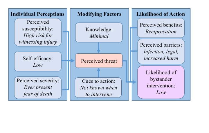
Introduction:  The paucity of trauma centers in the south side of Chicago leads to prolonged transport times and increased morbidity and mortality for those affected by penetrating trauma. An evidence-based community-driven Trauma First Responders Course (TFRC) could potentially mitigate this effect, but does not currently exist. Bystanders are present at 60-97% of traumas and are more likely to assist with prior training. However, the bystander effect remains a major barrier. We hypothesize that by utilizing the Health Belief Model as a framework, we can characterize the factors in our community that lead to bystander non-interference. Through this, neighborhood focus groups will facilitate effective course development and thus improved patient outcomes and community empowerment.

Methods:  The Health Belief Model determines the likelihood of an action by examining individual perceptions of susceptibility, self-efficacy, and severity of a health issue, then applying the modifying factors of knowledge and cues to action. The resulting perceived threat is then modulated by perceived benefits and barriers of performing the action. Written surveys and focus group questions were developed to specifically address each of these facets. Focus groups were conducted by 2 guides over an hour with 8-10 community members, including youth at highest risk for violence and key community leaders. Data were collected via surveys and recordings and analyzed quantitatively as well as qualitatively.

Results: The focus groups demonstrated consistency across many of the factors examined, see Figure. Participants felt the perceived susceptibility for witnessing an injury was high. Half stated they worry that they or someone they know will get hurt "a lot" or "all of the time" and that they are "unsure," "not confident," or "not at all confident" in their ability to render aid, demonstrating a sense of low self-efficacy. Only 39% of responders stated they had any form of first aid training, and less than 10% stated they had advanced training. A majority said they did not know when to intervene, which often stemmed from a concern that interference would lead to increased harm. There was significant fear of social or legal retaliation for helping a victim of violence, while 92% would want a stranger to help if they or someone they know was injured. Overall, the likelihood of bystander intervention was deemed to be low.

Conclusion: Critical factors have been identified through the structure of the Health Belief Model that contribute to bystander non-intervention in our community. These will need to be addressed during the development and implementation of an effective TFRC to empower community members to overcome the bystander effect.

11.20 Radiographic Evaluation of the Pregnant Trauma Patient: What Are We Willing to Miss?

E. Herfel1, E. Buggie2, M. Lieber2, J. Hill2  1OhioHealth Doctors Hospital,Obstetrics And Gynecology,Columbus, OH, USA 2OhioHealth Grant Medical Center,Trauma Services,Columbus, OHIO, USA

Introduction:  Trauma is the leading cause of non-obstetrical causes of death in pregnant patients. The use of radiographic imaging for evaluation of a pregnant trauma patient in the trauma bay is controversial, considering research has linked radiation exposure to inappropriate development in some children. However, in critical cases the benefits of using radiographic imaging to ensure maternal survival outweigh the risks of radiation exposure to the fetus. This study explored whether sparing fetal exposure to radiation by minimizing use of radiographic imaging put the mother at risk for a delayed diagnosis of injury. We hypothesize that minimizing the use of radiographic imaging in the initial assessment of pregnant trauma patients will not lead to a higher incidence of a delayed diagnosis or missed injury.

Methods:  We performed a seven year retrospective chart review at an urban level 1 trauma center reviewing pregnant patients and a 2:1 cohort of non pregnant patients matched for age, injury severity score (ISS) and injury type, all involved in blunt trauma. Collected data points include: number and type of imaging studies performed on initial presentation and those images that were delayed. Delayed imaging was defined as any imaging study obtained two hours or more after arrival in the trauma bay. A delayed diagnosis was defined as any injury identified by delayed imaging. The primary outcome was incidence of delayed diagnosis in the pregnant trauma patient compared to the non-pregnant patient.

Results: A total of 83 pregnant patients and 167 non-pregnant patients were examined. Average age was 23.7 years and average ISS was 2.7 in both groups. 95.2% of the pregnant population had at least one imaging study done versus 100% of the control group (p=0.004).  The pregnant population had an average of 4.3 images performed compared with an average of 6.8 images in the non pregnant cohort (p=<0.001). 18 (21.7 %) pregnant patients had delayed imaging and 58 (34.7%) control patients had delayed imaging (p=0.03). Only 1/18 of pregnant patients had a delayed diagnosis of a traumatic injury (transverse process fracture of the lumbar spine). 9/58 control patients had a delayed injury (p=0.17).

Conclusion: Our study demonstrates that bluntly injured pregnant trauma patients receive significantly fewer radiographic images upon presentation than their non-pregnant counterparts. However, only 1% of those pregnant patients had a delayed injury diagnosed. This was not significantly different from non-pregnant patients when matched for age and ISS. Though the ISS was low for both patient cohorts, this study suggests that mitigated radiographic imaging in the pregnant trauma patient is safe and does not result in delayed diagnosis of injury. 

11.19 Identification of Organ Failure Patterns for Early Stratification of Trauma Patients

D. Liu1, R. A. Namas1, Q. Mi1, O. Abdul-Malak1, J. Guardado1, B. Zuckerbraun1, J. Sperry1, M. Rosengart1, Y. Vodovotz1, B. Timothy1  1University Of Pittsburgh,General Surgery,Pittsburgh, PA, USA

Introduction: Multiple organ dysfunction syndrome (MODS) typically peaks within 5 days of injury and is associated with a complicated clinical course. However, the number of distinct organ failure patterns following injury in humans is unknown. Using MODScore parameter optimized to correlate with adverse in-hospital outcomes, we sought to establish the number of distinct pattern-specific patient clusters present in a highly characterized cohort of blunt trauma patients admitted to the ICU. Inflammatory networks based on measurements of 31 circulating inflammation biomarkers were characterized.

Methods: 376 patients admitted to the ICU and with sequential Marshall MODScores from days (D) 2-5 post-injury were studied. MODScores from D2-D5 were subjected to Fuzzy Clustering Analysis (FCA) to suggest trauma patient sub-groups. Eight widely accepted internal quality indices were calculated to determine the optimal number of trauma patient sub-groups using R. Inflammation biomarkers (31 cytokines and chemokines) were assayed (by Luminex™) in serial blood samples (3 samples within the first 24 h and then daily up to day 5 post-injury). Biomarkers were analyzed using Two-Way ANOVA (p<0.05). Dynamic network analysis (DyNA) was used to suggest dynamic connectivity and complexity among the inflammatory mediators.

Results: Six (75%) of the eight indices suggested that the optimal number of  clusters was 4: Group 1 (n=199, age=49 ± 1, male/female [M/F]=134/65, average MODScore=0.3); Group 2 (n=99, age=48 ± 2, M/F=70/29, average MODScore=2); Group 3 (n=53, age=47 ± 3, M/F=36/17, average MODScore=4); and Group 4 (n=25, age=46 ± 4, M/F=20/5, average MODScore=7). There were statistically significant differences among the four groups with regards to ICU LOS, total LOS, and days on mechanical ventilation being all greatest in Group 4, which in turn exhibited a higher incidence of nosocomial infection (76%) when compared to Groups 1, 2, and 3 (16%, 41%, and 49% respectively). Of the 31 circulating biomarkers measured, IL-6, MCP-1, IL-10, IL-8, IP-10, sST2, and MIG were differentially elevated upon presentation and over time among the groups. DyNA suggested a higher sustained degree of systemic interconnectivity in Group 4, which persisted up to D5 post-injury when compared to the other groups.

Conclusion: These results suggest that blunt trauma leads to 4 distinct organ failure patterns in patients who are admitted to the ICU and survive to discharge. The organ failure patterns are preceded by distinct patterns of inflammation biomarkers and followed by severity-specific differences in patients’ in-hospital outcomes.

11.18 The Impact of Severe Head Trauma on the Management of Isolated Open Tibia Fractures

N. N. Branch1, R. Wilson1  1Howard University College Of Medicine,Washington, DC, USA

Introduction: A consensus has yet to be established for the ideal timing of definitive fixation of open tibia fractures in the setting of traumatic brain injury (TIB).  Data regarding definitive fixation in patients with severe TBI has been inconclusive in studies of closed long bone fractures, however little is known regarding outcomes in the open fracture setting.  Therefore we sought to outline the perioperative complications in patients with severe head trauma who have undergone open reduction and internal fixation (ORIF) for isolated open tibia fractures (IOTF).

Methods: A retrospective analysis of the National Trauma Data Bank from 2007-2010 utilizing ICD-9 codes (823.10, 823.30, and 823.90) was conducted.  Cases > 18 years old with IOTF who underwent ORIF at a level I or level II trauma center were included.  Using Glasgow Coma Scale (GCS) totals patients were stratified by score (severe head injury < 8; minor head injury 8 >), with minor head injuries serving as the reference group.  Multivariate logistic regression was used to investigate postoperative complications.

Results: 9,331 isolated open tibia fractures during the study period underwent ORIF, of which 7,201 cases met inclusion criteria.  The majority were white (67%), males (74%), between 25-44 years old (42%) with private insurance (23%) and injured via motor vehicle collisions (25%). Mean days to ORIF was 2.98 sd. 7.4.  GCS mean 14.05 sd. 2.9 6.9% (n = 496) of cases had severe head injuries.  On multivariate analysis patients with severe head injuries 88% more likely to have fixation after hospital day 2, 81% increased odds of requiring transfusion, and more than three times more likely to develop acute respiratory distress syndrome (ARDS).  Odds of most infectious complications more than doubled. Development of any complication and perioperative cardiac arrest requiring CPR were also increased more than two fold, and patients had an increased relative risk for a longer than average length of stay. There was no additional risk of death. (Table 1).

Conclusion: Determining the timing of definitive fixation in IOTB in patients with severe head injuries remains a challenge.  These patients are at risk for infectious complications and an increased need for transfusion.  Notably these patients have more than threefold risk of ARDS possibly due to the severity of their head injury and requirements for prolonged ventilation.  While their management does not demonstrate an increased odds of death the risk of cardiac arrest or development of any complication warrants additional investigation for ways to reduce morbidity this small yet vulnerable population, and the role of GCS as a predictor of perioperative patient outcomes.

11.17 Troponin I in the Evaluation of Blunt Cardiac Injury Following Sternal Fracture

P. Hu1, R. Uhlich1, A. Witcher1, J. Kerby1, P. Bosarge1  1UAB,Acute Care Surgery/Surgery,Birmingham, AL, USA

Introduction:
Blunt cardiac injury (BCI) is often identified in the presence of sternal fractures in blunt trauma. Full evaluation and management of this injury remains controversial. Electrocardiogram (ECG) is the current standard in diagnosis and is recommended for any patient with possible BCI. The utility of troponins are less clear. It remains a matter of debate as to the type of troponin to be measured, the ideal timing, and required duration of monitoring.  We sought to determine the role of troponin I measurement in the evaluation of BCI following sternal fracture.

Methods:
Trauma patients admitted to an American College of Surgeons verified level I trauma center from 2011-2013 with a diagnosis of sternal fracture were identified from the trauma registry. A retrospective review of the medical record was performed to determine patient age, gender, length of stay, results of ECG, echocardiography, troponin I values, patient cardiac morbidity and mortality. The first three Troponin I levels collected during hospitalization were recorded. BCI was diagnosed by newly identified ECG abnormalities.

Results:
203 trauma patients were admitted with sternal fracture following blunt trauma. BCI was diagnosed in 89 patients by ECG. 101 patients had at least one troponin I level measured. Troponin I levels were undetectable in 15 patients with BCI compared to 14 without BCI. Mean troponin I levels were 0.328 ng/mL in patients with BCI compared to 0.055 ng/mL without. Troponin I greater than 0.1 ng/mL on admission has a positive predictive value of 89%. Negative predictive value of undetectable troponin I levels for BCI is 48%. Twelve  patients died within 30 days of hospital admission, however no deaths were due to acute heart failure, myocardial infarction, or cardiogenic shock.

Conclusion:
Troponin I levels offer little benefit following sternal fracture in excluding the possibility of blunt cardiac injury. While the presence of elevated troponins increases the likelihood of BCI, it does not appear to correlate with cardiac related morbidity or mortality.
 

11.16 Hemodynamically Abnormal Thoracoabdominal Trauma Should Undergo CT Prior To Definitive Therapy

J. Carney1, A. Strumwasser1, K. Matsushima1, D. Grabo1, D. Clark1, K. Inaba1, E. Benjamin1, L. Lam1, D. Demetriades1  1University Of Southern California,Surgery – Trauma/Acute Care Surgery,Los Angeles, CA, USA

Introduction:  The triage of hemodynamically abnormal trauma patients is debated. Controversial data suggests that hemodynamically abnormal patients can safely undergo CT prior to definitive therapy. We wished to investigate outcomes for hemodynamically abnormal thoracoabdominal trauma undergoing CT.  

Methods:  All hemodynamically abnormal (HR≥120, SBP<90 mmHg) patients arriving at our Level I trauma center in 2014 were reviewed.  Inclusion criteria were thoracoabdominal trauma patients that achieved hemodynamic normalization (SBP≥90 mmHg) and were eligible for CT.  Pregnant patients, pediatric patients (age<18), patients undergoing resuscitative thoracotomy, and isolated head, neck and extremity injuries were excluded.  Variables abstracted from the registry included patient demographics, injury mechanism, injury severity score (ISS), physical exam, E-FAST results, laboratory data, CT scan findings and operative details.  Primary outcomes included hospital length-of-stay (HLOS), ICU LOS, ventilator days and mortality.  Secondary outcomes included intraoperative data (procedure duration, fluids, blood loss), transfusion burden, incidence of venous thromboembolism (VTE), infectious complications, need for additional procedures, and total hospital cost.  Data was analyzed by unpaired Student’s t-test for continuous variables and Chi Square analysis for categorical variables with significance denoted at a p value of 0.05 or less.

Results:  A total of 201 patients met inclusion criteria. Thirty-five (17%) went directly to the OR at triage, 117 (59%) went to CT, and 49 (24%) were spared an operation. The CT and non-CT groups were well matched at baseline for age (34±2.0 vs. 38±1.2 years, p=0.1), injury burden (mean ISS-CT=18±2.7 vs. ISS-non-CT=18±0.9) and total resuscitation time (81±43.0 vs. 67±9.4 minutes, p=0.7).    No difference in time-to-normalization of lactate, HLOS, ICU LOS or mortality was observed (p>0.1 for each).  Patients undergoing CT prior to OR had increased recognition of intraabdominal injuries (89 vs. 74%, p=0.05), a significant reduction in negative explorations (2.3 vs. 8.6%, p<0.01) and a decreased need for PRBCs (2±0.6 vs. 7±2.5 units, p<0.01), FFP (1±0.3 vs. 4±1.3, p<0.01) and platelets (0.3±0.1 vs. 1±0.4 units, p=0.03) throughout admission.  Moreover, patients in the non-CT group had an increased need for surgical procedures (37.1 vs. 12.1%, p<0.01) after the index operation.  No differences were noted in any secondary outcome (p>0.1 for each).

Conclusion: Hemodynamically abnormal thoracoabdominal trauma achieving a SBP≥90 during resuscitation should undergo CT scanning prior to definitive therapy.  Imaging increases the identification of intraabdominal injuries, decreases negative explorations, decreases transfusion burden and decreases the need for additional surgical procedures without affecting morbidity, mortality or hospital cost.


11.15 Use of Routine Head CT After Initiation of Chemoprophylaxis in Patients with Intracranial Hemorrhage

C. L. Shelley1, P. M. Arnold2, K. Udobi1, J. Green1, A. W. Bennett1, S. Berry1, J. M. Howard1, T. McDonald1, M. Moncure1, R. Winfield1  1University Of Kansas Medical Center,Trauma Surgery,Kansas City, KS, USA 2University Of Kansas Medical Center,Neurosurgery,Kansas City, KS, USA

Introduction: Traumatic brain injury (TBI) is a risk factor for the development of venous thromboembolism (VTE). Administration of low dose unfractionated heparin (LDUH) or low molecular weight heparin (LMWH) is an effective preventive strategy for this problem, but must be weighed against the risk of progression of intracranial hemorrhage. Studies have documented safety of VTE prophylaxis in TBI; however, no series has included routine follow up head CT after chemoprophylaxis initiation. The purpose of this study was to assess progression of intracranial hemorrhage after VTE prophylaxis using routine 24-hour head CT, which is standard practice at our institution.

Methods: A retrospective review of our level 1 trauma center’s registry from January 1, 2010 to December 31, 2015 was performed. Adult patients with head CT demonstrating TBI, subsequent initiation of VTE chemoprophylaxis, and repeat head CT obtained within 24 hours of initiation of chemoprophylaxis were included. Patients with history of coagulopathy were excluded.

Results: 1,120 records were reviewed. 255 met inclusion and exclusion criteria. Median post-injury start date for chemoprophylaxis was 4 days, with 159 patients receiving LDUH and 96 receiving LMWH. No patient had neurologic decline prior to scheduled 24-hour scan. Progression of hemorrhage occurred in 9 patients (3.5%); 7 had hemorrhage at a prior intervention site, 1 had a new lesion identified, and 1 had increase in an existing injury. Progression was not significantly different between patients receiving LDUH (n=7) and LMWH (n=2) (x ²=0.95, p=0.33). No patient required surgical intervention as a result of progression.

Conclusion: There is a low rate of progression of TBI seen on routine CT after initiation of VTE prophylaxis. There is no difference in progression rates between patients receiving LDUH or LMWH, but progression is more likely if patients undergo neurosurgical intervention. Intervention for progression of TBI is unlikely after VTE prophylaxis is initiated.