59.06 Macrophage Transplantation Accelerates Repair of Cutaneous Wounds but not Cranial Defects

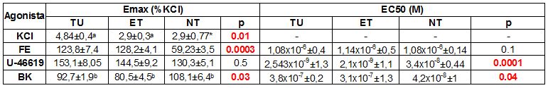
M. R. Borrelli1, M. S. Hu1, W. Hong1, G. G. Walmsley1, M. P. Murphy1, M. Lopez1, R. C. Ransom1, H. P. Lorenz1, M. T. Longaker1  1Stanford University,Plastic Surgery And Pediatric Regenerative Medicine,Palo Alto, CA, USA

Introduction:
An urgent biomedical need exists to repair both soft tissue and skeletal defects. With a growing incidence of comorbidities such as diabetes, peripheral vascular disease, and obesity, the number of chronic, non-healing wounds is increasing rapidly, costing the United States over $25 billion per year. Additionally, repair and reconstruction of the craniofacial skeleton represents a significant burden with over 50,000 craniotomies/craniectomies per year accounting for $585 million in medical care. We previously demonstrated that the supraphysiologic transplantation of macrophages delivered in a pullalan-collagen composite biomimetic hydrogel scaffold accelerated wound healing in wild type and diabetic mice through a mechanism related to angiogenesis. However, the effect of macrophage transplantation in skeletal defects is yet unknown. Herein, we perform supraphysiologic transplantation of macrophages into cranial defects to assess for improved healing.

Methods:
CD1 athymic nude mice at 60 days of age were anesthetized and unilateral full-thickness critical-size calvarial defects (4 mm in diameter) were created in the non-suture associated parietal bone. Mouse macrophages (1.0 x 10^6 cells), that were isolated from FVB-L2G mice and seeded onto hydroxyapatite-poly(lactic-co-glycolic acid) (HA-PLGA) scaffolds and incubated overnight, were placed into the cranial defects. Micro-CT evaluation was performed at 0, 2, 4, 6, and 8 weeks. Osseous healing was quantified using Adobe Photoshop. Three-dimensional in vivo imaging system (IVIS)/micro-CT was used to determine survival of macrophages in vivo.

Results:
Whereas supraphysiologic transplantation of macrophages accelerated healing of full-thickness cutaneous wounds, macrophage transplantation did not improve healing of critical-size calvarial defects through 8 weeks following creation of defects (p>0.05). This was confirmed with histology. Three dimensional IVIS/micro-CT demonstrated survival of macrophages through 8 weeks.

Conclusion:
Although macrophage transplantation accelerates wound healing via increased angiogenesis, supraphysiologic delivery to critical-size calvarial defects had no effect despite survival of transplanted macrophages. Further research may elucidate the exact mechanism that allows macrophage transplantation to accelerate healing of cutaneous wounds, but not cranial defects.
 

59.05 Reverse Thermal Gel: Our Candidate for an Early, Minimally Invasive Method to Patch Myelomeningocele

J. R. Bardill1, D. Park1, U. Shabeka3, A. I. Marwan1,2,3  3University Of Colorado Denver,Laboratory For Fetal Regenerative Biology,Aurora, CO, USA 1University Of Colorado Denver,Bioengineering,Aurora, CO, USA 2Children’s Hospital Colorado,Pediatric Surgery,Aurora, CO, USA

Introduction:  Myelomeningocele (MMC) results in lifelong neurological and functional deficits.  In-utero repair closes the defect, resulting in a 50% reduction in post-natal ventriculoperitoneal shunting. However, it is associated with maternal and fetal risks.  Novel fetoscopic approaches using 2-3 ports, pre-fabricated patches, and skin closure demonstrate promising results, however, they are performed late and have a prolonged operative time. Our novel, Reverse Thermal Gel (RTG) has posed a unique solution for an early, minimally invasive delivery of a patch.  So far, we have shown fast in-situ gelling properties and RTG stability after long term exposure in amniotic fluid. Moreover, RTG is non-toxic to cells and embryos in culture. Here, we hypothesize that RTG can serve as an in-vitro 3D cellular scaffold for mouse skin fibroblasts and will be successfully injected and localized to the MMC defect of mouse embryos with minimal inflammatory response.

Methods:  1. In-vitro 3D-RTG cellular scaffold:  Mouse skin fibroblasts were mixed with RTG-media solution, followed by gelling at 37°C to encapsulate the cells within the RTG.  The fibroblasts were cultured for 3 days and then viability was assessed using a live/dead stain and confocal microscopy.  2. RTG injection into mouse MMC defect embryos:  Grainyhead like-3 (Grhl3) mice express MMC phenotypes.  Pregnant E16.5 Grhl3 mice underwent laparotomy to expose uterine horns.  A RTG solution or saline was applied onto the MMC defect by transuterine injection.  On E19, the embryos were harvested to assess RTG coverage of the MMC defect.  3. Inflammatory response to RTG injection:  Defect tissue was stained for CD68 and F4/80 macrophage antibodies and imaged on a fluorescent microscope.

Results:  Fibroblast 3D-RTG culture live/dead staining reveals 90-95% live cells after 3 days. The RTG successfully applies to mouse MMC defects and forms a stable gel covering the defect.  A total of 6 MMC defect embryos are recovered at E19 harvest:  3 defect embryos demonstrate more than 50% coverage of the defect, while the other 3 show less than 50% coverage.  CD68 and F4/80 macrophage response demonstrates little to no difference in inflammation comparing RTG and saline injections in defect embryos.

Conclusion:  Using a mouse model, we demonstrate that by simple needle injection, a RTG can cover a mouse MMC defect and remain at the site of the defect until harvest with minimal inflammatory response.  However, RTG defect coverage decreases over time.  Moreover, the RTG has in-vitro­ fibroblast scaffolding properties, which could allow for native cellular integration into the patch and/or allow cellular treatments to be incorporated into the patch. Based on our results, we are currently modifying the RTG to enhance the adhesive properties. Overall, our work demonstrates that a RTG is a promising candidate for a minimally invasive approach to patch MMC and will be conducive to transitioning to a fetoscopic approach in a sheep model.

59.04 Adipose Derived Stromal Cell Enhancement of Cancer Growth Likely Due To Specific Gene Upregulations

D. M. Irizarry1, J. S. Flacco1, C. P. Blackshear1, C. F. Montenegro1, D. Nguyen1, N. Quarto1, A. Giaccia1, M. T. Longaker1,2, D. C. Wan1  1Stanford University,Plastic And Reconstructive Surgery,Palo Alto, CA, USA 2Institute For Stem Cell Research And Regenerative Medicine,Palo Alto, CA, USA

Introduction: Many oncologic surgeries create significant defects with a need for reconstruction. Further, radiation therapy induces fibrotic damage to skin. Fat grafting has been utilized to address these soft tissue deficiencies, and studies have suggested a regenerative effect on radiation-damaged skin. Inclusion of supplemental mesenchymal cells to fat grafts have also been shown to augment these effects. However, mesenchymal cells have been described in animal models to promote carcinoma growth, and with head and neck cancer, may enhance tumor invasion and formation of micrometastases. As cancer of the oral cavity is among the most common types of head and neck cancer, we chose to evaluate the effects of ASCs on the SAS HNSCC cell line.

Methods: SAS HNSCC cells were plated with either regular or ASC conditioned media. They were lifted and counted every other day for 6 days. For the in-vivo arm, 5X10⁵ GFP labeled SAS HNSCC cells were injected into the subcutaneous plane of the scalp of mice either alone, in addition to a 200uL fat graft, or in addition to a 200uL fat graft and 10,000 ASCs. CT scans were taken weekly for 4 weeks. To examine what factors released by ASCs might be responsible for promoting cancer proliferation, differential protein component analysis was performed on conditioned media from co-cultured ASCs and cancer cells and compared to media from cancer cells alone and ASCs alone.

Results: Cancer cells grown in ASC conditioned media show greater proliferation than those grown in regular media. At the 3 and 4 week timepoints, there was significantly greater tumor volume by microCT in mice receiving cancer with fat graft and ASCs compared to mice receiving cancer alone or in addition to a fat graft. Multiple upregulated factors were identified in the ASC-cancer co-culture medium when compared to cancer alone or ASCs alone, most notably epidermal growth factor was upregulated 1.4-fold, hepatocyte growth factor was upregulated 1.9-fold, and monocyte chemotactic protein was upregulated 5.0-fold. Interestingly, each of these three factors have been implicated in growth and/or metastatic development in head and neck cancer.

Conclusion: While the risk for head and neck cancer recurrence following fat grafting with or without ASC enrichment remains unclear, our preliminary data have shown that ASCs potentially stimulate cancer cell proliferation and tumor formation, and that this may be secondary to one or more likely multiple of the factors we have identified in our component screen. Our ongoing studies are currently testing this hypothesis. Although ASCs and fat grafting may have enormous regenerative potential, the oncologic ramifications of their use must therefore be carefully considered before widespread application.

59.03 Loss of Intestinal Alkaline Phosphatase Leads to Distinct Changes in Bone Phenotype

F. Kuehn1, F. Adiliaghdam1, S. R. Hamarneh1, R. Vasan1, E. Liu1, Y. Liu1, J. M. Ramirez1, F. C. Ko2, M. L. Bouxsein2, M. N. Wein2, M. B. Demay2, R. A. Hodin1  1Massachusetts General Hospital,Surgery,Boston, MA, USA 2Massachusetts General Hospital,Endocrinology,Boston, MA, USA

Introduction:  The gut is becoming increasingly recognized as the source of various systemic diseases, and recently it has been linked to bone metabolism and formation via the so-called gut-bone axis. The microbiome and gut-derived mediators such as lipopolysaccharides (LPS) are thought to impact bone metabolism and administration of beneficial bacteria (probiotics) such as lactobacillus has been shown to result in higher bone mineralization and bone strength. The gut brush border enzyme intestinal alkaline phosphatase (IAP) plays an important role in controlling calcium absorption, inhibiting LPS and other inflammatory mediators responsible for endotoxemia, and, also appears to preserve the normal of gut microbiota. Interestingly, IAP-deficient mice (AKP3-/-) also display a significant decrease in Lactobacillus, the genus shown to be beneficial to bone. Based on this background, we sought to determine the impact of IAP-deficiency on bone formation in mice.

Methods:  Transcript levels of IAP in mouse bone were measured using qPCR. Femurs of young (3 months) and old (21 months) IAP-KO and WT mice were analyzed by micro-CT scan and histopathology. Serum levels of alkaline phosphatase (AP) as a marker of active bone formation, and, serum calcium and phosphorus levels were measured in IAP-KO and WT mice. In-vitro target cell response upon exposure to blood serum from IAP-KO and WT mice was measured using primary bone marrow macrophages.

Results: IAP was not expressed in bone of WT mice. Transcript levels were similar to IAP-KO counterparts (IAP-WT vs. IAP-KO, 56.9 ± 17.7 vs. 41.4 ± 21.3 Relative Expression, p=0.4) and vanishingly low when compared to duodenum expression levels (bone vs. duodenum, 56.9 ± 17.7 vs. 25,430.3 ± 10,884.5 Relative Expression, p=0.01). Histological examination of 3-month old IAP-KO and WT did not show any differences, whereas older IAP-deficient mice showed a very distinct bone phenotype in histology and CT-scan (image 1). Younger KO mice did not display any abnormal levels in blood chemistry analysis but older IAP-KO animals showed an isolated increase in AP serum levels, significantly higher than in control animals (IAP-WT vs. IAP-KO, 80 ± 27.4 U/I vs. 453 ± 107.5 U/I, p =0.004). Normal GGT levels ruled out an underlying hepatic pathology causing the AP increase. Finally, to determine to the extent that systemic serum may contribute to inflammation in peripheral organs such as the bone marrow we tested the inflammatory response of target cells exposed to systemic blood serum from IAP-KO and WT mice. Serum from IAP-KO mice induced a significantly higher inflammatory target cell response than serum derived from WT counterparts (p<0.01).

Conclusion: Through its multiple functions, IAP seems to play a crucial role in connecting the gut to the bone. IAP-deficiency leads to chronic changes in bone formation, probably through dysbiosis and dissemination of pro-inflammatory mediators. 

59.02 Rapamycin Inhibits Primary and Recurrent Heterotopic Ossification in Genetically Susceptible FOP Mice

C. Hwang1, S. Ucer1, C. Pagani1, N. Patel1, A. Vaishampayan1, M. Sorkin1, M. T. Chung1, J. Li1, C. Breuler1, C. Priest1, A. N. Economides3, S. Agarwal1, Y. Mishina2, B. Levi1  1University Of Michigan,Section Of Plastic Surgery,Ann Arbor, MI, USA 2University Of Michigan,Department Of Biologic And Material Sciences, School Of Dentistry,Ann Arbor, MI, USA 3Regeneron Pharmaceuticals And Genetics Center,Tarrytown, NY, USA

Introduction: Heterotopic ossification (HO) is a debilitating formation of ectopic bone restricting joint mobility and causing chronic pain. Fibrodysplasia Ossificans Progressiva (FOP) is a congenital variant of HO caused by a genetic mutation in a bone morphogenetic receptor that causes severe, progressive lesions resulting in immobility and fatal mechanical respiratory failure at a premature age. With increased proclivity for osteogenesis at baseline, surgical extirpation is contraindicated in FOP patients due to universal recurrence. This disease presents a unique clinical challenge with no current treatments available. Previous research has shown mTOR inhibition by rapamycin to be a viable therapy for reducing trauma-induced HO. Mice of an FOP mouse model with a hyperactivating mutation in the ACVR1 receptor (ACVR1R206H/+) were examined to determine the effects of rapamycin on both primary and post-surgical HO.

Methods: Mice were designated for 2 distinct study arms. In the primary HO cohort, ACVR1R206H/+ P21 mice received bilateral hindlimb cardiotoxin (CTX) with Ad.cre injection and stratification to daily i.p. rapamycin (n=10 hindlimbs) v. PBS control (n=12). 3 weeks later, mice were scanned with in vivo µCT. Mice within the separate recurrence cohort received initial Ad.cre/CTX followed by baseline CT scans, Ad.cre reinjection bilaterally, and surgical removal of HO at the 3 week timepoint. After surgery, mice were randomized to daily rapamycin (5 mg/kg, n=12) or PBS (n=8) for 3 weeks with endpoint µCT. Contours were drawn manually around HO to compute total volumes at 800HU. Primary and recurrence cohorts were analyzed by Student’s t test and log-transform/ANOVA/Hochberg post-hoc respectively.

Results: Mice treated with rapamycin for 21 days showed 261-fold less ectopic bone when compared to PBS vehicle injection (p=0.002). In post-surgical mice, PBS injection showed statistically similar HO volumes to baseline volumes of pre-excision mice (p=0.054). However, rapamycin treatment reduced the recurrence of HO volume 11-fold (p=.044, Figure). Geometric means from de-transformed data for pre-excision v. PBS, pre-excision v. rapamycin, and PBS v rapamycin were 0.061, 1.398, and 22.961 respectively.

Conclusions: These studies demonstrate that rapamycin prevents primary development of HO and is also effective in preventing recurrence following surgical excision in a FOP mouse model. This study further corroborates rapamycin as a promising candidate for primary and post-surgical HO prophylaxis in children with FOP. Existing literature implicates similar molecular mechanisms among various etiologies of HO, suggesting a putative role for rapamycin even beyond FOP in post-traumatic and post-surgical HO patients.

59.01 Differences in vascular reactivity of arteries of colon tumors vs. non tumoral arteries in humans: role of phenylephrine, thromboxane and bradykinin.

N. Cupitra1, A. Múnera Duque2, JP León2, R. Narvaez-Sanchez1  1PHYSIS Group, Faculty of Medicine, University of Antioquia, Medellín, Antioquia, Colombia 2Department of Surgery, Faculty of Medicine, University of Antioquia, Medellín, Antioquia, Colombia

Cancer is one of the leading causes of death on the planet: in 2012, 8.2 million deaths were attributed to it, with the highest number of victims caused by lung cancer (19, 4%), hepatic (9.1%), gastric (8.8%) and colorectal (8.4%); (1). As an abnormal growth of cells, cancer needs vascular changes for growing and metastases. In the last two decades the tumor vascular system and its possible role in the development of cancer has been investigated, proposing it as a target for the control of nutrient supply, by modification of vascular tone, angiogenesis and neovascularization (2). Vascular reactivity (VR) could be a limiting factor for growth in cancer, since growing tissues require vasodilation and new vascularization. Promoting vasoconstriction in the tumor could reduce the supply of nutrients, while promoting vasodilation could increase therapies to reach more cancer cells. The present work aims to quantify differences in human VR of tumoral (TU, arteries which directly irrigate the tumor), extratumoral (ET, arteries of a tumoral patient, which do not irrigate tumor but the distal and proximal colon near the tumor) and non-tumoral arteries (NT, branches of mesenteric circulation of colectomized patients due to a non tumoral pathology), to the stimulus of potassium chloride (KCl), phenylephrine (FE), ET-1, U-46619 (analog of thromboxane A2), and bradykinin (BK).

Six patients with colon cancer and two with non tumoral pathology, from 30 to 80 years old, underwent hemicolectomy at “IPS Universitaria Leon XIII” Hospital, in Medellin, Colombia. All patients signed the respective informed consent. Mesenteric branches were collected and preserved in Krebs-Henseleit solution (SK, composition (mEq/liter): Na+, 143.5, K +, 5.4, Ca2 +, 5.1, Mg2+, 2.4, Cl-, 128, H2PO4-, 1.2, HCO3, 24.9, SO42-, 2.4, and glucose, 10) at 4° C where excess of peripheral tissue was removed. The arterial segment was cut into rings 3-5 mm long, which were mounted on stainless steel hooks in parallel through the lumen. One of the hooks was attached to a force transducer (ADInstruments Ltd, Model MLT0201, UK), and the other was held as a fixed support. The preparation was mounted in a 10 ml organ bath (Panlab/ Harvard, Model LE01046, Spain) containing SK. The tissue was maintained at 37° C and bubbled with carbogen (95% O2- 5% CO2). Each preparation was stabilized at 2 grams of basal tension along 90 minutes, before initiating stimulations with agonists, changing SK every 20 minutes. After stabilization the arterial ring was stimulated twice with KCl 4×10⁻² M recording the response for 10 minutes, and washing for 20 minutes between stimuli. Then U-46619 1×10⁻⁷ M and BK 1×10⁻⁵ M were applied to confirm the presence of endothelium. This verification was performed on all arterial rings. Then the evaluation was done applying in some rings KCl at a single dose KCl 4×10⁻² M, or FE at cumulative dose of 5×10⁻⁸ M to 1×10⁻⁵ M, or U-46619 cumulative dose of 1×10-13 M to 1×10⁻⁷ M, or, precontracting with U-46619, BK at cumulative dose of 1×10⁻⁹ M to 1×10⁻⁵ M. The maximal relative response (“Emax%KCl”, maximal contraction normalized in respect to maximal contraction to KCl) and sensitivity (EC50, concentration which generates 50% of response) were recorded by the Powerlab PL-3504 polygraph (ADInstruments, Australia). Analyzes were performed with t-Student and ANOVA with significance level p <0.05. The EC50 was calculated by non-linear regression of the concentration-response curves. The statistical package GraphPad prism 6 Statistics was used. Experimental results are presented as means ± SEM of the Emax and EC50:

The greater response of TU but not of NT to KCl suggests histological differences that we have not specified yet. The increase in Emax to FE and EC50 U-46619 of TU and ET with respect to NT, suggests that the tumor microenvironment favors the contractile response in the arteries that irrigate it: it has been reported that oxidative stress activates kinases that promote proliferation and resistance to anticancer agents, and induces endothelial dysfunction that worsens vasodilation. The beta-adrenergic receptors participate in the initiation and progression of the tumor; but more evidence is lacking about the role of alpha-adrenergic signaling and thromboxane A2 in vascular control in cancer. The higher sensitivity to U-46619 in TU and ET suggests an increase in the activity of the thromboxane-A2 dependent mechanisms, coinciding with what is reported in greater activation of thromboxane-prostanoid receptors. Our relaxation analysis did not show differences of VR to BK in Emax nor in EC50 between TU and ET, contradicting a possible endothelial damage, at least in TU and ET. These preliminary results require contrasting with non-tumoral arteries, measuring differential expression of receptors, doing histology evaluating integrity of the vascular wall, and quantifying probable endothelial dysfunction by specifying affected mechanisms, using specific blockers.

41.20 Rapid Detection of Clostridium difficile Toxins in Serum by Raman Spectroscopy.

S. Koya1, J. V. Martin2, S. Yurgelevic1, D. M. Liberati2, M. Brusatori1, C. Huang1, G. W. Auner1, L. N. Diebel2  1Wayne State University,Smart Sensors And Integrated Microsystems (SSIM)/ Michael And Marian Ilitch Department Of Surgery/School Of Medicine,Detroit, MI, USA 2Wayne State University,Michael And Marian Ilitch Department Of Surgery/School Of Medicine,Detroit, MI, USA

Introduction:
Clostridium difficile infection (CDI) is due to the effects of toxins, toxin A (TcdA) and toxin B (TcdB) on the host. Severe CDI is associated with systemic signs of infection. Animal models of CDI demonstrate a strong correlation between systemic toxemia and the occurrence of severe disease. However current technologies have low sensitivity to detect C. difficile toxemia in human subjects. Raman spectroscopy (RS) is an upcoming technology that is used to detect bacteria and their toxins. We speculate that RS may be a sensitive method to detect clinically relevant concentrations of C. difficile toxins in serum. 

Methods:
Serum samples were spiked with varying concentrations of TcdA, TcdB and both. RS was performed on air dried serum drop on a mirror polished stainless-steel slide. Raman spectra were obtained, background corrected, vector normalized and analyzed by Partial Least Square Linear Discriminant Analysis (PLS-LDA). PLS-LDA model accuracy was measured by cross-validation and bootstrap method.

Results:
At 1 pg/ml concentration, toxin spiked serum was distinguished from control serum 100%. PLS-LDA model performed well with cross-validated error rate ranging from 8 to 15% and bootstrap error rate ranging from 4 to 9%. Similar results were obtained for other concentrations of toxins (100 pg/ml, 1 ng/ml). 

Conclusion:
Raman spectroscopy has the potential to rapidly detect C. difficile toxins in serum at clinically relevant concentrations and may be useful as a diagnostic tool to modify therapy and and predict outcome in critically ill CDI patients.  

41.19 Supercomputing Precision MODS: Insights from an in silico cohort of of 2 Billion simulated patients

C. Cockrell1, G. An1  1University Of Chicago,Surgery,Chicago, IL, USA

Introduction:  There remain considerable challenges in the ability to reconstruct the behavior of human systemic inflammation and multiple organ dysfunction syndrome (MODS) from available human biomarker/mediator data,: this reconstruction is a necessary step to be able to rationally engineer putative precision therapies for sepsis/MODS. Alternatively, the increasing ability to capture physiological data in the clinical setting can provide depictions of physiological phenotypes, but offer no insight or pathway toward the design of mediator-based interventions. Other fields of science have used computational modeling and simulation to help contextualize multi-dimensional data in order to describe system function and link multiples scales of behavior. Advances in supercomputer-aided modeling can provide this capability to biomedical research, including in the area of sepsis.

Methods:  We analyzed an extension of a prior agent-based model (ABM) of multiple organ dysfunction (MODS) (An, Theoretical Biology and Medical Modelling 2008, 5:11) using our developed methods of Probabilistic Basins of Attraction (PBoAs) and Stochastic Trajectory Analysis (STAs) (Cockrell and An, Journal of Theoretical Biology 2017 (430):157-168). 2 billion patients were simulated representing a 90 day hospital course due to microbial sepsis arising from the following initial insults: 1) pneumonia, 2) urosepsis and 3) primary septicemia. Parameter space was coarse-grained for pulmonary, cardiac, renal, hepatic and gastrointestinal physiology, and linked to SOFA scores when applicable. Linear classifiers were applied to determine if simulated patients could be classified on biomarker state and physiological score within the 1st 72 h with samples q6 h.

Results: Boundary conditions for parameter sets were identified corresponding to plausible patterns of MODS arising from different sources of initial infection, but with a probability distribution of atypical MODS patterns for each type of initial infection. PBoAs of specific organ systems demonstrated tipping points at which individual organ system dysfunction extended to MODS, and displayed a hierarchical effect separating implemented organ physiology/function from blood-borne cytokine mediated endothelial dysfunction. Characterization of biomarker system state was unable to separate survivors/non-survivors (AUROC .61), but the addition of physiological state improved this (AUROC .91).

Conclusion: Supercomputing simulations of MODS can be used to generate higher-order physiological behavioral manifolds that provide a path towards linking cellular-molecular mechanistic models with clinically-available physiological data. Modeling and simulation of this type can be used to provide a bridge between sparse biomarker/mediator data and more readily accessible physiological signal data, and aid in the engineering of truly predictive models and precision therapies.


41.18 Single Dose Toxicity of Adenosine, Lidocaine, and Magnesium (ALM) Solution in a Murine Model

D. Rattigan1, J. Wang1, J. Marcotte1, R. Irons1, K. Cahill1, P. Zheng1, J. Gaughan1, S. Brown1, J. Carpenter1, G. Dobson2, J. P. Hazelton1  1Cooper University Hospital,Camden, NJ, USA 2James Cook University,College Of Medicine And Deintistry,Townsville, QUEENSLAND, Australia

Introduction:
Several common critical clinical pathologies both in military and civilian scenarios require rapid triage and prompt resuscitation. Traditional resuscitation for clinical scenarios such as sepsis and hemorrhagic shock include large volume infusions of isotonic crystalloid solution. more recent data has demonstrated that  low volume resuscitation techniques are a safe alternative with fewer subacute sequelae (1-3). Adenosine, lidocaine, and magnesium (ALM) in a high concentration solution is employed as a non-depoloraizing cardioplegic agent that reduces arrhythmias and reperfusion injury after cardiac resuscitation (4-7). In several animal studies, low concentration ALM solution has shown promising applications in various shock states (4-17), however, the baseline toxicity has never been characterized in healthy animals. The purpose of our study was to determine the baseline toxicity of this resuscitation fluid in a mouse model.

Methods:
In this study, 100 Healthy adult mice were given an intravenous bolus of increasing concentrations of ALM to establish the median lethal dose as well as any nonlethal toxicity. Mice that did not expire immediately were observed for several hours for any gross clinical toxicity and then for two weeks for any subacute toxicity. After two weeks, surviving animals were euthanized and the brain, heart, lungs, liver and kidneys were harvested for histology and apoptosis assay to determine if there was any occult toxicity.

Results:
Doses up to 75 times the previously studied therapeutic resuscitative dose of 1 cc/kg of 1/3/2.5mM ALM solution were well tolerated by all mice while higher doses demonstrated obvious clinical toxicity or mortality with the median lethal dose being a concentration 29.68-fold and a volume 1.9-fold that of the highest therapeutic volume ever studied in animals (4cc/kg) giving a therapeutic index of 55.65. TUNEL assay revealed no apoptotic cells in brain or heart tissue but did show some activity in kidney cells starting at doses 10 times the therapeutic concentration.

Conclusion:
The previously studied dosing of low concentration ALM is safe in healthy small animals with no obvious adverse clinical consequences and has a wide therapeutic index.  Studies in larger animals with more invasive hemodynamic and laboratory monitoring are required to ensure no subclinical toxicity prior to any human clinical trials.

41.17 Circulating DNA in Sepsis

D. Holden1, T. Murphy1, T. O. Baslanti1, Z. Wang1, G. Ghita1, S. Brakenridge1, A. Bihorac1, P. Efron1, F. Moore1, L. L. Moldawer1  1University Of Florida,College Of Medicine,Gainesville, FL, USA

Introduction: Nuclear DNA (ncDNA) and mitochondrial DNA (mtDNA) are damage associated molecular patterns (DAMPs) that are known to stimulate a pro-inflammatory response through pattern recognition receptors such as Toll-like receptors.  The association of circulating ncDNA and mtDNA with the persistence of organ dysfunction and the development of chronic critical illness (CCI) after sepsis are poorly understood. 

Methods: In this prospective observational cohort of critically ill surgical patients that developed sepsis, we measured circulating ncDNA and mtDNA levels on 0.5, 1, 4, 7, 14, and 21 days after sepsis by ddPCR. DAMP levels were then compared based on clinical outcomes, including the development of CCI (≥14 days ICU LOS with ongoing organ dysfunction) in comparison to those with rapid recovery (RAP), as well as by 1-year mortality. 

Results: We found that ncDNA and mtDNA were significantly higher in sepsis patients than in healthy controls. Additionally, ncDNA levels were significantly higher at 0.5, 1, 4 and 7 days after sepsis in patients that subsequently developed CCI as compared to RAP (Figure; p<0.05). ncDNA was correlated with serum lactate (Pearson: 0.85) and aspartate aminotransferase (Pearson: 0.97) consistent with shock severity and end organ tissue damage.  ncDNA levels peaked at 4 days after sepsis in both groups.  There were no significant differences in mtDNA levels between CCI and RAP at all time points. Elevated mtDNA levels at days 14 and 21 in hospitalized patients were significantly associated with 1 year survival (Figure 1). 

Conclusion: Elevated nuDNA levels early after sepsis are associated with persistent organ dysfunction and the development of CCI.  In the population of patients who experience prolonged hospital stays, the patients who have higher levels of circulating mtDNA do better, and more of these patients are alive at one year.  Since these same patients who have higher circulating mtDNA levels, do not have elevated circulating ncDNA levels, this means that the circulating mtDNA is not resulting from tissue death and cell necrosis.  Therefore for the patients who following sepsis, both experience prolonged hospital stays AND go on to survive greater than 1 year, there is something actively occurring in their bodies that is contributing to the release of circulating mtDNA.  This active release may be part of neutrophil NETosis, and it may be that patients with prolonged hospital stay who are capable of efficiently producing NETs and the associated circulating mtDNA have greater 1 year survival.


41.16 IN-VITRO MODEL OF MINOR AORTIC INJURY DEMONSTRATES SHEAR FORCES FACILITATING AORTIC CRACK PROPAGATION

J. Rabin1, A. Siddiqui2, J. Gipple2, Z. N. Kon3, B. Taylor3, T. M. Scalea1, H. W. Haslach2  1R Adams Cowley Shock Trauma Center,Surgery,Baltimore, MD, USA 2University Of Maryland,Mechanical Engineering,College Park, MD, USA 3University Of Maryland,Cardiac Surgery,Baltimore, MD, USA

Introduction:
Non-operative management is considered an appropriate treatment strategy for minor aortic injury, while blood pressure control and anti-impulse therapy are routinely utilized to help minimize injury progression. However a universal medical regimen for low grade intimal injuries has not been adopted and risks of injury progression not well described. This study investigates an in-vitro model of minor aortic injury to help identify risks of injury progression and factors associated with the mechanical failure of injured aortic tissue.   

Methods:
With IRB approval, ascending aortic tissue was obtained from the operating room after aortic aneurysm repair or heart transplant procurement, stored at 4 degrees Celsius in buffered solution and tested within 48 hours. Minimal aortic injury was modeled by creating a small partial thickness radial notch on the intimal surface of aortic tissue rings. These aortic rings were circumferentially expanded on a custom testing device under video acquisition until maximum diameter or rupture. The test video and aortic tissue were then analyzed to determine point of failure and the crack propagation length & angle. 

Results:
A total of 8 rings were obtained from 3 aneurysmal and 2 healthy aortas. Aneurysmal aortic tissue was obtained from a 71 year old female and 68 & 49 year old males. Healthy tissue was obtained from 2 males under the age of 30. All specimens demonstrated circumferential crack propagation from the root of the notch (fig 1) with an average angle 96.3±8.2 degrees between radius and crack. Length of propagation was longer in the aneurysmal tissues (6.60±4.19 mm vs 2.27±1.20 mm). There was no difference in initiation of crack propagation between healthy and aneurysmal tissue which occurred at an average of 1.8 times initial diameter.   

Conclusions:
Dilation of minimally damaged or injured aortic rings is associated with crack propagation and injury progression, which contributes to the aortic tissue's mechanical failure. This crack progression is consistent with an applied shear force and deformation within the layers of aortic tissue. Such shear forces are routinely generated through normal circumferential expansion that occurs with each pulsation, as the aortic wall stretches and relaxes, with the magnitude of these aortic shear forces affected by both pulse and blood pressure. The crack propagation demonstrated in this model illustrates the mechanical response to increased levels of applied shear which appears to compromise the structural integrity of the tissue and increases the risk of mechanical failure and aortic rupture. This suggests that strategies to reduce shear stress such as β- blockade, also be implemented in patients with minor aortic injury.  


41.14 Ceramide Contributes to Neutrophil Dysfunction in a PICS Murine Model

L. K. Winer1, A. M. Pugh1, E. Gulbins3, C. C. Caldwell1, V. Nomellini2  1University Of Cincinnati,Division Of Research, Department Of Surgery,Cincinnati, OH, USA 2University Of Cincinnati,Division Of Trauma, Critical Care, And Acute Care Surgery, Department Of Surgery,Cincinnati, OH, USA 3University Of Duisburg-Essen,Department Of Molecular Biology,Essen, GERMANY, Germany

Introduction:  Critically ill patients who survive the acute phase of sepsis can progress to persistent inflammation, immunosuppression and catabolism syndrome (PICS), and are at risk for secondary infection, organ failure, and death. Characteristics of PICS include increased circulating myeloid cells, decreased lymphocytes, and muscle loss. We recently reported that 8 days after cecal ligation and puncture (CLP), mice concurrently display the aforementioned features of PICS and are susceptible to lung infection. During lung infection, bronchial alveolar lavage analysis reveals impaired neutrophil accumulation and increased bacterial loads in PICS mice. However, the mechanisms by which PICS weakens the host response to infection remain unclear. Previous studies suggest that elevated levels of the membrane lipid ceramide induce cellular dysfunction. Here, we test the hypothesis that ceramide modulates neutrophil reactive oxygen species (ROS) production and chemotaxis during PICS. 

Methods:  Male CF-1 mice underwent 33% cecal ligation with a single 25-gauge needle puncture. Neutrophil dihydrorhodamine (DHR), a surrogate for ROS production, was measured by flow cytometry. Neutrophil chemotaxis was conducted on transwell plates with KC as the chemoattractant. Separate cohorts of healthy cells were treated with 10 μ M of C16-ceramide for 30 minutes and washed prior to DHR and chemotaxis assays. A student t-test was used for statistical comparison, and p-values ≤ 0.05 were considered statistically significant.

Results: We performed DHR and chemotaxis assays to (1) characterize neutrophils during PICS and (2) measure the effect of ceramide on neutrophil activity. We found that neutrophils isolated from PICS mice demonstrate a nearly 50% DHR increase (p<0.05, n=17) and a greater than 50% reduction in chemotaxis (p<0.05, n=17) compared with healthy controls. These effects were mimicked by ex vivo addition of C16-ceramide to healthy neutrophils, which increased DHR by nearly 10% (p<0.05, n = 6). Ceramide also decreased neutrophil chemotaxis by more than 30% (p<0.05, n = 7). Altogether, both PICS and ceramide increase neutrophil ROS and suppress chemotaxis. 

Conclusion: This study demonstrates that during PICS, there is a marked increase in neutrophil ROS production and impaired neutrophil chemotaxis. This combination of factors may underlie the decreased bacterial clearance and increased mortality previously observed in PICS mice. When applied to healthy mouse neutrophils, exogenous C16-ceramide produces a similar pattern of DHR expression and chemotaxis as that seen in vitro. We therefore speculate that C16-ceramide is associated with PICS neutrophil dysfunction, and may represent a therapeutic target in critical illness.
 

41.13 Angiopoietin Dysregulation Following Severe Pediatric Trauma and Its Correlation with Endotheliopathy

R. P. Richter2, P. J. Hu1, R. M. Uhlich1, M. Shroyer3, J. D. Kerby1, J. F. Pittet4, R. T. Russell3, J. R. Richter1  1University Of Alabama at Birmingham,Acute Care Surgery,Birmingham, Alabama, USA 2University Of Alabama at Birmingham,Pediatric Critical Care Medicine,Birmingham, Alabama, USA 3University Of Alabama at Birmingham,Pediatric Surgery,Birmingham, Alabama, USA 4University Of Alabama at Birmingham,Critical Care Anesthesiology,Birmingham, Alabama, USA

Introduction:  The mechanisms and effects of vascular endotheliopathy in pediatric trauma are poorly understood. Angiopoietins are known mediators of endothelial cell (EC) integrity and are dysregulated during inflammatory states. Angiopoietin-1 (Agpt-1) helps maintain endothelial cell homeostasis. Agpt-2 is released after direct or indirect EC injury and promotes EC destabilization, thereby disrupting vascular endothelial integrity. The balance of these cytokines, quantified as Agpt-2-to-Agpt-1 ratio (Agpt-2:Agpt-1), reflects overall endothelial health and stability. We have recently established that plasma levels of syndecan-1 (Syn-1), an essential proteoglycan in the endothelial glycocalyx and a biomarker of endotheliopathy, correlate with mortality in pediatric trauma patients. However, at present, the behavior of the angiopoietins and their association with Syn-1 are not known in pediatric trauma. The objective of the current study was to (1) measure plasma levels of Agpt-1 and Agpt-2 following pediatric trauma and (2) correlate Agpt levels with plasma levels of Syn-1. As Agpt-2 levels positively correlate with illness severity in adult trauma, we hypothesize that Agpt-2 and the Agpt-2:Agpt-1 ratio significantly increase in injured patients compared to controls and have a positive correlation with Syn-1.

Methods:  We performed a prospective observational study comparing 45 pediatric trauma patients with 9 healthy pre-operative pediatric control patients at a level 1 pediatric trauma center from 2013 to 2016. Agpt-1 and Agpt-2 levels were measured in trauma patients upon hospital arrival and 24 hours after admission, from which Agpt-2:Agpt-1 ratios were derived. Syn-1 levels were measured at admission. The University of Alabama at Birmingham Institutional Review Board approved the study.

Results: Median age of trauma patients was 9 years (interquartile range, IQR, 6, 13), and the median injury severity score was 22 (IQR 16, 29). Compared to controls, Agpt-1 rose in trauma patients, though not significantly (p=0.335), and decreased to levels near control by 24 hours (p=0.112, Table). Agpt-2 rose significantly at admission (p=0.047) compared to controls and remained elevated after 24 hours (p=0.049), translating to a significantly elevated Agpt-2:Agpt-1 ratio at 24 hours (p=0.005). Syn-1 positively correlated with Agpt-2 levels at admission (r2=0.219, p=0.001) and 24 hours (r2=0.156, p=0.008) but not with Agpt-1 or the Agpt-2:Agpt-1 ratio.

Conclusion: Our findings reflect dysregulation of Agpt-1 and Agpt-2 after pediatric trauma. The correlation of Agpt-2 with Syn-1 suggests that Agpt-2 may be an important mediator of endotheliopathy after pediatric trauma and a potential therapeutic target.


41.12 Effects of Propranolol and Clonidine on Bone Marrow Cytokines following Trauma and Chronic Stress

E. Miller1, T. J. Loftus1, J. Millar1, K. Kannan1, I. Alamo1, J. Plazas1, P. Efron1, A. Mohr1  1University Of Florida,Department Of Surgery,Gainesville, FL, USA

Introduction: Attenuating the neuroendocrine stress response with propranolol and clonidine has demonstrated efficacy in abrogating persistent injury-associated anemia. Several hematopoietic cytokines may contribute to this process. We hypothesized that propranolol and clonidine would decrease bone marrow expression of high mobility group box 1 (HMGB1) and increase bone marrow expression of interleukin 1 (IL-1), interleukin 10 (IL-10), stem cell factor (SCF), and B-cell lymphoma-extra large (Bcl-xL).

Methods: Male Sprague-Dawley rats were allocated to naïve control, lung contusion followed by hemorrhagic shock and daily chronic stress (LCHS/CS), LCHS/CS +propranolol, or LCHS/CS +clonidine (n=6-7/group). Chronic stress was performed by placement in a restraint cylinder for two hours daily until sacrifice and bone marrow collection on day seven. Bone marrow expression of HMGB1, IL-1a, IL-1b, IL-10, SCF, and Bcl-xL was assessed by polymerase chain reaction. Ratios of cDNA/B-actin were reported as mean ±standard deviation, ap<0.05 vs. naïve, bp<0.05 vs. untreated counterpart.

Results: Raw bone marrow cytokine expression values are listed in the table. Bone marrow HMGB1 expression was significantly increased following LCHS/CS compared to naïve animals, and was significantly decreased by propranolol (47% decrease, p=0.040) and clonidine (7% decrease, p=0.037). IL-1a was significantly decreased following LCHS/CS, and was significantly increased by propranolol (2,833% increase, p <0.001) and clonidine (1,842% increase, p<0.001). IL-1b was significantly decreased following LCHS/CS, and was significantly increased by propranolol (18,654% increase, p<0.001), and clonidine (10,073% increase, p<0.001). IL-10 was significantly decreased following LCHS/CS, and was not significantly affected by propranolol or clonidine. SCF was significantly decreased following LCHS/CS, and was significantly increased by propranolol (629% increase, p<0.001) and clonidine (468% increase, p<0.001). Bcl-xL was not significantly affected by LCHS/CS, and was significantly increased by propranolol (59% increase, p=0.006) and clonidine (77% increase, p<0.001).

Conclusion: Attenuating the neuroendocrine stress response with propranolol and clonidine modulates the bone marrow cytokine response to severe trauma and chronic stress, favoring effective erythropoiesis. Future research should assess the therapeutic value of propranolol and clonidine to prevent persistent injury-associated anemia among critically ill trauma patients.


41.11 Pulsed Ultrasound of the Speen Reduces Inflammatory Response in Rats with Severe Sepsis

E. J. Charles1, B. Miao1, R. G. Sawyer1, Z. Yang1  1University Of Virginia,Surgery,Charlottesville, VA, USA

Introduction: The spleen is the largest secondary immune organ that mediates immunological processes and inflammatory responses. Our previous study using rats with severe septic shock demonstrates that the spleen plays a pivotal role in mediating inflammatory responses and contributes to shortened survival duration. Recent studies have elucidated a role for pulsed ultrasound (pUS) to modulate spleen-mediated inflammatory responses and attenuate renal ischemia-reperfusion injury.  We hypothesized that pUS treatment of the spleen improves survival in rats with severe septic shock by inhibiting inflammatory responses.

Methods: Male Sprague Dawley rats (n=3-4/group) underwent cecal ligation and incision (CLI) to induce severe sepsis or sham celiotomy.  After 2 hours, rats were treated with peritoneal lavage (300 mL normal saline) and intraperitoneal cefazolin (0.1 mg/g body weight). Three experimental groups were randomized: no further treatment (Control), splenectomy (Splx), or pUS treatment during washout, and compared to sham rats. pUS was administered with 7 MHz frequency and 1.2 bursting mechanical index for 1 second, and repeated every 6 seconds for 2 minutes (total exposure = 20 seconds). Plasma was obtained in all groups 4 hours after laparotomy.

Results: Control rats had significantly higher plasma levels of TNF-α (19.5±5 vs. 0±0 pg/mL, p=0.0007) and MCP-1 (6962.6±1088.2 vs. 127.7±59 pg/mL, p=0.0001) compared with Sham.  Both splenectomy and pUS treatment at 2 hours significantly reduced circulating levels of TNF-α (Splx: 1.8±0.7 pg/mL, p=0.002; pUS: 6.9±1.3 pg/mL, p=0.02) and MCP-1 (Splx: 2397.7±446.6 pg/mL, p=0.006; pUS: 3680.5±736.3 pg/mL, p=0.03) compared with Control (Figure).  Circulating levels of TNF-α and MCP-1 were not significantly different between Splx and pUS rats.  Phosphorylated Akt in the splenic tissue was significantly higher in pUS compared with Control (56.4±4.4 vs. 42.5±1.4 % of total Akt, p=0.048).  Additionally, pUS rats had significantly lower levels of splenic tissue IL-10 (p=0.048) and decreased leukocytosis (p=0.03) compared with Control rats.

Conclusion: Both pUS treatment of the spleen and splenectomy significantly reduce circulating levels of pro-inflammatory TNF-α and MCP-1 in rats with severe septic shock. pUS is a non-pharmacologic and noninvasive treatment that may improve survival in patients with severe sepsis by modulating the spleen and inhibiting the inflammatory response.


41.10 Source Control Mitigates Hypothermia and Prevents Lymphopenia in Septic Mice

A. M. Pugh1, L. K. Winer1, V. Nomellini1,2, C. C. Caldwell1  1University Of Cincinnati,Division Of Research, Department Of Surgery,Cincinnati, OH, USA 2University Of Cincinnati,Division Of Trauma, Critical Care, And Acute Care Surgery,Cincinnati, OH, USA

Introduction: Hypothermia is shown to predict persistent lymphopenia in critically ill patients. Although less than 20% of septic patients present with hypothermia, this patient population demonstrates twice the mortality of febrile septic patients. The acute response to sepsis has been extensively investigated with the cecal ligation and puncture (CLP) murine model. However, this model lacks surgical source control and antibiotic administration. Our objective was to develop a sepsis model that included source control and could predict mortality. We hypothesize that a CLP-excision (CLP-E) murine model will mitigate hypothermia and lymphopenia after the acute phase of sepsis.


Methods:  Performed CLP on outbred mice with a 50% ligation of the cecum coupled with a through-and-through puncture using a 22-gauge needle. The acute phase of sepsis was determined by the onset of hypothermia, the nadir occurring between 3-4 hours post-CLP. Thus, 4 hours post-CLP, the necrotic cecum was excised, peritoneal wash was performed, and 2.5 mg/kg imipenem was administered. Monitored temperature using Anipills inserted during the CLP. Flow cytometry was used to characterize peritoneal neutrophils and enumerate splenic T cells.


Results: We first hypothesized that source control would improve survival. We demonstrated that CLP-E mortality was 12% compared to 67% in the CLP model. We observed that the persistent hypothermia in CLP mice normalized only after source control (mean = 33.9 vs 36.2?C, p<0.05). Additionally, peritoneal bacterial load was analyzed to verify source control. There was a 2-log decrease in bacterial load after source control in the CLP-E model. We next hypothesized that the activation of neutrophils taken from the infected foci could predict mortality. Peritoneal neutrophil CD11b intensity was significantly different between the mice that survived and those that did not (mean = 1.341×10⁵ vs. 0.7216×10⁵ MFI; p<0.05). A receiver operator characteristic (ROC) curve was then generated using neutrophil CD11b intensity as a predictor of mortality (AUC = 0.85). Lastly, we hypothesized that amelioration of hypothermia in CLP-E mice would prevent lymphopenia observed in the CLP model. Splenic CD4 T cell levels were depleted 32% in CLP mice compared to the untouched, while CLP-E CD4 counts were equivalent to the control group. Splenic CD8 T cell levels were not significantly different between CLP and untouched mice. However, there was a 42% increase in CLP-E mice.


Conclusion: In conclusion, we developed a murine sepsis model that includes surgical and antibiotic source control with the ability to predict mortality. Obtaining source control normalized core body temperature and prevented the lymphopenia seen in septic mice.


41.09 A CIRP-derived Peptide Attenuates Inflammation and Tissue Injury after Gut Ischemia-Reperfusion

J. T. McGinn1, W. Yang1,2, F. Zhang2, M. Aziz2, J. M. Nicastro1, G. Coppa1, P. Wang1,2  1Northwell Health,Surgery,MANHASSET, NEW YORK, USA 2The Feinstein Institute For Medical Research,Center For Immunology And Inflammation,MANHASSET, NEW YORK, USA

Introduction: Gut ischemia-reperfusion (I/R) can occur in shock and mesenteric occlusive diseases. I/R injury is the result of a maladaptive inflammatory response in addition to the ischemic insult. Gut I/R can cause local tissue damage, as well as remote organ damage, particularly lung injury. Extracellular cold-inducible RNA-binding protein (CIRP) functions as a damage-associated molecular pattern (DAMP) and has been demonstrated to be responsible for the damage occurring after I/R. A short peptide derived from CIRP, named C23, has demonstrated efficacy in blocking extracellular CIRP action by binding to CIRP’s binding site on Toll-like receptor 4 (TLR4)-myeloid differentiation factor 2 (MD2) complex. We hypothesize that C23 acts as a CIRP antagonist and reduces inflammation and tissue injury induced by gut I/R.

Methods: Male C57BL/6 mice (20-30 g) were subjected to 60-min of intestinal ischemia by clamping the superior mesenteric artery, after which the clamps were removed to allow reperfusion. Immediately after reperfusion, either normal saline (vehicle) or C23 peptide treatment (8 mg/kg body weight) was injected intraperitoneally into the animals. Four hours after reperfusion, blood, lungs and gut were collected for various analyses (Table 1). Animals without surgery served as the control group.

Results:After I/R, the serum levels of the organ injury markers LDH and AST were increased in the vehicle-treated animals, while C23 treated animals exhibited a significant reduction in LDH and AST by 48% and 53%, respectively. The serum levels of the proinflammatory cytokine TNF-a were elevated by 25-fold in the vehicle-treated mice, while this was decreased by 72% in C23-treated group. Similarly, TNF-a protein levels in the gut and IL-6 mRNA levels in the lungs were reduced by 69% and 86%, respectively, in the C23-treated group in comparison to the vehicle-treated group. Moreover, the expression of MIP-2 and the level of MPO in the lungs were dramatically increased after I/R and reduced by 91% and 25%, respectively, in the C23-treated group. Additionally, the expression of COX2 in the lungs after I/R was also decreased by 57% with C23 treatment as compared to vehicle group.

Conclusion:Treatment with the short peptide C23, an antagonist for extracellular CIRP activity, not only decreases inflammation at the local level in the gut but also systemically and remotely in the lungs after gut I/R. C23 treatment also shows a reduction of organ injury induced by I/R. Therefore, C23 peptide could be an effective therapeutic candidate in gut I/R injury.  


41.08 Formalizing analysis of intentional trauma with a population-level iterated Prisoner’s Dilemma Model

O. Khanolkar2, G. An1  1University Of Chicago,Surgery,Chicago, IL, USA 2University Of Illinois At Chicago,College Of Medicine,Chicago, IL, USA

Introduction:  In 2016 Chicago experienced a near-historic level of intentional violence, and level that has unfortunately persisted; extensive data analysis of the various factors thought to affect violence was unable to explain why this occurred. This failure is due in part to the lack of a formal unifying framework that can integrate these multiple factors in a dynamic fashion. The fields of Game Theory and Behavioral Economics have provided frameworks to understand how population level phenomena arise from the behavior of individual actors. A classic Game Theoretic model is the Iterated Prisoner’s Dilemma (IPD), used to examine the evolution of cooperation and cheating (proxy for violence). A strategy termed “generous tit-for-tat” is the most evolutionarily successful approach to the IPD, which incorporates a parameter that essentially consists of forgiveness. We parameterize this model to incorporate socio-economic-racial factors into a conflict propensity/resolution matrix and implemented in a virtual population through agent-based modeling.

Methods:  Computational agents utilized the “generous tit-for-tat” IPD strategy where the “generosity” parameter” (GP) adapted based on prior encounters. The IPD reward matrix varied across an environmental scarcity metric corresponding to economic development, and GP varied on “racial” group identification. Two classes of agents represented community members and police. All simulations were initialized with the same GP, run to dynamic equilibrium, and the following metrics collected: GP population distributions, # of cheating/conflicts. An additional set of experiments simulated conflict resolution behavioral modification by boosting the GP among targeted agents (both random individuals and among police).

Results: There were 4 main findings: 1) Scarcity drove adaptation to decreased GP and increased cheating/conflict; 2) Scarcity drove convergence of decreased GP between the community and police; 3) “Racial” group identification exacerbated convergence of decreased GP between the community and police; and 4) Behavioral Modification had a temporary effect and required re-application to have a global effect, consistent with reported clinical findings.

Conclusion: This initial abstract Game Theoretic computational model demonstrated plausible behavior consistent with real world observations. The diverging GP based on scarcity is consistent with the concept that “rational” behavior in the real world needs to be appropriately contextualized; the formal demonstration of the baseline similarity of the agents offers potential that such a framework can be used to build empathy for disadvantaged communities, and between those communities and police. Formal integrative methods such as this prototype offer the possibility of developing evaluative frameworks that can better parse the generative factors leading to violence and aid in the design, development and optimization of potential interventions.


41.07 Pursuit of Biomarkers for Radiation Exposure Using a Two Model Approach

J. S. Vazquez1,2, J. L. Clifford3, M. C. Bravo4, T. Orfeo4, M. McLawhorn2, L. T. Moffatt2, K. E. Brummel-Ziedins4, J. W. Shupp1,2  1MedStar Washington Hospital Center,The Burn Center, Department Of Surgery,Washington, DC, USA 2Firefighters’ Burn And Surgical Research Laboratory,Washington, DC, USA 3U.S Army Center For Environmental Health Research,Fort Detricj, MD, USA 4University Of Vermont,Department Of Biochemistry,Colchester, VT, USA

Introduction:
Mass casualty incidents could potentially create polytrauma injuries with extensive ionizing radiation (IR) exposure. Studies have demonstrated that radiation-induced coagulopathy (RIC) and associated hemorrhage occurring as part of the acute radiation syndrome is one of the principle causes of death in humans from radiation exposure. This phenomenon can occur at doses near the LD50, where RIC can result in bleeding, microvascular thrombosis, and multi-organ failure. In order to gain detailed insight into the effects of IR on biological systems we conducted a two-model approach: 1) radiation dose response and time course experiments in mice involving a skin transcriptome analysis and 2) exposure of whole human blood ex-vivo to IR.

Methods:

Groups of C57 black mice (n=5/cohort) were exposed to whole body X-ray at total doses of 0, 1, 3, 6, or 20Gy. Skin biopsies were obtained at serial time points post irradiation (Day 0, 4, 7, 14, 21, 28) and RNA isolated for microarray analysis.  Ingenuity Pathways Analysis (IPA) software was used to identify gene networks with genes differentially expressed >2-fold between 20Gy and 0Gy groups at days 0, 4, and 7. Healthy human blood was exposed ex-vivo to IR at a dose of 5Gy and analyzed immediately post-exposure or at 40 min post-exposure by rotational thromboelastometry (ROTEM). Both groups were compared to non-irradiated blood as a control. Thrombin generation assays (TGA) using plasma isolated from exposed blood were performed.

Results:
Sammon plots showed that gene expression in mice clustered along both time and dose parameters. We observed a clear separation of the mice receiving the highest dose of radiation (20Gy), and within this group a separation with time following radiation exposure. An initial comparison was made for genes exhibiting a 2-fold or greater difference in expression between 0 and 20Gy exposure, with a P value < 0.01. IPA software revealed a number of significantly altered gene functional networks, including the hematological system as well as networks related to dermatological disease and conditions, molecular transport, cell death and survival, lipid metabolism, and others. A particular gene of interest is ADTRP (androgen-dependent tissue factor pathway inhibitor (TFPI) regulating protein) which was downregulated in the fourth ranked gene network at Day 0. The TGA and ROTEM analyses demonstrated that the ex vivo human blood samples were more procoagulant post irradiation.

Conclusion:
The mouse model reveals distinct dose and time dependent differences in gene expression. ADTRP appears to be downregulated which would lead to a decreased expression and activity of the natural anticoagulant TFPI. Decreased TFPI may account for the procoagulant profile seen in our ex-vivo human blood exposed to IR. ADTRP may be a formidable candidate marker of radiation exposure and RIC and is deserving of further studies.

41.06 Heme oxygenase-1 promoter polymorphisms influences nosocomial infections following trauma

P. Waltz1, T. Cyr1, R. Namas1, Y. Vodovotz1, R. Shapiro1, B. Zuckerbraun1  1University Of Pittsburgh,Surgery,Pittsburgh, PA, USA

Introduction: HO-1 is an important protein to restore inflammatory and immune homeostasis following stress.  Promoter polymorphisms in the HO-1 gene influence the level of HO-1 expression in different organs and tissues to affect response to inflammation and disease.  Two specific polymorphisms have been suggested to exert functional importance, including a (GT)n dinucleotide length polymorphism and the T(-413)A single nucleotide polymorphism (SNP).  The length polymorphism is better studied with a clear understanding that longer (GT)n repeats exhibits lower HO-1 transcriptional activity.  The purpose of these studies was to test the hypothesis that HO-1 polymorphisms associated with higher production of HO-1 protein levels decreases the incidence of nosocomial infection in trauma patients.

Methods:  DNA collected prospectively and isolated from 403 trauma patients admitted to the intensive care unit was analyzed retrospectively for both polymorphisms.  Demographics, injury severity score, shock index, clinical outcomes including the development of nosocomial infection and multiple organ dysfunciton, as well as cytokines andchemokine analysis was performed.

Results:  Polymorphism distribution was similar to that previously described.  ~96.5 percent of patients that had a low expressing profile in one polymorphism type, had a high expressing polymorphism in the other type.  Nosocomial infection was seen in 22.3% of the population.  Individuals that had high expressing HO-1 polymorphisms in both types (AA/GTshort) demonstrated a 0% nosocmial infection rate (0/5 patients) while low expressing polymorphisms in both types (TT/GTlong) hd a 60% nosocmial infection rate (6/10) P<0.05.  In patients that presented in shock with higher shock indices, there was a higher rate of nosocmial infeciton.  The SNP polymorphisms (AA, AT, TT) were associated with a trend of increasing NI based upon the SNP.

Conclusion:  HO-1 polymorphisms associated with higher levels of HO-1 expression were associated with decreased nosomial infections following trauma.  Further study is warranted, but these polymorphism may help to understand risk and individualize care moving forward.