41.05 Coconut Water Reduces Hepatic Ischemia/Reperfusion Injury and Secondary Lung Injury

K. N. Wright1, A. Motameni1, J. Lakshmanan1, B. Zhang1, B. G. Harbrecht1  1University Of Louisville,Department Of Surgery,Louisville, KY, USA

Introduction:  Our laboratory previously demonstrated that coconut water decreased cytokine-induced iNOS mRNA and protein accumulation, nitrite production, and improved hepatocyte viability in our established model of in vitro hepatic inflammation. In the current study, we investigated the influence of coconut water on liver injury and inflammation and secondary lung injury after hepatic ischemia and reperfusion (IR). 

Methods:  Mice were randomized to drink either coconut water or standard tap water (n=6/group) for seven days prior to warm hepatic IR, where the portal vein was occluded for 60 minutes followed by reperfusion for 6 hours. Following reperfusion, liver, lungs, and serum were collected. Control sham animals were fed coconut water or standard tap water for seven days prior to laparotomy without hepatic IR. qRT-PCR was used to determine relative IL-6, IL-10, TNF-α, and iNOS mRNA levels. Alanine aminotransferase activity was determined by colorimetric assay (n=4/group). Necrosis and inflammation were assessed by whole mount hematoxylin and eosin (H&E) staining. Neutrophil infiltration was assessed by immunohistochemistry staining for myeloperoxidase.

Results: Liver injury after IR, as quantified by ALT, was attenuated in CW IR mice compared to control IR mice (783±140 U/mL vs 1892±108 U/mL, p<0.0005). CW also decreased expression of pro-inflammatory cytokines IL-6, TNF-α , and iNOS mRNA in mice liver tissue after hepatic IR as compared to control sham and CW IR mice (p<0.05). CW decreased expression of IL-6 in mice lung tissue after hepatic IR as compared to control sham and CW IR mice (p<0.05), but had no effect on expression of iNOS and TNF-α. Expression of IL-10 in lung tissue in CW sham, control IR, and CW IR mice were increased compared to control sham (p<0.05). Liver H&E staining showed decreased focal necrosis after IR in mice treated with CW. Lung H&E staining showed decreased cellular infiltrate after IR in mice treated with CW (Figure 1). Liver tissue from control IR mice showed a statistically significant increase in the number of neutrophils (55±8 per mm2) compared to control sham (21±4 per mm2) and CW IR mice (22±4 per mm2) (p<0.005). Lung tissue from control IR mice showed a statistically significant increase in the number of neutrophils (197±38 per mm2) compared to control sham (100±27 per mm2) and CW IR mice (101±18 per mm2) (p<0.05).

Conclusion: CW decreases inflammation and necrosis in liver and lung tissue of mice after hepatic ischemia and reperfusion by decreasing pro-inflammatory cytokines and neutrophil infiltration. CW could potentially be used in the clinical setting in the critically ill patient.

41.04 Effects of the Duration of Aortic Balloon Occlusion on Outcomes of Traumatic Cardiac Arrest in Swine

J. Xu1,2,3, P. Shen1,2,4, S. Xia1,2, S. Liu1,2, Z. Li3, M. Zhang1,2  1Department Of Emergency Medicine,Second Affiliated Hospital, Zhejiang University School Of Medicine,Hangzhou, ZHEJIANG, China 2Institute Of Emergency Medicine,Zhejiang University,Hangzhou, ZHEJIANG, China 3Department Of Emergency Medicine,Yuyao People’s Hospital, Medical School Of Ningbo University,Yuyao, ZHEJIANG, China 4Department Of Intensive Care Medicine,The First Hospital Of Jiaxing,Jiaxing, ZHEJIANG, China

Introduction: Early investigations demonstrated that aortic balloon occlusion (ABO) improved cardiac and cerebral perfusion during resuscitation and the success of resuscitation in non-traumatic cardiac arrest. Recent investigations demonstrated that ABO was effective in controlling traumatic hemorrhage, however, a prolonged occlusion could result in irreversible organ injury. Here we investigated the effects of different durations of ABO on outcomes of traumatic cardiac arrest. We hypothesized that ABO would improve resuscitation success, and a 30-min ABO would be better to produce post-resuscitation organ protection compared with 60-min ABO in a pig model of traumatic cardiac arrest.

Methods: In 27 male pigs weighing 33 ± 4 kg, 45% of estimated blood volume was removed within 20 mins. The animals were then subjected to 5 mins of untreated ventricular fibrillation and 5 mins of cardiopulmonary resuscitation (CPR). Coincident with the start of CPR, the animals were randomized to receive either 30-min ABO (n=7), 60-min ABO (n=8) or control (n=12). Meanwhile, fluid resuscitation was initiated by the infusion of normal saline with 1.5 times of hemorrhage volume in one hour, and finished by the reinfusion of 50% of the shed blood in another one hour. The resuscitated animals were observed for 24 hrs. Continuous and categorical variables were compared with one way analysis of variance and Fisher’s exact test, respectively.

Results:During CPR, coronary perfusion pressure was significantly greater in the 30-min and 60-min ABO groups than the control group (36.7±2.2 and 36.6±2.9 vs. 27.7±3.0 mmHg, both P<0.005). The success rate of resuscitation was higher in animals received ABO compared to the control group (7/7 and 8/8 vs. 9/12, P= 0.26 and 0.24). After resuscitation, ejection fraction was significantly increased and cardiac troponin I was significantly decreased in the 30-min ABO group than the 60-min ABO and control groups (6-hr ejection fraction, 66±4 vs. 58±6 and 59±5 %, both P<0.05; 6-hr cardiac troponin I, 343±34 vs. 421±73 and 433±46 pg/ml, both P<0.05). Neurologic dysfunction and cerebral injury were also significantly alleviated in the 30-min ABO group compared to the other two groups (24-hr neurological deficit score, 124±20 vs. 173±31 and 190±31, both P<0.005; 24-hr neuron specific enolase, 20.2±3.3 vs. 27.1±2.7 and 28.5±1.1 ng/ml, both P<0.005). Additionally, serum creatinine, blood urea nitrogen, intestinal fatty acid binding protein and diamine oxidase were lower in the 30-min ABO group than the other two groups although statistically insignificantly different.

Conclusion:In a pig model of traumatic cardiac arrest, ABO augmented the efficacy of resuscitation. A 30-min ABO was superior to 60-min ABO in improving post-resuscitation cardiac and neurologic outcomes without exacerbating the injuries of kidney and intestine.

41.03 Immune Genomic Expression Correlates with Discharge Location and Poor Outcomes in Trauma Patients

J. E. Vidosh1, D. E. Trimble1, M. P. Klueh1, E. Otles1, C. Wu1, L. M. Frydrych1, J. Cuschieri2, M. J. Delano1  1University Of Michigan,Department Of Surgery, Division Of Acute Care Surgery,Ann Arbor, MI, USA 2University Of Washington,Department Of Surgery,Seattle, WA, USA

Introduction: Patients who survive hospitalization following traumatic injury are discharged to either a skilled nursing facility (SNF), rehabilitation center (rehab), or home. Mortality is significantly higher in patients discharged to a SNF compared to rehab or home. The biological mechanisms associated with this increased mortality are unknown; however, the ability to identify patients with an increased mortality risk early during their course is crucial. We hypothesize that distinct genomic expression patterns exist in blood neutrophils, monocytes, and lymphocytes and that these patterns can predict discharge disposition early after injury.

Methods: Inflammation and the Host Response to Injury multicenter database was utilized to review data of 167 blunt trauma patients ≥18 years old. Patient discharge dispositions were identified as SNF, rehab, or home. Patient demographic and clinical data were compared using ANOVA. Affymetrix Glue Grant Human Transcriptome (GG-H) Arrays™ obtained at 7 standardized time points over 28 days were used to assess blood neutrophil, monocyte, and lymphocyte genomic expression. Microarray data was normalized across all subsets using Robust Multi-array Average™ (RMA) software. Pseudo-time ANOVA was incorporated by BRB Array Tools™ version 4.2.1 to compare the genomic changes using a FDR of 0.05%.

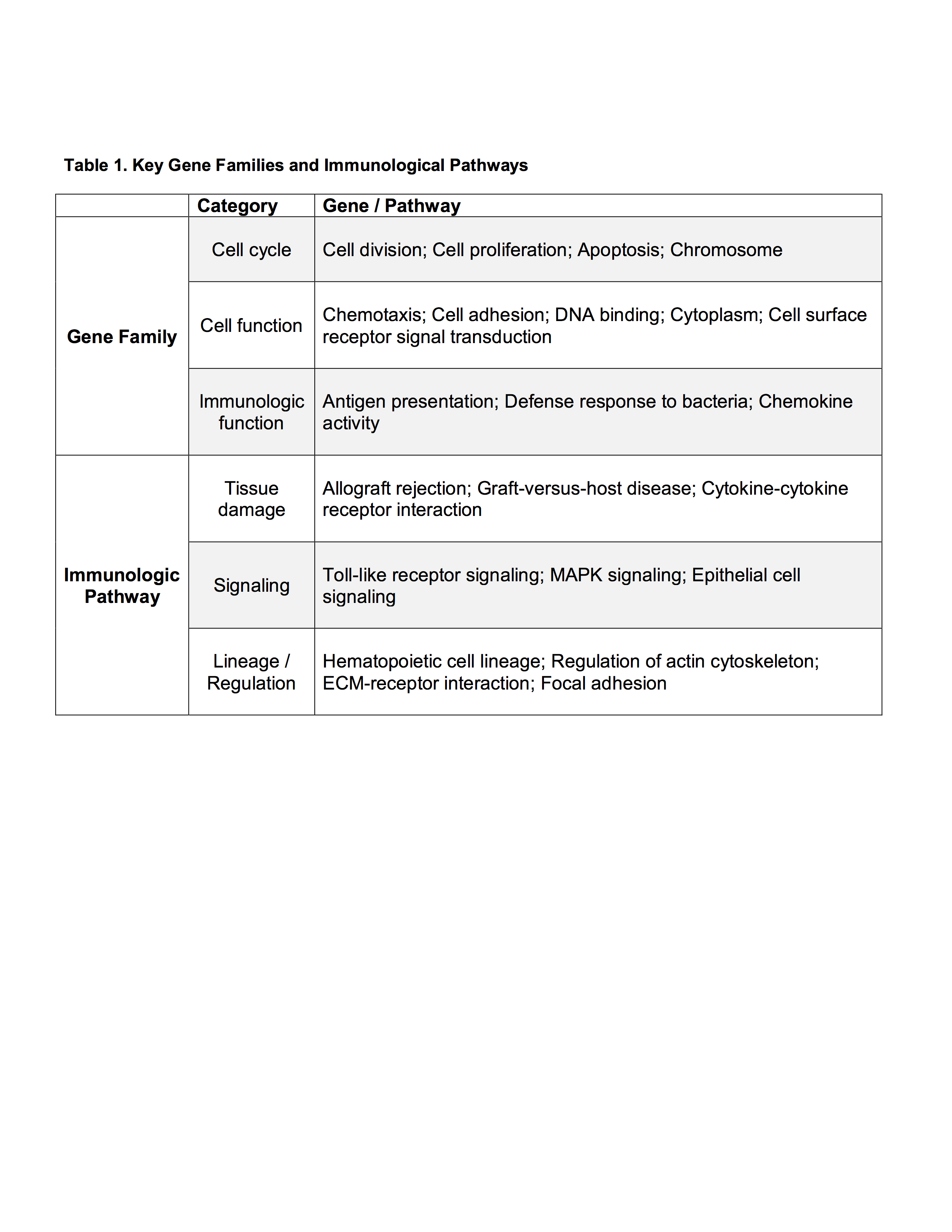
Results: When comparing discharge dispositions, SNF patients experienced greater rates of long-term organ failure (p<0.001). Analysis of genomic expression between discharge groups at 12 hours of admission demonstrated modest differences in lymphocytes and neutrophils, while significant differences were found in monocytes. Monocyte genomic differences persisted over 28 days. At 12 hours, 1187 monocyte genes significantly differed in expression. Over the 28 days, 1058 genes significantly differed. The most important gene families and immunological pathways represented are included in Table 1. The most represented ontological groups are those involved in wound healing and tissue regeneration, which may provoke SNF-associated organ failure.

Conclusion: Monocyte genomic expression differs significantly among severely injured trauma patients depending on discharge disposition, while only modest differences are seen in neutrophil and lymphocyte genomic expression. Monocyte genomic patterns can be detected within 12 hours of admission, persist over time, and are associated with an increased risk of organ failure. Although early genomic expression patterns can identify trauma patients with a poor clinical trajectory requiring SNF placement, more research is necessary to determine if early interventions can alter the clinical course, affect discharge disposition, and improve outcomes.

41.02 Paradoxical Suppression of a Subset of Circulating Mediators in the Most Severely Injured Patients

A. J. Lamparello1, R. A. Namas1, I. Billiar1, Y. Vodovotz1, T. R. Billiar1  1University Of Pittsburg,Pittsburgh, PA, USA

Introduction:
Trauma results in an increase in circulating inflammatory mediators referred to as a “cytokine storm.”  The magnitude of this increase correlates with complications and is thought to be dependent on the severity of injury.  However, a relatively limited number of inflammatory mediators have been characterized in human trauma.  Here, we measured the dynamic changes in 31 cytokines and chemokines in a large cohort of blunt trauma patients and analyzed the differences as a function of injury severity.

Methods:
We performed a retrospective study using a cohort of 472 blunt trauma survivors.  Plasma was sampled three times within the first 24 hours (including admission) and then daily from days 1 to 7 post-injury.  The samples were assayed using Luminex multiplex assay.  Three cohorts were analyzed as a function of injury severity.  Injury severity score (ISS) was calculated for each patient upon discharge and categorized as mild (ISS: 1-15, n=180), moderate (ISS: 15-24, n=170), or severe (ISS: ≥25, n=122).  Statistical difference (p < 0.05) between the groups was determined either by Student t test or χ2 test as appropriate.  Two-way analysis of variance was used to determine statistical significance of differing levels of inflammatory mediators.

Results:
Patients with severe injury had a significantly longer ICU length of stay (LOS), total LOS, days on mechanical ventilation, and rate of nosocomial infections compared to patients with mild or moderate injury based on ISS scores.  As expected, a number of inflammatory mediators exhibited elevations on admission and over time that positively correlated with injury severity (e.g., IL-6, IL-7, IL-17, sIL-2Rα, GM-CSF, IP-10/CXCL10, MIG/CXCL9, and MCP-1/CCL2).  In striking contrast, circulating levels of a subset of mediators, although significantly elevated in the mild and moderate injury groups (relative to healthy controls), were significantly suppressed in the most severely injured patients.  These mediators, which included IL-33, IL-9, IL-21, IL-22, IL-23, and IL-25, were lower during the first 24 hours post-admission and remained low throughout hospital stay.  Of note, several of these mediators have known tissue protective and reparative actions (e.g., IL-9, IL-22, and IL-33).

Conclusion:
Our novel findings identify a subset of mediators that are suppressed in the most severely injured patients.  These observations in injured humans provide potentially important insights into mechanisms associated with immune dysfunction following trauma.
 

41.01 The Uniquely-Human CHRFAM7A Gene Alters Ligand Binding to the α -7 Nicotinic Acetylcholine Receptor

T. Chan1, E. Williams1, B. Eliceiri1, A. Baird1, T. Costantini1  1University Of California – San Diego,Division Of Trauma, Surgical Critical Care, Burns And Acute Care Surgery/Department Of Surgery,San Diego, CA, USA

Introduction: The α-7 nicotinic acetylcholine receptor (α7nAChR) is required for the anti-inflammatory activity of the vagus nerve and is thought to be essential to resolve inflammation after severe injury.  CHRFAM7A is a uniquely-human gene that encodes a human-specific subunit of the α7nAChR and a putative negative inhibitor of ligand binding. We previously demonstrated that CHRFAM7A expression is highly variable in human leukocytes suggesting a source of human variability in vagal anti-inflammatory responsiveness. We therefore hypothesized that expression of CHRFAM7A would alter ligand binding to the α7nAChR and give insight into human variability in the anti-inflammatory response to injury and infection.

Methods:  Transgenic mice were engineered to ubiquitously express the uniquely-human gene CHRFAM7A under control of the EF-1α promoter. CHRFAM7A gene expression was confirmed by PCR, quantitative RT-PCR and immunoblotting with an antibody raised to a peptide sequence unique to CHRFAM7A. Muscle tissue and peritoneal macrophages were harvested from these transgenic mice and ligand binding to α7nAChR compared to sibling-matched wild-type C57 mice.  Immunostaining of the neuromuscular junction was performed using α-bungarotoxin (α-BTX), a ligand specific for the α7nAChR. Macrophage α-BTX binding was measured using flow cytometry and immunohistochemistry.

Results: Human CHRFAM7A gene and protein expression was measured in transgenic mice but was undetectable in cells from wild-type animals. α-BTX co-stained with neurofilament at the neuromuscular junction in wild-type mice, however, α-BTX staining was absent in muscle from CHRFAM7A transgenic mice, demonstrating a loss of α7nAChR function. Similarly, CHRFAM7A expression in macrophages decreased α-BTX binding.

Conclusion: CHRFAM7A prevents binding of α-BTX to the α7nAChR. The variability of CHRFAM7A expression seen in humans may, therefore, contribute to human variability in the α7nAChR-dependent anti-inflammatory response to injury and infection. By extension, human CHRFAM7A expression may be responsible for the limited clinical effectiveness of vagal therapeutics that rely on a functional α7nAChR.


23.10 Ablation of Cystathionine-Gamma-Lyase Promotes Colitis-Associated Carcinogenesis

K. Thanki1, M. Nicholls1, M. Maskey1, C. Phillips1, P. Johnson1, J. R. Zatarain1, K. Modis1, S. Qiu3, I. V. Pinchuk2, M. R. Hellmich1, C. Chao1  1University Of Texas Medical Branch,Surgery,Galveston, TX, USA 2University Of Texas Medical Branch,Internal Medicine,Galveston, TX, USA 3University Of Texas Medical Branch,Pathology,Galveston, TX, USA

Introduction: Hydrogen sulfide (H2S) is an important gasotransmitter that is anti-inflammatory at physiological levels. H2S is produced, in large part, by two enzymes in the reverse transsulfuration pathway: cystathionine-γ-lyase (CSE) and cystathionine-β-synthase (CBS). In mouse models of acute colitis, the activities of CSE and CBS are upregulated. Pharmacological inhibition or gene knockout (KO) of CSE worsens inflammation and delays tissue repair.  However the role of CSE in chronic inflammatory conditions, such as ulcerative colitis (UC) and UC-associated carcinogenesis (UCAC) is unknown.

Methods: C57BL/6 CSE knockout (CSE KO) mice and wild-type (WT) controls (N = 148) were given a single injection of the mutagen azoxymethane (AOM) and then randomized to receive either 1, 2 or 3 cycles of the colitis-inducer dextran sodium sulfate (DSS). Each cycle of colitis was induced by administration of 2% DSS in drinking water for a period of seven days followed by 14 days of regular drinking water. Tissues (colon, liver, and lymph nodes) were harvested at 21, 42, 63 and 80 days post AOM injection. Total tumor burden and inflammation were quantified by gross and histological examination using an established scoring system measuring inflammatory infiltrate, mucosal structure (crypt height, epithelial cell loss) and ulceration. CSE mRNA and protein levels from human normal mucosal and UC specimens were determined using qPCR and western blotting, respectively.

Results: Colonic mucosa from UC patients (n=10) exhibited a 2.9-fold decrease in CSE mRNA (qPCR, p<0.05) and protein expression when compared to normal mucosa (n=8). CSE KO mice demonstrate comparable inflammation to WT mice in response to repeated DSS treatment. KO mice demonstrate accelerated UCAC and disease progression as reflected by increased number and area of tumors (i.e., tumor burden) at 21 days (Fig 1A,B). At all other later time points, there were no significant differences in tumor burden. The AOM treatment alone did not cause carcinogenesis in either WT or CSE KO mice.

Conclusion: Our data shows that the colon mucosa from UC patients expresses significantly less CSE, an important producer of H2S. In a mouse model of UCAC, we show that absence of CSE does not protect colon mucosa from inflammation but does accelerate the time to colitis-associated tumor formation, and increases total tumor burden. Theses data suggest that CSE does not impact the inflammatory response but is important in normal mucosal restitution after injury, protecting the colon from UCAC. 


23.08 Priming with IL-7/15 to Generate Metabolically Fit CD8+ T Cells in the Tumor Microenvironment

S. Patel1, T. Hoki1, T. Yamauchi1, K. A. Collins1, C. A. Eppolito1, A. J. Francois1, J. V. Welch1, J. A. DiTursi1, K. Odunsi1,2,3, F. Ito1,4,5  1Roswell Park Cancer Institute,Center For Immunotherapy,Buffalo, NY, USA 2Roswell Park Cancer Institute,Department Of Gynecologic Oncology,Buffalo, NY, USA 3Roswell Park Cancer Institute,Department Of Immunology,Buffalo, NY, USA 4State University Of New York At Buffalo,Department Of Surgery, University At Buffalo Jacobs School Of Medicine And Biomedical Sciences,Buffalo, NY, USA 5Roswell Park Cancer Institute,Department Of Surgical Oncology,Buffalo, NY, USA

Introduction:
Current approaches to adoptive cell therapy (ACT) with antigen-specific T cells are limited by the difficulty of obtaining sufficient numbers of T cells against targeted antigens with effective in vivo characteristics. Whereas interleukin (IL)-2 has been widely used for generation of antitumor T cells in vitro clinically, dose-dependent effects of IL-2 on differentiation of T cells are associated with decreased proliferative and self-renewal capacity in vivo. IL-7 and IL-15 are also common γ  chain cytokines that play pivotal roles in homeostasis, proliferation, and maintenance of memory CD8+ T cells. Accumulating evidence largely from examining hematological malignancies indicates that the combined use of IL-7 and IL-15 (IL-7/15) can produce T cells that confer superior antitumor immunity in vivo. However, antitumor efficacy of IL-7/15-primed T cells in an orthotopic tumor model has not been rigorously evaluated.

Methods:
Pmel-1 T-cell receptor transgenic CD8+ T cells were activated with the cognate antigen gp100 expressed on B16 melanoma in IL-7/15 for 6 days. IL-2 was used for a control. Phenotype and function as well as metabolic profile of IL-2- and IL-7/15-primed T cells were evaluated. To determine in vivo antitumor efficacy, C57BL/6 mice bearing subcutaneous B16F10 melanoma were treated with adoptive transfer of IL-2- or IL-7/15-primed Pmel-1 T cells, followed by systemic administration of IL-2, and vaccination with gp100, anti-CD40 antibody, and toll-like receptor (TLR) agonist to augment antitumor efficacy of transferred T cells.

Results:
Cell expansion was significantly higher when T cells were activated in IL-7/15 at day 6 compared with ones in IL-2. IL-7/15-primed T cells consisted of a higher proportion of less-differentiated CD44+CD62L+ T cells, and secreted significantly more IL-2 against the target antigen compared to IL-2-primed T cells while both had comparable effector function such as specific lysis of targets and IFNγ  production in vitro. Furthermore, IL-7/15-primed T cells had higher mitochondrial spare respiratory capacity than IL-2-primed T cells, suggesting that IL-7/15-primed T cells have capacity to produce more ATP in case of a sudden increase in energy demand. In line with this, adoptively-transferred IL-7/15-primed T cells expressed significantly higher Ki67 than IL-2-primed T cells in the tumor microenvironment (TME). Significantly delayed tumor growth and improved survival were observed in mice treated with IL-7/15-primed T cells compared to IL-2-primed T cells.

Conclusion:
Taken together, our studies suggest that IL-7/15 modulates the metabolic programming of T cells to promote more robust and efficient CD8+ T cells that can proliferate in the TME. In particular, IL-7/15-primed T cells have higher self-renewal and spare respiratory capacity with potent effector function that correspond to significantly improved survival in an orthotopic tumor model.
 

23.09 Interrogation of Immune Checkpoints in Pancreatic Cancer Organoids Reveal Novel Cytotoxic Therapies

K. Hirai1, M. Lin1, S. Hoque1, M. Choi1, Y. Zhang1, G. V. Georgakis1, A. R. Sasson1, M. Gao1, J. Kim1  1Stony Brook University Medical Center,Stony Brook, NY, USA

Introduction:  We have detected the immune checkpoints programmed cell death ligand 1 (PD-L1) and programmed cell death 1 (PD-1) on pancreatic cancer cells and tissues, even in the absence of immune cells.  In this study we sought to determine the functional role of PD-L1/PD-1 in pancreatic cancer cells and tissues and to determine the cytotoxic efficacy when targeting the PD-L1/PD-1 axis with current FDA approved drugs. 

Methods:  To evaluate the function of immune checkpoints in pancreatic cancer, we used the established human pancreatic cancer cell lines AsPC-1, MIAPaCa-2, and PANC-1. With IRB approval and written informed consent, we established pancreatic adenocarcinoma patient derived organoids (PDO) from operative tissues. Initially, we exposed all pancreatic cancer cells to exogenous PD-L1 or PD-1 (1μg/mL) and examined the activation of the mitogen activated protein kinase pathway. To assess the specificity of the PD-L1-PD-1 interaction, we stably transfected PD-1 short hairpin RNA into PANC-1 cells and also used pembrolizumab, a clinically active anti-PD-1 monoclonal antibody. We utilized pancreatic cancer cells and PDOs to assess the cytotoxic efficacy of current FDA approved inhibitors of the PD-L1/PD-1 axis.

Results: Exposure of exogenously administered PD-L1 or PD-1 increased levels of ERK phosphorylation in all 3 pancreatic cancer cell lines. Using stably transfected PD-1 shRNA in PANC-1 cells, we observed that ERK phosphorylation was attenuated when cells were exposed to PD-L1. Additionally, pretreatment of the 3 pancreatic cancer cell lines to pembrolizumab (anti-PD-1) inhibited ERK phosphorylation. Altogether, these studies indicate that the specific PD-L1-PD-1 interaction results in activation of MAPK signaling. Using pancreatic cancer cells and successfully created PDOs, we sought to determine treatment efficacy using FDA approved immune checkpoint inhibitors [nivolumab, NIV (anti-PD-1); pembrolizumab, PEM (anti-PD-1); and atezolizumab, ATE (anti-PD-L1)]. We utilized daratumumab (DARA) as a negative monoclonal antibody control. In PDOs, we also assessed trametinib (TRAM), a small molecule MAPK inhibitor. We completed a cytotoxicity assay (CellTiter-Glo) in triplicate and observed considerable PDO cytotoxicity with NIV (34.1%), PEM (52.0%), and ATE (39.4%). However, greatest PDO death was observed when anti-PD1 drug (PEM) was combined with TRAM (83.2%).

Conclusion: The immune checkpoints PD-L1 and PD-1 are expressed on pancreatic cancer cells and activate oncogenic signaling pathways. Drug therapies combining immune checkpoint inhibitors with MAPK antagonists have tremendous potential for novel therapeutic advances in pancreatic cancer.


23.07 Immunologic Changes in Regionally Treated Melanoma

J. A. Perone1, T. Tamesa1,2, M. Tsutsui2, R. E. Alvarado1, S. Moore1, P. Dolber1, I. Pinchuk1, K. Olino1, D. Tyler1  1University Of Texas Medical Branch,Galveston, TX, USA 2Durham Veterans Affairs Medical Center,Durham, NC, USA

Introduction:
Advanced melanoma remains a highly lethal disease with poor long-term survival in the majority of patients. Although systemic immunotherapy has improved outcomes, there is much room for improvement.  One strategy to help augment systemic immunotherapy has been regional therapeutics.  Regional therapies offer variable immunogenic effects on the microenvironment, usually with minimal toxicity, that could synergize with systemic treatments to improve antitumor responses. To better define the spectrum of immune effects produced by regional therapy, we picked four different chemotherapeutic agents to explore the range of immune mediated events that occur in the context of variable mechanisms of cell death.

Methods:
B16 F10.9-OVA melanoma cells were injected intradermally in the left leg of C57BL/6 female mice to create a solitary leg tumor.  At tumor volumes of ~50mm3 animals were randomized and isolated limb infusion (ILI) was performed using the maximally tolerated dose of saline, melphalan, doxorubicin, temozolomide, or oxaliplatin.  Following survival studies, immunologic evaluation was performed by harvesting Inguinal (ILN) and popliteal lymph nodes (PLN) on days 3 and 10 post ILI and immunologic evaluation was performed utilizing RT-PCR and flow cytometry, staining for CD4, CD8, FoxP3, Tbet, PD-1, and PD-L1. 

Results:
While regional chemotherapy was exceptionally effective at killing tumor, we were surprised to see the induction of immunosuppressive profiles in the microenvironment at days 3 and 10.  Increased expression of PD-L1 on CD4+ and CD8+ Tcells was observed not only in inguinal nodes but also in popliteal nodes and tumors on day 10 following treatment with either melphalan or doxorubicin (Figure 1).  Immunosuppression associated with temozolomide and oxaliplatin was primarily manifested as an increase in tregs(CD4+FoxP3+) in both the ILN and PLN between days 3 and 10, as well as an increase in exhausted Tcells (CD8+PD1+) and suppressor Tcells (CD8+FoxP3+) (Figure 2).  PCR analysis of tumors treated with doxorubicin also showed similar increase in tregs compared to naïve animals.

Conclusion:
Regional chemotherapy used in ILI creates an immunosuppressive environment around treated tumors that varies depending on the chemotherapeutic agent utilized.  Characterization of the immunosuppressive profile induced by regional treatment allows development of novel strategies, such as combining systemic anti-PD1 treatment with melphalan-based ILI, to augment both regional and systemic anti tumor response. 
 

23.06 Blocking NETs Reduces Macrophage Infiltration and Progression of Steatohepatitis to Liver Cancer

D. J. Van Der Windt1, V. Sud1, P. Varley1, J. Goswami1, H. Zhang1, H. Yazdani1, P. Loughran2, M. I. Minervini4, H. Huang1, R. L. Simmons1, A. Tsung1  1University Of Pittsburgh,Surgical Oncology,Pittsburgh, PENNSYLVANIA, USA 2University Of Pittsburgh,Center For Biologic Imaging,Pittsburgh, PENNSYLVANIA, USA 3University Of Pittsburgh,Department Of Medicine,Pittsburgh, PENNSYLVANIA, USA 4University Of Pittsburgh,Department Of Pathology,Pittsburgh, PENNSYLVANIA, USA

Introduction:
Nonalcoholic steatohepatitis (NASH) is a rapidly increasing precursor of hepatocellular carcinoma (HCC) whether or not cirrhosis is evident, underlining the importance of inflammation as a hallmark of cancer development. Neutrophils are increasingly recognized as regulators of the protumorigenic inflammatory environment. Neutrophils can expel their chromatin structures along with pro-inflammatory proteins into the extracelluar environment leading to the formation of neutrophil extracellular traps (NETs). We have previously reported that blocking NETs can reduce the development of HCC in an experimental model of NASH. Here we sought to further characterize the role of NETs in the inflammatory environment in NASH that can give rise to HCC.

Methods:
NASH mice were created by exposing C57Bl/6 mice to streptozotocin (200ug I.P. <5d from birth) and high fat diet. The development of NASH was evaluated over time by serum and tissue levels of inflammatory cytokines, flow cytometry of liver non-parenchymal cells, and histology. NET blockade was achieved with injections of DNase1 (100U I.P. 3x/wk), or by using peptidylarginine deiminase 4 knockout (PAD4 KO) mice (that are genetically unable to form NETs) 

Results:
Neutrophil infiltration was seen in NASH mice as early as 5 weeks by flow cytometry (6.6±0.8% of CD45+ non-parenchymal cells in NASH vs. 3.6±0.5% in control mice, p<0.05). NASH mouse livers exhibited NET formation by immunofluorescence and western blot for citrullinated histone, a specific NET marker. In vitro, the common free fatty acids, palmitic and linoleic acid (but not oleic acid), were able to stimulate neutrophils to form NETs, suggesting a mechanism for NASH to stimulate NETs. In NASH mice in vivo, neutrophil infiltration was followed by an influx of infiltrating macrophages at 8 weeks with increased levels of IL-6 and TNFa. NET blockade with DNase1 reduced macrophage infiltration from 6.2±1.3% to 3.2±0.7% (p<0.05), and reduced inflammation in the liver (IL-6 was reduced from 22.2±6.7 to 5.8±1.9 pg/mL, TNFa expression was reduces 4-fold, all p<0.05), resulting in a decrease in NASH activity scores (p<0.05). In the long term, the alterations in the liver inflammatory environment resulted in significantly fewer tumors in DNase1 treated NASH mice and PAD4KO NASH mice, compared to wild type NASH mice (1.3±1.1 and 2.2±1.8, vs. 8.7±3.4, p<0.05). 

Conclusion:
In steatohepatitis, neutrophil extracellular traps (NETs) attract macrophages, which are a known effector cell in NASH. NET blockade reduces macrophage infiltration and significantly alters the chronic inflammation, thereby reduces the risks of developing HCC.
 

23.05 CRISPR/Cas9 Genome Editing of Tumor-specific CD8+ T cell-derived Induced Pluripotent Stem Cells

T. Yamauchi1, H. Saito2,3, T. Hoki1, F. Ito1,2,4,5  2University Of Michigan,Surgery,Ann Arbor, MI, USA 3Kanazawa Medical University,Biochemistry,Kanazawa, ISHIKAWA, Japan 4State University Of New York At Buffalo,Surgery,Buffalo, NY, USA 5Roswell Park Cancer Institute,Surgical Oncology,Buffalo, NY, USA 1Roswell Park Cancer Institute,Center For Immunotherapy,Buffalo, NY, USA

Introduction: Current approaches to adoptive T cell therapy are limited by the difficulty of obtaining sufficient numbers of T cells against targeted antigens with effective in vivo characteristics.  This limitation can be overcome by using induced pluripotent stem cells (iPSCs) that could provide an unlimited source of autologous T cells. We and others have shown that iPSC-derived regenerated T cells have potent antitumor efficacy in vitro and in vivo. Recently, the type II bacterial CRISPR (clustered regularly interspaced short palindromic repeats)/Cas (CRISPR-associated) system was developed as an efficient and versatile technology for genome editing in eukaryotic cells and whole organisms. The potential of iPSCs can be further enhanced by genome engineering and then used to study individual gene function, track cells or endogenous proteins with a knock-in reporter, and correct genetic defects for gene therapy. 

Methods: We reprogrammed T-cell receptor (TCR) transgenic CD8+ T cells into pluripotency, and established a syngeneic mouse model for evaluating in vitro and in vivo antitumor reactivity of regenerated T cells from iPSCs bearing a rearranged TCR of known antigen specificity. Plasmids encoding rtTA and tet-O-Cas9-2A-mCherry were obtained and co-transduced using the lentiviral system. The Cas9 expression was analyzed after the doxycycline treatment, and clonally-derived Cas9-iPSCs were obtained.

Results: Pluripotency of TCR transgenic T cell-derived iPSCs (TiPSCs) were confirmed with immunostaining of embryonic stem cell (ESC) markers, RT-PCR (reverse transcription-polymerase chain reaction) analysis of pluripotency-associated transcription factors, and whole genome expression profiling by microarray analysis that demonstrated a high degree of similarity in their gene expression patterns and close correlation values with ESCs, but distinct from parental CD8+ T cells. Cytogenetic analysis revealed derived TiPSCs maintained normal karyotype. The TiPSCs differentiated to embryoid bodies in vitro, and upregulation of marker genes for all three germ layers was detected by immunostaining. Their differentiation capacity was further confirmed by teratoma formation in immune-deficient mice in vivo. Moreover, we confirmed that the TiPSCs retained the same rearranged configuration of TCR chain genes as the original TCR transgenic T cells. TiPSCs were, then, subjected to the lentivirus-mediated transduction of tetracycline-inducible Cas9 vectors. The transduction efficacy was confirmed by the mCherry fluorescence and the RT-PCR against the Cas9 sequence.

Conclusion: Our results indicate successful reprogramming of antigen-specific T cells and lentivirus-mediated transduction of tetracycline-inducible Cas9 vectors into TiPSCs, which will allow us to generate an unlimited number of phenotypically defined, functional and expandable genome-edited autologous antigen-specific T cells.

23.04 CD40 signaling is required for expansion of terminally-differentiated CX3CR1+ CD8+ T cells

F. Ito1,2,3, T. Hoki2, C. A. Eppolito2, A. J. Francois2, K. Odunsi2,4,5, T. Yamauchi2  3State University Of New York At Buffalo,Surgery,Buffalo, NY, USA 1Roswell Park Cancer Institute,Surgical Oncology,Buffalo, NY, USA 2Roswell Park Cancer Institute,Center For Immunotherapy,Buffalo, NY, USA 4Roswell Park Cancer Institute,Gynecologic Oncology,Buffalo, NY, USA 5Roswell Park Cancer Institute,Immunology,Buffalo, NY, USA

Introduction: Successful immunotherapeutic treatment of chronic infectious diseases and cancer requires the generation of a strong cellular immune response. Combined CD40 antibody and toll-like receptor (TLR) (CD40/TLR) stimulation has been found to mediate potent cellular immunity in the context of tumor immunology and cancer immunotherapy. However, the mechanisms of enhanced antitumor efficacy by CD40/TLR stimulation remain elusive.

Methods: To investigate phenotype and function of CD40/TLR vaccine-stimulated antigen-specific T cells, we used MC38 colon adenocarcinoma and B16 melanoma models. To evaluate endogenous T cell response, MC38 tumor-bearing C57BL/6 mice were treated with mutated-neoantigen peptide, agonistic CD40 antibody, and poly I:C (TLR3). To assess phenotype and function of adoptively-transferred T cells, we used pmel-1 T cell receptor (TCR) transgenic CD8+ T cells specific for the gp100 melanocyte differentiation antigen expressed on B16 melanoma. In vitro–activated Pmel-1 CD8+ T cells were adoptively transferred into C57BL/6 mice bearing subcutaneous B16 melanomas. Systemic administration of IL-2 and vaccination with the gp100, anti-CD40 antibody, and Imiquimod (TLR7) were used to enhance antitumor immunity of transferred T cells. Tissues including blood, spleen, and tumors were harvested for further analysis. In both tumor models, control mice were treated with no vaccination, CD40 antibody alone or TLR agonist alone.

Results: In both tumor models, mice treated with combined CD40/TLR vaccination had significantly decreased tumor growth and improved survival compared to no vaccination, CD40 antibody alone or TLR agonist alone. Interestingly, vaccination with the cognate antigen and CD40/TLR not only expanded antigen-specific CD8 T cells in both tumor models, but also facilitated them to express the chemokine receptor, CX3CR1. CX3CR1+ CD8+ T cells were found to express higher levels of killer-cell lectin like receptor G1 (KLRG1), TNF-related apoptosis-inducing ligand (TRAIL), perforin, and granzyme, suggesting terminally-differentiated subset. The generation of CX3CR1+ CD8+ T cells was greatly facilitated by CD40 antibody while TLR agonist increases the expansion of total number of antigen-specific CD8 T cells. Importantly, total number and frequency of CX3CR1+ CD8+ T cells were significantly decreased in tumor-bearing CD40 knockout (KO) mice, indicating that host expression of CD40 is required for generation and expansion of CX3CR1+ CD8+ T cells.

Conclusion: Effective vaccination with the cognate antigen and CD40/TLR accompanies generation of tumor-specific terminally-differentiated CX3CR1+ CD8+ T cells dependent on CD40 signaling.

23.03 Depletion of Gut Microbiome Lowers Cancer Burden in Murine Models by Modulating the Immune System

V. Sethi1, B. Giri1, B. Garg1, M. Tarique1, S. Lavania1, L. Hellmund1, Z. Malchiodi1, S. Kurtom1, A. Farrantella1, S. Banerjee1, S. Ramakrishnan1, S. Roy1, A. Saluja1, V. Dudeja1  1University Of Miami,Department Of Surgery,Miami, FL, USA

Introduction: Microbiome of the gut forms an important ‘hidden organ’ of our body, changing with diet, disease state, use of antibiotics, and even age. While literature abounds with associational studies of changed gut microbiome with disease states like inflammatory bowel disease, diabetes, colitis etc., precious little is known on how, if at all, the gut microbiome modulates cancer and metastases. The aim of this current study was to analyze this obscure cancer-gut microbiome relationship. 

Methods: The gut microbiome of C57BL/6 mice was depleted by daily oral administration of broad-spectrum antibiotics having minimal systemic absorption. Various cancers were modelled on antibiotic-given mice as well as control mice and tumor burden was compared between the two groups. These models included subcutaneous implantation of pancreatic cancer cells derived from KPC (Kras LSL.G12D/+; p53 R172H/+ ;Pdx::Cre) mice and melanoma cells derived from Braf-Pten (Tyr::CreER; Braf V600E/+;Ptenlox5/lox5  ) mice . Additionally hepatic metastases were induced by intrasplenic injections of KPC pancreatic cancer cells, B16-F10 melanoma cancer cells and MC38 colon cancer cells. Tumors were immunophenotyped through flow cytometry and immunostained for various antigens. Various ex-vivo cytotoxicity experiments were performed with cancer cells and splenocytes. To confirm the role of  adaptive immunity, similar experiments were performed in immunodeficient Rag1 knockout mice having a Rag1tm1Mom mutation.

Results:  Depletion of the gut microbiome significantly decreased primary cancer and metastases burden in all the studied murine models. Tumors in antibiotics-gavaged mice showed increased cleaved caspase 3 staining. Ex-vivo, splenocytes from antibiotics-gavaged mice caused a much higher cytotoxicity of cancer cells than control splenocytes. The role of immune system was confirmed when antibiotic administration in immunodeficient Rag1 KO mice failed to show a reduced cancer burden compared to control mice. 

Conclusion: Gut microbiome depletion causes reduced cancer burden by modulating the immune system. 


23.02 Defining CD8+ T-cell Subsets that are Rescued by PD-1/PD-L1 Blockade in the Tumor Microenvironment

T. Yamauchi1, T. Hoki1, C. A. Eppolito1, A. Francois1, K. Odunsi1,2,3, F. Ito1,4,5  1Roswell Park Cancer Institute,Center For Immunotherapy,Buffalo, NY, USA 2Roswell Park Cancer Institute,Gynecologic Oncology,Buffalo, NY, USA 3Roswell Park Cancer Institute,Immunology,Buffalo, NY, USA 4State University Of New York At Buffalo,Surgery,Buffalo, NY, USA 5Roswell Park Cancer Institute,Surgical Oncology,Buffalo, NY, USA

Introduction: Cancer immunotherapies that target the T-cell immune checkpoints, such as programmed cell death-1 (PD-1) and its ligand (PD-L1) have shown unprecedented success for the treatment of a variety of malignancies including melanoma. Although a significant number of cancer patients benefit from immune checkpoint inhibitors (CPIs), many fail to have clinical responses.   A better understanding of the mechanisms that regulate CD8+ T-cell responses in the tumor microenvironment is required to improve immunotherapies that restore function in exhausted CD8+ T cells.  Although heterogeneity of effector CD8+ T cells in the tumor microenvironment (TME) has been recognized, their functions and roles are ill-defined.

Methods: We have evaluated phenotypical and functional heterogeneity of tumor-infiltrating lymphocytes (TILs) after adoptive transfer of ex vivo primed pmel-1 T cell receptor (TCR) transgenic CD8+ T cells specific for the gp100 melanocyte differentiation antigen expressed on B16 melanoma. In vitro–activated Pmel-1 CD8+ T cells were adoptively transferred into C57BL/6 mice bearing subcutaneous B16 melanomas that had been established for 11-14 days. Systemic administration of IL-2 and vaccination with anti-CD40 antibody and toll-like receptor (TLR) agonist were used to enhance antitumor immunity of transferred T cells. Tumor and spleen were harvested for functional analysis of adoptively transferred T cells.

Results: We found that the chemokine receptor, CX3CR1 identified three distinct effector CD8+ T-cell subsets, CX3CR1 negative (-), intermediate (int), and high (hi) in blood, spleen and the TME.  A CX3CR1hi subset contained terminally-differentiated CD8+ T cells that expressed higher levels of killer-cell lectin like receptor G1 (KLRG1), TNF-related apoptosis-inducing ligand (TRAIL), perforin, and granzyme. Significantly more CX3CR1int CD8+ T cells expressed CD25 compared to other subsets, suggesting this is the subset that is rapidly proliferate and preferentially generate terminally-differentiated T cells. Unexpectedly, despite their terminally differentiated status, a CX3CR1hi CD8+ T-cell subset expressed significantly lower levels of co-inhibitory receptors, PD-1, LAG3, and TIGIT compared to CX3CR1 and CX3CR1int CD8+ T-cell subsets in the TME. In line with this, proliferation and cytokine production of CX3CR1 and CX3CR1int CD8+ T-cell subsets were significantly decreased in the TME compared to CX3CR1hi CD8+ T-cell subset. Importantly, PD-1/PD-L1 blockade significantly improved effector functions of CX3CR1 and CX3CR1int CD8+ T-cell subsets in the TME.

Conclusion: The chemokine receptor, CX3CR1 defines distinct effector CD8+ T-cell subsets in periphery and in the TME. Tumor-infiltrating CX3CR1 and CX3CR1int CD8+ T-cell subsets express high levels of co-inhibitory receptors, PD-1, LAG3, and TIGIT, and their effector functions are improved by PD-1/PD-L1 blockade. 

23.01 SMAD4 Loss in Patient-Derived Colorectal Cancer Tumoroids Confirms Chemoresistance.

B. Szeglin1, C. Wu9,11, I. Wasserman2, S. Uppada3, X. Chen6, K. Ganesh8, A. Elghouayel7,11, J. Shia5, A. Barlas10, P. B. Paty11, M. R. Weiser11, J. G. Guillem11, G. M. Nash11, K. Manova-Todorova10, P. Dhawan3, R. Beauchamp4, N. E. Kemeny8, J. Garcia-Aguilar11, C. L. Sawyers9, J. Smith9,11  1Albert Einstein College Of Medicine,Bronx, NY, USA 2Icahn School Of Medicine At Mount Sinai,New York, NY, USA 3University Of Nebraska Medical Center,Department Of Biochemistry And Molecular Biology,Omaha, NE, USA 4Vanderbilt University Medical Center,Section Of Surgical Sciences,Nashville, TN, USA 5Memorial Sloan-Kettering Cancer Center,Department Of Pathology,New York, NY, USA 6University Of Miami Miller School Of Medicine,Department Of Bioinformatics And Biostatistics,Miami, FL, USA 7College Of William And Mary,Williamsburg, VA, USA 8Memorial Sloan-Kettering Cancer Center,Department Of Medical Oncology,New York, NY, USA 9Memorial Sloan-Kettering Cancer Center,Human Oncology And Pathogenesis Program,New York, NY, USA 10Memorial Sloan-Kettering Cancer Center,Department Of Molecular Cytology,New York, NY, USA 11Memorial Sloan-Kettering Cancer Center,Colorectal Service, Department Of Surgery,New York, NY, USA

Introduction:
Loss of SMAD4, the central node of the TGF-β  superfamily, occurs in 10-20% of colorectal cancer (CRC) cases. SMAD4 loss in the context of activated Wnt signaling may play a role in disease progression and resistance to standard 5-fluorouracil (5-FU) based chemotherapy, but the underlying mechanisms are poorly understood. Development of relevant CRC models to better study SMAD4 biology and associated chemoresistance is needed.

Methods:
Fresh CRC specimens were obtained at time of resection. Tumors were dissociated to individual cells and seeded within a Matrigel matrix in our 3D tumoroid cell culture model. SMAD4 mutant versus SMAD4-wildtype (wt) tumoroids were injected subcutaneously into immunocompromised mice. The mice were treated with systemic 5-FU and tumors weighed at necropsy. In addition, CRISPR/Cas9 was used to knockdown (kd) SMAD4 in patient-derived tumoroids ex vivo.  SMAD4 expression was restored in SMAD4 mutant SW480 CRC cells. The SMAD4-kd tumoroids, SMAD4-wt tumoroids, and CRC cells with restored SMAD4 expression were treated with 5-FU or FOLFIRI (5-FU, leucovorin, and irinotecan) to determine dose-response differences. In a parallel, exploratory analysis, microarray expression data from 250 CRC patients was used to generate a SMAD4 signature (FDR < 10-7). The Illumina BaseSpace Correlation Engine was used to correlate this signature with compounds that could be used in synergy with 5-FU based chemotherapy in the context of SMAD4 loss.

Results:
Engrafted SMAD4-deficient tumors did not respond to 5-FU treatment, while SMAD4-retained tumors demonstrated decreased tumor weight compared to vehicle (p < 0.02). SMAD4-kd tumoroids treated with either 5-FU or FOLFIRI ex vivo were significantly more resistant to treatment than SMAD4-wt tumoroids (p < 0.01). Conversely, restoration of SMAD4 expression in CRC cells mutant for SMAD4 was associated with significant response to 5-FU based therapy (p < 0.01).  Finally, the SMAD4 signature implicated 3-3-diindolylmethane, a putative Wnt pathway inhibitor, as a lead candidate for use in the context of SMAD4 deficiency and 5-FU based chemotherapy.

Conclusion:
We demonstrate that loss of SMAD4 is associated with chemoresistance to 5-FU and FOLFIRI treatment in in vivo and ex vivo CRC tumoroid models, thereby establishing relevant biological systems to study patient-specific resistance mechanisms. Furthermore, in silico analysis of a SMAD4 gene expression signature reveals 3-3-diindolylmethane as a possible therapeutic compound to target activated Wnt signaling in the context of SMAD4 loss in CRC patients undergoing 5-FU based chemotherapy. 
 

2.20 Preclinical Evaluation of Novel Retinoic Acid Derivatives in Neuroblastoma

A. J. Lazenby1, A. P. Williams1, L. L. Stafman1, J. Aye1, V. R. Atigadda2, J. Stewart1, D. D. Muccio4, C. Grubbs3, E. A. Beierle1  2University Of Alabama at Birmingham,Dermatology,Birmingham, Alabama, USA 3University Of Alabama at Birmingham,Surgery,Birmingham, Alabama, USA 4University Of Alabama at Birmingham,Chemistry,Birmingham, Alabama, USA 5University Of Alabama at Birmingham,Pharmacology And Toxicology,Birmingham, Alabama, USA 6University Of Alabama at Birmingham,Pediatrics,Birmingham, Alabama, USA 1University Of Alabama at Birmingham,Pediatric Surgery,Birmingham, Alabama, USA

Introduction:  Neuroblastoma (NB), a tumor derived from neural crest cells, is the most common extracranial solid tumor in children. 13-cis-retinoic acid (RA) is a differentiating agent currently utilized in therapy for high risk NB, but its use is limited by toxicities related to cholesterol and lipid metabolism. A synthetic rexinoid, 9-cis-UAB30 (UAB30), has been developed which has a favorable toxicity profile, demonstrating no significant toxicities in animal or human studies. Our lab has shown that UAB30 decreased NB cell proliferation and tumor growth in murine NB models. While UAB30 is promising, a reduction in drug dosage would be ideal to decrease the risk of potential side effects. Therefore, the aim of this project was to initiate pre-clinical evaluation of 9 new, synthetic rexinoid compounds (UAB111, UAB113, UAB114, UAB115, UAB116, UAB125, UAB126, 5-Me-UAB30 and 7-Me-UAB30) designed to have greater potency than that of UAB30 or RA. We hypothesized that these compounds would affect NB survival and differentiation in a manner comparable to RA and UAB30.

Methods:  Four NB cell lines were utilized: 2 MYCN non-amplified (SK-N-AS, SH-EP) and 2 MYCN amplified [SK-N-BE(2), WAC2]. The effect of the rexinoids on cell viability and differentiation were evaluated using alamarBlue® assay and assessment of neurite outgrowth, respectively. Cells were treated for 72 hours at increasing concentrations ranging from 0 to 100 µM. Data were compared using Student’s t-test or ANOVA as appropriate and reported as mean ± SEM with p≤0.05 considered statistically significant.

Results: The 9 new rexinoids were tested against compounds previously studied, RA and UAB30, in all four NB cell lines. The SK-N-AS cell line was the most sensitive to the novel compounds, showing significantly decreased viability after treatment with 5 of them (UAB111, UAB113, UAB114, UAB116, 7-Me-UAB30) compared to RA or UAB30 (Figure, upper panel).  The remaining cell lines, SK-N-BE(2), SH-EP and WAC2 demonstrated results similar to each other and had the most significant changes in viability with UAB116 and 7-Me-UAB30 (Figure, lower panel), so these 2 rexinoids were utilized for further studies. Neurite outgrowths are a measure of NB cell differentiation. Treatment with UAB116 or 7-Me-UAB30, resulted in a statistically significant increase in neurite outgrowths in both the SH-EP and SK-N-AS cell lines, indicating differentiation.

Conclusion: Treatment with these synthetic rexinoids, particularly UAB116 and 7-Me-UAB30, decreased NB cell viability significantly more than RA or UAB30. In addition, they led to NB cell differentiation. These data indicate that UAB116 and 7-Me-UAB30 should be further evaluated as potential novel therapeutics for NB.

2.19 The Role of Intestinal TLR4 in the Development of Small Bowel Resection Associated Metabolic Syndrome

C. M. Courtney1,2, L. K. Barron1,2, E. J. Onufer1,2, J. Guo1, B. W. Warner1,2  1Washington University,Pediatric Surgery,St. Louis, MO, USA 2St Louis Children’s Hospital,Pediatric Surgery,St Louis, MO, USA

Introduction:  Toll-like receptor 4 (TLR4) signaling plays a crucial role in inflammatory cytokine responses and the metabolic consequences of massive small bowel resection (SBR). Our laboratory has pioneered a murine model that demonstrates altered body composition, impaired glucose tolerance, and hepatic steatosis after 50% SBR. Interestingly global TLR-4 knockout mice do not develop this metabolic phenotype after SBR. While the role of hepatic TLR4 has been studied in unoperated mice with similar phenotypes, the role of intestinal epithelial TLR4 is presently unknown. Our aim was to determine if a direct relationship exists between intestinal TLR4 expression and the novel resection associated metabolic phenotype.

Methods:  Wild type (WT) and intestinal TLR4 knock out (iTLR4-KO) mice underwent 50% proximal small bowel resection and were maintained on standard liquid diet for 10 weeks. Weights were obtained weekly. At 10 weeks, mice were fasted overnight and glucose tolerance testing (GTT) was performed. Body composition was analyzed via echo magnetic resonance. At the time of sacrifice, liver was fixed in formalin, embedded paraffin and stained with hematoxylin and eosin. Slides were analyzed at 40x magnification (5 random fields/animal) using NIS Elements V4.3 software. Lipid area was calculated as measured lipid/ total parenchymal area. Body weights and glucose tolerance were compared using 2-way repeated measures ANOVA.  Fasting blood glucose, body composition, and hepatic steatosis were compared using student’s t-test. 

Results: When compared to wild type (WT) mice, iTLR4-KO mice demonstrated similar weight loss and recovery after SBR. Body composition between the groups was also similar at 10 weeks after operation with WT mice demonstrating 15% ± 0.02 fat mass and 84% ± 0.02 lean mass and iTLR4-KO mice with 17% ± 0.002 fat mass and 82% ± 0.002 lean mass (n=4 WT, n=8 iTLR4-KO). When comparing fasting blood glucose (FBG), the groups are not significantly different with WT FBG measuring 121.8 ± 9.077 (n=4) and iTLR4-KO FBG measuring 108.1 ± 5.894 (n=7). After 2mg/g intraperitoneal glucose load, iTLR4-KO glucose tolerance was not significantly different from WT mice (Figure 1A). Additionally, hepatic steatosis was not significantly different between the groups at 10 weeks post operatively (Figure 1B).

Conclusion: Specifically knocking out intestinal TLR 4 does not appear to alter the metabolic phenotype follow SBR. Future studies isolating other locations of TLR4 signaling are needed to help gain an understanding of the link between massive loss of small bowel and the resulting metabolic syndrome. 


2.18 Is Gastroschisis A Lymphatic Disease? Study In A Fetal Rabbit Model

F. Scorletti1, M. Oria1, F. Y. Lim1, J. L. Peiro1  1Cincinnati Children’s Hospital Medical Center,The Center For Fetal, Cellular And Molecular Therapy,Cincinnati, OH, USA

Introduction: Gastroschisis (GS) patients have high morbidity secondary to intestinal dysmotility and malabsorption that require parenteral nutrition for a long period.  Main alterations in the extruded bowel include edema and inflammation caused by amniotic fluid contact. Sufficient lymphatic clearance plays a crucial role in clearing the inflammation process, while lymphatic failure is believed to intensify the intestinal edema. Our aim was to investigate the intestinal lymphatic system alteration in a rabbit GS experimental model.

Methods: IACUC#2013-0294. Gastroschisis was surgically created on fetal New-Zealand rabbits at E24 and harvested on day E30 (term=31days). The siblings were collected as controls (n=10 per group). Body and intestinal weights (BW, IW), their ratio (IBR) and dry test were performed. RNA was isolated and pellets were partially re-extracted, precipitated, DNAseI-digested and cleaned. A 1-µg cDNA sample was used to set up RT-qPCR using primers for VEGF-C, VEGFR3 and GAPDH. Histology, immunofluorescence (IF) and western blotting (WB) were assessed to VEGF-C (Vascular Endothelial Growth Factor), VEGFR-3 (receptors), SMA (Smooth Muscle Actin) in GS eviscerated bowel and controls.

Results: IW, IBR and water (in dry test) were significantly increased (p<0.005) in the exposed GS intestine [1.391 (±0.208) versus 1.029 (±0.156); 0.041 (±0.004) versus 0.036 (±0.003); 96.08 (±1.13) versus 80.36 (±6.26), respectively]. VEGF-C and VEGFR3 gene expression were down-regulated in exposed GS especially at early stages respect time matched controls (p<0.05). Whereas, VEGFR-3 and SMA were increased (p<0.05) in GS pups versus controls. VEGFR-3 staining showed a different distribution of lymphatic vessel receptors decreasing on muscular and subserosa layers to be increased on intestinal villi.

Conclusion: Exposed bowel in GS showed more water content suggesting edema. VEGF-3/VEGFR-3 is the primary trophic signal for the proper development and maintenance of the intestinal lymphatic vessels. We hypothesize that these changes in expression and structure of this pathway can be intent to decrease the permeability of lymphatic vessels to canalize the excess of edema. Those disturbances could alter the integrity of the intestinal lymphatic vessel network. The failure to adequately clear lymph from the intestine wall may sustain and intensify intestinal inflammation and edema. Additionally, impaired lymphatic system development may also explain the poor absorption in babies with gastroschisis.


2.17 Impact of Toll-like Receptor 4 Stimulation on Human Neonatal Neutrophil Transcriptomic Response

S. L. Raymond1, R. B. Hawkins1, T. J. Murphy1, J. C. Rincon1, J. A. Stortz1, M. Lopez2, R. Ungaro1, H. V. Baker2, J. L. Wynn3, L. L. Moldawer1, S. D. Larson1  1University Of Florida College Of Medicine,Department Of Surgery,Gainesville, FL, USA 2University Of Florida College Of Medicine,Department Of Molecular Genetics And Microbiology,Gainesville, FL, USA 3University Of Florida College Of Medicine,Department Of Pediatrics,Gainesville, FL, USA

Introduction: Neonates rely predominantly on the innate immune system to recognize and combat life-threatening bacterial infections. Neutrophils, a key component of the innate immune system, therefore play a crucial role in neonatal survival. In neonatal murine models, toll-like receptor (TLR) agonists have shown promise in improving survival to polymicrobial sepsis by enhancing neutrophil recruitment. To better understand the transcriptomic response in human neonates to TLR 4 stimulation, we measured neutrophil transcriptomics using microfluidic approaches that require less than 0.5 ml of blood.

Methods: Whole blood samples from six young adults (21-45 years old), six term neonates (gestational age >37 weeks), and six preterm neonates (gestational age <37 weeks) were collected and incubated for 2 hours with and without TLR 4 stimulation by lipopolysaccharide (final concentration of 100 ng/ml).  CD66b+ cells were subsequently captured by positive selection on antibody coated microfluidic devices, and genome-wide expression analyses were performed. Ingenuity Pathway Analysis software was utilized to predict biological functions of significant genes. Significant pathways were determined using a –log p-value greater than 1.3 and a z-score greater than 2 or less than -2.

Results: Supervised analysis identified 1517 unique genes whose expression significantly differed between age groups in response to TLR 4 stimulation (p<0.001, FC>1.5). Biological function predictions suggest that both adults and neonates had a significant overexpression of genes involved in immune response of cells, cell-mediated response, and inflammatory response following TLR 4 stimulation. Adults and term neonates also had overexpression of genes associated with antimicrobial and antiviral response. Preterm neonates failed to upregulate these same pathways but had a significant overexpression of genes involved in the recruitment and response of neutrophils which was not observed in adults.

Conclusion: Microfluidic techniques were utilized on whole blood to identify unique neutrophil transcriptomic profiles among different age groups in response to TLR 4 stimulation. These novel data provide important insights into the neonatal neutrophil response.


2.16 Etoposide Loaded Silk Wafers Slow Neuroblastoma Tumor Growth

J. C. Harris3, B. Yavuz1, J. Zeki2, J. Coburn4, N. Ikegaki2, D. Levitin1, D. Kaplan1, B. Chiu2,3  1Tufts University,Biomedical Engineering,Boston, MA, USA 2University Of Illinois Chicago,Pediatric Surgery,Chicago, IL, USA 3Rush University Medical Center,Surgery,Chicago, IL, USA 4Worcester Polytechnic Institute,Biomedical Engineering,Worcester, MA, USA

Introduction: Etoposide is used to treat high-risk neuroblastoma patients. In this study we created sustained release, etoposide-loaded silk wafers to treat murine orthotopic neuroblastoma tumors.  We hypothesized that intra-tumoral implantation of etoposide wafers could decrease tumor growth.

Methods: Silk wafers were made using previously described techniques and loaded with 100 μg etoposide. In vitro testing was performed to evaluate the etoposide release profile from the wafer, and the dose-dependent toxicity was determined using human neuroblastoma KELLY cells. KELLY cells were used to create orthotopic tumor in mice. When tumor volume reached >100mm3 on ultrasound (1) Etoposide 3% uncoated (EtoU-3%), (2) Etoposide 6% uncoated (EtoU-6%), (3) Etoposide 6% glycerin coated (Eto20x-6%) or (4) control wafer was implanted into the tumor. Tumor size was longitudinally measured using ultrasound following implantation. Paraffin-embedded tumor sections were stained with H&E.

Results: Etoposide killed 50% of the KELLY cells in vitro at 1 μg/mL. EtoU-3% released 72% of the loaded drug in 24 hours and 92.6% in 2 days. Drug release from EtoU-6% and Eto20x-6% continued for 30 days and 45 days, respectively. Tumors treated with EtoU-6% took 8.6 ± 1.7 days to reach 500mm3, EtoU-3% took 8.1 ± 1.7 days, 6% control wafers took 5.7 ± 1.0 days, and 3% control wafers 4.4 ± 1.0 days. Significantly slower tumor growth was seen with EtoU-6% and EtoU-3% compared to their respective controls (p=0.01, p=0.02), but no difference between EtoU-6% and EtoU-3% (p=0.63). Tumors treated with EtoU-6% took 8.2 ± 3.0 days to reach 500mm3, Eto20x-6% took 5.9 ± 1.9 days, and control wafers 1.6 ± 0.6 days. EtoU-6% and Eto20x-6% significantly slowed tumor growth compared to control wafer (p=0.01, p=0.02) but no difference in tumor growth between EtoU-6% and Eto20x-6% (p=0.23). H&E staining demonstrated tumor necrosis adjacent to the wafer in tumors implanted with EtoU-3%.

Conclusions: Silk wafers can be loaded with etoposide and used for intra-tumoral treatment of neuroblastoma. All formulations of etoposide-loaded silk wafer slowed tumor growth compared to controls, and the additional silk coating provided extended release. Silk wafer is a versatile implantable drug delivery system for neuroblastoma treatment.