22.08 Role of Cardiac Progenitor Cells in Macrophage Polarization During Fetal Cardiac Regeneration post-MI

C. Zgheib1, M. H. Hodges1, M. Allukian2, J. Xu1, K. L. Spiller3, J. H. Gorman2, R. C. Gorman2, K. W. Liechty1  1Laboratory For Fetal And Regenerative Biology, Department Of Surgery, School Of Medicine, University Of Colorado Denver – Anschutz Medical Campus And Colorado Children’s Hospital,Aurora, COLORADO, USA 2The University Of Pennsylvania School Of Medicine,Surgery,Philadelphia, PA, USA 3Drexel University, School Of Biomedical Engineering,Philadelphia, PA, USA

Introduction: Myocardial infarction (MI) remains the leading cause of death in the US. In contrast to the fibrotic response observed in adults, we have previously shown that the fetal response to MI is regenerative and is associated with increased angiogenesis and decreased inflammation. This cardiac regeneration requires both recruitment of cardiac progenitor cells (CPC) to replace lost myocardium as well as restoration of blood supply to support these recruited cells. Macrophages are also known to contribute to this repair process; however, the contribution of macrophages to tissue repair depends on their phenotype: M1 macrophages are pro-inflammatory and initiate angiogenesis; M2a macrophages are pro-fibrotic and contribute to blood vessel maturation; and M2c macrophages are pro-angiogenic, anti-inflammatory, and contribute to tissue remodeling. However, the relationship between CPCs and macrophages during the regenerative process has yet to be defined. Thus, we propose that CPCs promote fetal cardiac regeneration after MI by regulating the polarization of macrophages.

Methods: To test this hypothesis, 20% apical infarcts were created in fetal sheep by LAD ligation. Subsets of these infarcts were injected with a lentivirus containing the SDF-1α inhibitor (SDFi) transgene or an empty vector control. Hearts were harvested 3 or 30 days following infarction. Real time PCR was used to analyze the expression of macrophage phenotypes markers: IL-1β (M10, CD206 (M2a), and CD163 (M2c). 

Results: Our results revealed a significant upregulation of IL-1β, CD206, and CD163 gene expression in the fetal hearts 3 days post-MI; this upregulation returned to baseline at 30 days. In contrast, fetal hearts treated with SDFi demonstrated a significant increase in the expression of all macrophage markers 30 days post-MI, when compared to both untreated fetal hearts and treated hearts 3 days post-MI. These results suggest an early increase in the expression of M1, M2a, and M2c macrophage markers in the fetal hearts 3 days post-MI, with reversal of this upregulation following myocardial regeneration 30 days post-MI. Blocking CPC recruitment by inhibition of SDF-1α was associated with a persistent elevation in M1, M2a, and M2c gene expression, consistent with prolonged inflammation, fibrosis, and remodeling.

Conclusion: Our results suggest that following MI, CPCs promote fetal cardiac regeneration via the modulation of macrophage phenotype. Strategies to manipulate macrophage phenotype may represent a therapeutic target to promote regeneration in adult hearts after MI.

22.04 Macrophages Differentially Regulate Collagen Expression in Fetal versus Adult Cardiac Fibroblasts

M. M. Hodges1, C. Zgheib1, J. Xu1, J. Hu1, K. W. Liechty1  1University Of Colorado Denver,Department Of General Surgery, Laboratory For Fetal And Regenerative Biology,Aurora, CO, USA

Introduction:  Ischemic heart disease remains the leading cause of death worldwide.  Despite tremendous advances in the medical management of ischemic cardiomyopathy, the 5-year mortality rate following a diagnosis of heart failure exceeds 50%. In our novel, ovine model of myocardial regeneration after myocardial infarction (MI), we have previously shown that the fetal response to MI is regenerative, whereas the adult response to MI is reparative and is associated with decreased angiogenesis, increased inflammation, and increased fibrosis. Macrophages have been shown to play key roles in regulation of angiogenesis and inflammation; however, the impact that macrophages have on the development of fibrosis and scar formation after MI has yet to be described. We hypothesize that macrophages differentially regulate gene expression of collagen, matrix metalloproteinases (MMP-2 and MMP-9), and transforming growth factors (TGFβ-1 and TGFβ-3) in fetal and adult cardiac fibroblasts.

Methods: To test this hypothesis, cardiac fibroblasts were isolated from the left ventricle of adult and fetal sheep. After isolation, these cardiac fibroblasts were co-cultured for 24 hours with macrophages from the RAW 264.7 cell line.  Quantitative polymerase chain reaction was used to quantify the gene expression of collagen 1α2, collagen 3α1, MMP-2, MMP-9, TGFβ-1, and TGFβ-3.

Results: When compared to adult cardiac fibroblasts, fetal cardiac fibroblasts have significantly upregulated expression of both col1α2 and col3α1, as well as significantly upregulated expression of MMP-2, MMP-9, TGFβ-1, and TGFβ-3.  After co-culture with macrophages, adult cardiac fibroblasts demonstrated significant upregulation of collagen 1α2, which was associated with a significant upregulation in TGFβ-3 and downregulation in MMP-9 gene expression. Conversely, after co-culture with macrophages, fetal cardiac fibroblasts demonstrate a significant downregulation in both col1α2 and col3α1, as well as a significant downregulation in MMP-2, MMP-9, TGFβ-1, and TGFβ-3. These results suggest a differential response of fetal and adult cardiac fibroblasts to co-culture with macrophages.  

Conclusion: Our results suggest that macrophages may play an integral role in regulating the differential responses of the fetus and adult following MI. Additionally, our results suggest that fetal cardiac fibroblasts and adult cardiac fibroblasts are characterized by baseline phenotypic differences that may further contribute to the regenerative response observed in the fetus after MI, compared to the reparative response observed in the adult after MI. A more in-depth understanding of both the phenotypic differences between adult and fetal cardiac fibroblasts, as well as the varying responses following co-culture with macrophages will enable a more complete understanding of the role macrophages play in regulating fibrosis after MI.

22.02 Both HMGB1 and DNA Released from Ischemic Myocardium Are Required to Cause Reperfusion Injury

E. J. Charles1, Y. Tian2, J. H. Mehaffey1, D. Wu1, I. L. Kron1, Z. Yang1  1University Of Virginia,Surgery,Charlottesville, VA, Virgin Islands, U.S. 2Tianjin Medical University General Hospital,Cardiovascular Surgery,Tianjin, , China

Introduction: Damage-associated molecular patterns such as high mobility group box 1 (HMGB1) and mitochondrial DNA (mtDNA) may play critical roles in mediating myocardial ischemia-reperfusion (IR) injury.  We hypothesized that HMGB1 and cell-free mtDNA collectively released from ischemia myocardium would lead to activation of splenic leukocytes and cause reperfusion injury.

Methods: Levels of HMGB1 and cell-free mtDNA were measured in plasma and cardiac perfusate obtained from C57BL6 mice (n=4-8/group) that underwent sham surgery or 10, 20, or 40-minute occlusions of the left coronary artery (LCA) without reperfusion.  Perfusate was obtained via antegrade coronary perfusion with phosphate-buffered saline. Levels were measured using SYTOX® Green florescence and Western blot. Separate C57BL6 mice (n=4-8/group) underwent 20 minutes of LCA occlusion followed by 60 minutes of reperfusion.  Resultant myocardial infarct size was measured with TTC and Phthalo blue staining.  Treated groups received recombinant HMGB1 (0.1μg/g/2μl; rtHMGB1), mtDNA obtained from naive C57BL6 mouse livers (0.5ug/g/2μl; mtDNA), or both rtHMGB1 and mtDNA (H+D) 5 minutes prior to reperfusion via external jugular vein injection.  An additional group of mice underwent splenectomy prior to LCA occlusion and received both rtHMGB1 and mtDNA (H+D+Splx).

Results: Plasma levels of HMGB1 and mtDNA were low and not significantly different between mice undergoing sham or LCA occlusion without reperfusion.  However, cardiac perfusate levels were significantly increased in ischemic hearts after 40 minutes of LCA occlusion (p<0.05 vs. other groups).  Infarct size as a percentage of left ventricular risk region after 40 minutes of LCA occlusion without reperfusion was 23.9±5.4%, compared to 0.0% after 20-minute occlusion (p<0.05). In mice undergoing 20 minutes of LCA occlusion followed by 60 minutes of reperfusion, injection of rtHMGB1 or mtDNA did not exacerbate infarct size compared to control mice (p>0.99).  However, H+D mice had significantly larger infarct size (21.2±4.9%), compared with control, rtHMGB1, and mtDNA (all p<0.01, Figure 1). This increased infarct size was attenuated by splenectomy (H+D+Splx: 5.3±2.1%, p=0.02 vs H+D).  There were no significant differences between groups in size of risk region as a percentage of left ventricular mass.

Conclusion: The release of both HMGB1 and mtDNA together from ischemic myocardium is critical to cause reperfusion injury and leads to increased infarct size.  Blocking the effects HMGB1 and/or mtDNA on splenic leukocyte activation may provide a therapeutic option for attenuating myocardial IR injury.


21.08 Inflammatory Cytokine Regulation of Extracellular Matrix Results in Attenuated Renal Fibrosis

X. Wang1, P. Duann1, C. Lu1, C. Moles1, H. Li1, M. Fahrenholtz1, M. Rae1, Y. Dhamija1, J. Cheng2, S. Balaji1, S. Keswani1  1Baylor College Of Medicine,Surgery,Houston, TX, USA 2Baylor College Of Medicine,Medicine,Houston, TX, USA

Introduction:

Renal fibrosis is a pathological characteristic of chronic kidney disease (CKD), which affects nearly 700 million patients globally, and is a product of aberrant inflammation and extracellular matrix (ECM) deposition. Patients with CKD are associated with a three-fold or higher mortality rate compared to the general population. We have previously shown a novel role for interleukin-10 (IL-10) in dermal fibrosis, beyond its accepted anti-inflammatory role. In this role, IL-10 regulates the ECM, specifically hyaluronan (HA), and TGFβ isoforms, which are crucial for regenerative tissue repair. However, the roles of IL-10 and HA in renal fibrosis are not completely elucidated. We hypothesize that IL-10 might regulate HA and TGFβ expression in the kidney, and attenuate renal fibrosis in murine unilateral urethral obstruction (UUO) model. 

Methods:
Primary renal fibroblasts (FB) were isolated from 8-10 week-old male C57BL/6J (WT) mice. IL-10 (200 ng/ml) with or without hyaluronidase (HYAL, 1.5 unit/ml) was added to cultures. HA matrices were analyzed by particle-exclusion assay at 24h. Gene expression of HA synthases 1, 2, and 3 (HAS1-3), hyaluronidases 1 and 2 (HYAL1-2) and TGFβ-1 were assessed by qPCR at 1, 2, 3 and 6 h. 8 weeks C57BL/6J (WT) and IL-10 KO male mice were injected with lenti-IL-10/ lenti-GFP (1×10¹⁰ IU) under the kidney capsule. Three days after the injection, unilateral ureteral obstruction (UUO) was performed. UUO/sham kidneys and serum were collected at 14 days after UUO for RNA, ELISA, and immunohistochemical (IHC) analysis. n=3/treatment group; p-values by ANOVA.

Results:
In vitro, IL-10 resulted in an upregulation of HAS-1,2, and 3 expression at 2h after treatment, and a significant downregulation of HYAL 1, 2 and TGFβ-1. IL-10 resulted in a 1.88-fold increase in HA-rich matrix formation at 24h, and the effect was abolished by HYAL treatment (p<0.05). In vivo, IL-10 KO mice demonstrated more fibrosis than WT mice. Lenti-IL-10 treatment resulted in less dilated tubules and decreased kidney fibrosis, as well as reduced α-SMA expression as compared to lenti-GFP treated kidneys in both WT and IL-10 KO mice. The HA level in serum was 1.7-fold higher in lenti-IL-10 treated mice as compared to lenti-GFP treated (p<0.05) (Fig.1).

Conclusion:
Our data demonstrates that IL-10 regulates HA metabolism and TGFβ expression of renal FB in vitro, and is effect of IL-10 is validated in the UUO model. The endogenous IL-10 is essential for normal kidney integrity against excessive fibrosis with UUO injury. This previously unreported mechanism for IL-10 regulation of ECM in the kidney may have a significant impact for future therapies to ameliorate kidney fibrosis.

21.04 Protease Tailored Flourogenic Substrates Outperform CEA in Identifying Mucinous Pancreatic Cysts

D. A. Dominguez1, S. Ivry3, S. Hatcher3, E. Gilbert2, S. Kumar2, W. Park6, M. Schmidt7, R. Brand4, A. O’Donoghue5, K. Kirkwood2, C. Craik3  1UCSF East Bay,Department Of General Surgery,Oakland, CA, USA 2UCSF,Department Of General Surgery,San Francisco, CA, USA 3UCSF,Department Of Pharmaceutical Chemistry,San Francisco, CA, USA 4University Of Pittsburgh,Department Of Gastroenterology,Pittsburgh, PA, USA 5UCSD,Department Of Pharmacy And Pharmaceutical Sciences,San Diego, CA, USA 6Stanford University,Department Of Gastroenterology,Palo Alto, CA, USA 7Indiana University,Department Of Surgery,Indianapolis, IN, USA

Introduction:  Risk stratification of pancreatic cystic lesions remains an area of great clinical uncertainty.  Measurement of cyst fluid CEA is used to predict which cysts are mucinous, and therefore may have malignant potential, but its accuracy limits its usefulness.  Due to this lack of definitive pre-operative characterization, some patients will die from undiagnosed cancers, while others undergo unnecessary pancreatic resections with significant morbidity.  Dysregulation of protease expression and activity has been previously reported in mucinous pancreatic cyst fluid.  We tested the hypothesis that differences in proteolytic activity between mucinous (MUC) and non-mucinous (NON-MUC) cysts could be used to improve our ability to identify pre-malignant pancreatic tumors.

Methods:  We first analyzed cyst fluid from a cohort of human pancreatic neoplasms (MUC, n=16; NON-MUC, n=7) using multiplex substrate-profiling by mass spectrometry (MSP-MS), which is an unbiased, comprehensive technology for analyzing patterns of proteolytic activity. We found that aspartyl protease activity was unique to MUC pancreatic cysts. Next, using shotgun proteomic analysis, we identified 2 proteases (Prot1, Prot2), that were selectively expressed in MUC cyst fluid.  Specific cleavage profiles were used to design a selective fluorescent substrate for each protease. Substrates were then used to assess the activity of each protease in a larger patient cohort (MUC, n=71; NON-MUC, n=39); ROC curves were generated for each substrate.  Performance was compared with CEA values (CLIA lab) using >= 192 ng/mL as a cut off for MUC cysts. 

Results:  Receiver operator characteristic (ROC) curves for Prot1 and Prot2 exhibited an area under the curve (AUC) of 0.98 and 0.82, respectively. Prot1 demonstrated a sensitivity of 93% and specificity of 100%.  Prot2 had a sensitivity of 70% and a specificity of 92%.  Testing required < 0.2 mL cyst fluid.  CEA had an AUC of 0.86, and a sensitivity and specificity of 65% and 94%, respectively.

Conclusion:  MSP-MS is a powerful technology to examine dysregulated proteolysis in complex biological fluids.  The resultant fluorogenic substrates outperformed CEA, the highest of which had a sensitivity of 93% and a specificity of 100%, for the detection of MUC pancreatic cysts.  Our fluorescent assay has the potential to be a rapid, inexpensive, and highly accurate predictor of MUC pancreatic cysts. 


19.06 Release of HMGB1 by Platelets Promotes DVT Formation

M. R. Dyer1, Q. Chen1, A. Gutierrez1, B. Zuckerbraun1, M. Neal1  1University Of Pittsburgh,Surgery,Pittsburgh, PA, USA

Introduction:  Deep vein thrombosis (DVT) is a common and highly morbid complication following trauma. The damage-associated molecular pattern molecule, high-mobility group box-1 (HMGB1) has been shown to be a key mediator of the inflammatory response following trauma. In pro-inflammatory states neutrophils can be stimulated to release their nuclear content including DNA, histones, and granule contents to form what is referred to as neutrophil extracellular traps (NETs). NETs have been shown to be pro-thrombotic and are implicated in the development of DVT. Recently platelet-derived HMGB1 was found to be a key mediator of microvascular thrombosis and formation of NETs in the lung following trauma. We hypothesized that HMGB1 released from activated platelets would promote DVT formation via NET production.

Methods:  We created transgenic mice lacking HMGB1 specifically on platelets (HMGB1 PF4), and subjected them to IVC ligation to induce DVT formation, with comparison to control (HMGB1 Flox). Recombinant HMGB1 or RNase free DNase was administered via tail vein injection 30 minutes prior to IVC ligation in some experiments. Clots were harvested at 24 and 72 hours following IVC ligation, quantified by length and weight, and analyzed for NET expression by immunofluorescence (IF). Blood was obtained by cardiac puncture to determine HMGB1 circulating levels.

Results: HMGB1 levels are increased following DVT formation compared to sham (32 vs. 10.8 ng/ml) and platelets are the predominant source of HMGB1 during DVT (HMGB1 Flox 44.3 vs. HMGB1 PF4 16.3 ng/ml, p<0.05). Recombinant HMGB1 increased clot burden at 24 hours compared to control (25.3 vs 11.6 mg, p<0.05). HMGB1 PF4 mice have decreased thrombus formation at 24 hours (HMGB1 Flox 12.9 vs. HMGB1 PF4 7.4 mg, p<0.05) and 72 hours (HMGB1 Flox 32.6  vs. HMGB1 PF4 19.5 mg, p<0.05). IF staining of harvested clots for citrullinated histone H3, a marker of NETs, demonstrated significantly less NETs in HMGB1 PF4 mice (HMGB1 Flox 2291 vs. HMGB1 PF4 126, p<0.05), indicating a role for platelet HMGB1 in NET formation. Treatment with DNase, which degrades NETs, eliminated the difference in clot burden between HMGB1 PF4 and HMGB1 Flox mice (9 vs. 11 mg, p=0.18).

Conclusion: HMGB1 is increased following DVT and platelets are the significant source of circulating HMGB1. Platelet released HMGB1 increases thrombus burden via formation of NETs in a murine model of DVT. These data provide a potential critical link between inflammation and post-trauma thrombotic complications.


19.05 Protease Tailored Flourogenic Substrates Outperform CEA in Identifying Mucinous Pancreatic Cysts

D. A. Dominguez1, S. Ivry3, S. Hatcher3, E. Gilbert2, S. Kumar2, W. Park6, M. Schmidt7, R. Brand4, A. O’Donoghue5, K. Kirkwood2, C. Craik3  1UCSF East Bay,Department Of General Surgery,Oakland, CA, USA 2UCSF,Department Of General Surgery,San Francisco, CA, USA 3UCSF,Department Of Pharmaceutical Chemistry,San Francisco, CA, USA 4University Of Pittsburgh,Department Of Gastroenterology,Pittsburgh, PA, USA 5UCSD,Department Of Pharmacy And Pharmaceutical Sciences,San Diego, CA, USA 6Stanford University,Department Of Gastroenterology,Palo Alto, CA, USA 7Indiana University,Department Of Surgery,Indianapolis, IN, USA

Introduction:  Risk stratification of pancreatic cystic lesions remains an area of great clinical uncertainty.  Measurement of cyst fluid CEA is used to predict which cysts are mucinous, and therefore may have malignant potential, but its accuracy limits its usefulness.  Due to this lack of definitive pre-operative characterization, some patients will die from undiagnosed cancers, while others undergo unnecessary pancreatic resections with significant morbidity.  Dysregulation of protease expression and activity has been previously reported in mucinous pancreatic cyst fluid.  We tested the hypothesis that differences in proteolytic activity between mucinous (MUC) and non-mucinous (NON-MUC) cysts could be used to improve our ability to identify pre-malignant pancreatic tumors.

Methods:  We first analyzed cyst fluid from a cohort of human pancreatic neoplasms (MUC, n=16; NON-MUC, n=7) using multiplex substrate-profiling by mass spectrometry (MSP-MS), which is an unbiased, comprehensive technology for analyzing patterns of proteolytic activity. We found that aspartyl protease activity was unique to MUC pancreatic cysts. Next, using shotgun proteomic analysis, we identified 2 proteases (Prot1, Prot2), that were selectively expressed in MUC cyst fluid.  Specific cleavage profiles were used to design a selective fluorescent substrate for each protease. Substrates were then used to assess the activity of each protease in a larger patient cohort (MUC, n=71; NON-MUC, n=39); ROC curves were generated for each substrate.  Performance was compared with CEA values (CLIA lab) using >= 192 ng/mL as a cut off for MUC cysts. 

Results:  Receiver operator characteristic (ROC) curves for Prot1 and Prot2 exhibited an area under the curve (AUC) of 0.98 and 0.82, respectively. Prot1 demonstrated a sensitivity of 93% and specificity of 100%.  Prot2 had a sensitivity of 70% and a specificity of 92%.  Testing required < 0.2 mL cyst fluid.  CEA had an AUC of 0.86, and a sensitivity and specificity of 65% and 94%, respectively.

Conclusion:  MSP-MS is a powerful technology to examine dysregulated proteolysis in complex biological fluids.  The resultant fluorogenic substrates outperformed CEA, the highest of which had a sensitivity of 93% and a specificity of 100%, for the detection of MUC pancreatic cysts.  Our fluorescent assay has the potential to be a rapid, inexpensive, and highly accurate predictor of MUC pancreatic cysts. 


03.13 Reducing Hypercatecholaminemia After Trauma And Its Impact On The Erythropoietin Receptor

I. G. Alamo1, K. B. Kannan1, T. J. Loftus1, H. Ramos1, P. A. Efron1, A. M. Mohr1, A. M. Mohr1  1University Of Florida,Trauma,Gainesville, FL, USA

Introduction: Following severe injury, a norepinephrine (NE)-mediated hypercatecholamine state develops that is associated with dysfunctional erythroid maturation and manifests as a persistent injury-associated anemia. Previously, the reduction of the effects of NE, by propranolol (BB) or clonidine, has led to improvements in erythroid bone marrow function. The erythropoietin receptor (EPOr) is necessary for the maturation of early to late erythroid progenitor cells. In a clinically relevant rodent of lung contusion (LC)/hemorrhagic shock (HS)/chronic stress (CS), we hypothesize that both BB and clonidine improve erythroid maturation by modulating the expression of EPOr. 

Methods: Propranolol (BB) a non-selective beta adrenergic blocker, was given to competitively block NE binding following LCHS and LCHS/CS ± BB (10mg/kg) in male Sprague Dawley rodents. To centrally deplete NE release, clonidine, an alpha 2-agonist, was also given to rats following LCHS and LCHS/CS ± clonidine (75ug/kg). Animals underwent two hours of daily restraint stress until the day of sacrifice on day 7. Bone marrow EPOr mRNA and EPOr protein expression as well as bone marrow BFU-E and CFUE-E colony growth were assessed. Data was presented as mean±SD; *p<0.05 vs. untreated counterpart by t-test.

Results:Daily administration of BB following LCHS/CS improved bone marrow EPOr mRNA and EPOr protein expression by 94 and 96% (Figure). Similarly, the use of clonidine following LCHS/CS increased EPOr mRNA and EPOr protein expression by 90 and 96% (Figure). When BM EPOr expression was restored in the LCHS/CS+BB and LCHS/CS+clonidine groups, bone marrow BFU-E and CFU-E colony growth was similar to that of naïve animals (Figure). 

Conclusion:The addition of chronic stress to severe trauma negatively impacts bone marrow EPOr expression and erythroid colony growth. However, both daily propranolol and clonidine restore EPOr expression, which improves BFU-E and CFU-E colony growth.  Modulation of the hypercatecholaminemia state following severe trauma and chronic stress may provide a therapeutic alternative to improve erythroid maturation.  
 

03.12 Persistent Injury-Associated Anemia: The Role of the Bone Marrow Microenvironment

J. K. Millar1, K. B. Kannan2, T. J. Loftus2, I. G. Alamo2, J. Plazas3, P. A. Efron2, A. M. Mohr2  1University Of Florida,College Of Medicine,Gainesville, FL, USA 2University Of Florida,Department Of Surgery And Center For Sepsis And Critical Illness Research,Gainesville, FL, USA 3University Of Florida,Gainesville, FL, USA

Introduction: Following severe injury, a hypercatecholamine state develops that is associated with bone marrow dysfunction characterized by increased mobilization of hematopoietic progenitor cells to the peripheral blood and decreased growth of bone marrow progenitor cells. Bone marrow dysfunction clinically manifests as a persistent injury-associated anemia. Since conditions within the bone marrow microenvironment define the development of erythroid progenitor cells, we sought to determine the influence of lung contusion (LC), hemorrhagic shock (HS), and chronic stress (CS) on the hematopoietic cytokine response.


Methods: Bone marrow was obtained from male Sprague-Dawley rats (n=6/group) sacrificed seven days after lung contusion and hemorrhagic shock (LCHS) or LCHS followed by daily chronic restraint stress (LCHS/CS). End point polymerase chain reaction was performed for interleukin-1β (IL-1β), interleukin-10 (IL-10), stem cell factor (SCF), transforming growth factor-β (TGF-β), High Mobility Group Box-1 (HMGB-1), and B-cell lymphoma-extra large (Bcl-xL). Data were presented as mean ±standard deviation; *p<0.05 vs. naïve by t-test.  


Results: Seven days following LCHS and LCHS/CS, expression of hematopoietic cytokines (IL-1β, IL-10, SCF, and TGF-β) was decreased (Table). Seven days following LCHS and LCHS/CS, expression of HMGB-1, a mediator of granulocyte colony stimulating factor release, was significantly increased (Table). There were no statistically significant differences in cytokine expression between LCHS and LCHS/CS models. Bcl-xL was not affected by LCHS or LCHS/CS (naïve: 44 ±12, LCHS: 44 ±12, LCHS/CS: 37 ±1).     


Conclusions: The bone marrow microenvironment was significantly altered following severe trauma.  Hematopoietic cytokines were downregulated and the pro-inflammatory cytokine, HMGB-1, was upregulated.  Modulation of the bone marrow microenvironment may represent a therapeutic strategy following severe trauma to alleviate persistent injury-associated anemia.     

03.11 Alternative RNA Splicing in Critical Illness: Gene Expression Evidence

S. F. Monaghan1, K. J. Cygan2, J. Lomas-Neira1, C. Chun1, Y. Chen1, W. G. Cioffi1, W. G. Fairbrother2, A. Ayala1  1Brown University School Of Medicine,Surgery,Providence, RI, USA 2Brown University,Center For Computational Molecular Biology,Providence, RI, USA

Introduction: Previous work has focused on the regulation of transcription of DNA into RNA as it relates to critical illness. However, the next step in protein production, RNA splicing, has not received much attention. Over 90% of human genes with multiple exons have alternative splicing events. With such a high rate of variations from the transcribed gene to the protein, splicing must be under exquisite control; particularly when cellular resources are limited during critical illness. Here we hypothesize RNA splicing is altered in critical illness as evidenced by changes in the gene expression of RNA splicing protein. 

Methods: A murine model of acute respiratory distress syndrome (ARDS) was induced by subjecting mice to hemorrhagic shock followed by cecal ligation and puncture. Mice undergoing severe critical illness from this model were compared to sham controls. Blood and lung samples were collected and RNA was purified from the samples using the Globinclear (Ambion) and MasterPure RNA purification (Epicentre) kits. Next generation RNA sequencing was performed (Genewiz). Analysis of the RNA sequencing data was done using the STAR aligner and the GenomicAlignments package for R/Bioconductor. Genes were categorized as RNA splicing proteins using gene ontology (GO) terms. Alpha was set at 0.05 while adjusting for the number reads obtained.

Results:Gene expression in mice with induced critical illness (ARDS) were compared to sham controls. More genes in the blood (17%) had significantly different expression levels between the groups (critically ill mouse vs sham control) when compared to the genes in the lung tissue (4%, p=0.0001). 618 genes had significant changes in expression in both the blood and lung tissue. Using GO terms, 17 RNA splicing genes were significantly changed in the blood and only one gene was identified in the lung (see table, in order of amount of change). Of the 17 genes with significantly different expression 5 showed an increased in expression while 12 showed a decrease in expression. Although fewer genes identified had more expression, they had a cumulative 86 fold change in expression compared to a 46 fold change in expression of the 12 genes that decreased. The one gene identified in the lung showed small decrease in expression.  

Conclusion:Numerous genes involved in RNA splicing show significant changes in expression, particularly in the blood, in an animal model system of critical illness. Since resources are limited in critical illness, the alterations in the expression of these RNA splicing proteins suggests there will be a change in the RNA processing during times of stress. Future work will assess changes in alternative splicing in critical illness and attempt to understand this process. 


03.04 Pancreatic Cancer Reverses the Gut-protective Effects of EGF Overexpression in Sepsis

J. D. Lyons1, Z. Liang1, N. Klingensmith1, K. Fay1, C. Chen1, C. Coopersmith1  1Emory University,Surgery,Atlanta, GA, USA

Introduction:
Sepsis increases intestinal epithelial cell (IEC) death, decreases IEC proliferation, and worsens gut barrier function. The trophic hormone epidermal growth factor (EGF), given either systemically or by local tissue over-expression, has been repeatedly shown in mouse models to limit gut damage during sepsis and improve survival. Pre-existing malignancy, however, is known to worsen sepsis survival and alter patterns of gut damage in septic shock, possibly by inducing changes in circulating lymphocyte populations. We sought to determine if EGF remained beneficial in septic mice with pre-existing cancer.

Methods:

Experiments were performed on age-matched fabpi-EGF mice (animals with enterocyte-specific expression of EGF), wild-type (WT) mice, and RAG1-/- mice (animals that lack mature lymphocytes). Mice were given subcutaneous injections of a pancreatic cancer cell line (Pan02), observed for 3 weeks to allow tumor growth, and then received intra-tracheal injections of Pseudomonas aeruginosa to model sepsis from bacterial pneumonia. Mice were either sacrificed at 24 hours or followed for 7-day survival. IEC death by apoptosis was quantified histologically, counting morphologically apoptotic cells on hematoxylin-eosin staining. IEC proliferation was quantified by injecting mice with the thymidine analogue bromodeoxyuridine (BRDU) prior to sacrifice and staining for BRDU-positive cells in intestinal crypts. Gut permeability was assayed by detecting serum concentrations of fluorescent dextran (FD4), which was orally gavaged prior to sacrifice. Means from each group were compared via T-test with Welch’s correction, and survival studies were compared via Log-Rank test. P < 0.05 determined significant throughout.

Results:
Although gut EGF expression has been shown to be enterocyte-protective in healhy animals subjected to sepsis modles, Fabpi-EGF mice with cancer paradoxically had increased rates of septic IEC death compared to mice with cancer alone (29.6±15.8 apoptotic cells/100 intestinal crypts v. 14.6±6.3, n=7-8, p=0.049). Gut permeability was also surprisingly significantly increased in septic cancer mice that expressed EGF as compared to septic mice with only cancer (5990±3500 ng FD4/ml serum v. 2840±1230, n=6-10, p=0.02). Rates of IEC proliferation did not differ between groups (258.3±36.5 BRDU-positive cells/100 crypts v. 249.9±43.3, n=6-9, p=0.69). Furthermore, there was no difference in 7-day survival between fabpi-EGF mice with cancer and mice with cancer alone (35% vs 17%, n=35-36, p=0.09) though EGF therapy has previously exerted a strong survival benefit. RAG1-/- mice with cancer had significantly worsened gut cell death after sepsis than RAG1-/- mice without cancer (38.8±25.5 apoptotic cells/100 crypts v. 7.3±5.4, n=4-5, p=0.049), suggesting cancer may impact IEC death processes in sepsis independent of lymphocytes.

Conclusion:
Gut-specific expression of EGF, while protective in healthy animals, loses its ability to improve survival and appears to actually worsen septic gut damage when mice have pre-existing cancer. How this reversal comes about is unclear, but our data suggest cancer’s impacts on IEC death after sepsis may be independent of cancer’s known effects on immune cell populations. 
 

02.14 Xanthohumol Increases DR5 Expression and Enhances Growth Suppression with TRAIL in Neuroblastoma

S. Engelsgjerd1, S. Kunnimalaiyaan1, T. Gamblin1, M. Kunnimalaiyaan1  1Medical College Of Wisconsin,Surgical Oncology/Surgery,Milwaukee, WI, USA

Introduction:  High-risk neuroblastoma (NB) is a lethal childhood cancer. Published data have reported the anti-proliferative effect of Xanthohumol (XN), a prenylated chalcone, in various cancer types suggesting that XN could be a useful small molecule compound against cancer.  The effect and mechanism of XN on NB cell proliferation is unknown. This study hypothesizes that XN will inhibit NB growth, and the effects of XN on cellular proliferation as well as the mechanism of action in NB cell lines were examined. The TNF-Related Apoptosis Inducing Ligand (TRAIL) is an endogenous ligand that is expressed in monocytes, macrophages, dendritic cells, natural killer (NK) cells, and activated T cells. TRAIL mediates apoptosis through binding of transmembrane receptors, death receptor 4 (DR4) and/or death receptor 5 (DR5). Cancer cells are frequently resistant to TRAIL-mediated apoptosis, and the cause of this may be decreased expression of death receptors. This study hypothesizes that XN increases DR5 expression in NB cells thus sensitizing them to TRAIL.

Methods:  The effect of XN in human NB cell lines NGP, SH-SY-5Y, and SK-N-AS was determined via MTT assay. Cell confluency assay by cell live imaging was also carried out for SK-N-AS and NGP cell lines after XN treatment. Cell lysates were analyzed through Western blotting for pro- and anti-apoptotic markers as well as death receptor (DR5). Synergistic analysis of XN and TRAIL in SK-N-AS cells was performed via MTT assay.

Results: XN treatment causes a statistically significant decrease in the viability of NB cells with IC50 values of approximately 12 µM for all three cell lines. Inhibition of cell proliferation via apoptosis was evidenced by an increase in pro-apoptotic markers, including cleaved PARP, cleaved caspase-3, and Bax, and a reduction in anti-apoptotic markers BcL-2 and survivin. Importantly, XN treatment increased expression of DR5. Furthermore, statistically significant synergistic reduction was observed following pretreatment with XN (41%) compared to either TRAIL or XN alone (10%) in SK-N-AS cells.

Conclusion: XN treatment reduces NB cell growth via apoptosis in a dose-dependent manner. Treatment is associated with an increase in DR5 expression. Most importantly, enhanced growth reduction was observed in combination with TRAIL. This is the first study to demonstrate that XN alters the expression of DR5 as well as the synergistic effect of XN on TRAIL in NB. This study provides a strong rationale for further preclinical in vitro and in vivo analysis of XN in combination with TRAIL. 


02.16 Tumor Derived Microparticles and Exosomes Induce Invasion in Pancreatic Cancer

I. A. Naqvi1, R. Gunaratne1, J. Yeh4, D. Pisetsky5, B. Sullenger2, R. White3  1Duke University Medical Center,School Of Medicine,Durham, NC, USA 2Duke University Medical Center,Department Of Surgery,Durham, NC, USA 3University Of California – San Diego,Department Of Surgery,San Diego, CA, USA 4University Of North Carolina At Chapel Hill,School Of Medicine,Chapel Hill, NC, USA 5Duke University Medical Center,Department Of Medicine,Durham, NC, USA

Introduction:  Pancreatic cancer (PC) has the worst prognosis of the major cancers. Metastatic progression is one of the main reasons PC is so deadly. Microparticles (MPs) and exosomes (EXOs) are cell-derived lipid particles derived from the plasma membrane and endoplasmic reticulum, respectively. Recent work has shown that intercellular communication via MPs and EXOs may play a role in tumor progression. We investigated the functional effects of tumor-derived MPs and EXOs on a pancreatic cancer cell line derived from a genetically engineered mouse model (KPC).

Methods:  KPC cells were cultured in EXO-free media at 100% confluence for 72 hours after which cell supernatant was collected and centrifuged at 20,000g to isolate MPs and further at 120,000g to isolate EXO. Effects of MPs and EXO on cellular invasion were quantified by Transwell Matrigel-Invasion assay. Briefly, cells were plated in the upper chamber of a Transwell chamber with either MPs, EXO, or vehicle control and allowed to invade for 24 hours. The Transwell membrane was then fixed and stained with crystal violet. Invaded cells were quantified using Image-J software. The effects of MPs and EXO on intracellular signaling were investigated via western blot.

Results: We observed that treating KPC cells with tumor cell-derived MPs and EXOs significantly increased their invasiveness in the Transwell Matrigel-Invasion Assay Figure 1A and 1B). We also observed that treating with MPs and EXOs caused increased phosphorylation of both p65 NFkB and p-38 MAPK, both known to be important mediators of tumor progression. 

Conclusion: Our results are consistent with recent work showing that MPs and EXOs may be playing a critical role in cancer progression and metastasis. Ongoing work is focused on inhibiting the effects of MPs and EXOs on cancer cells and understanding the molecular underpinnings of the effects induced by MP and EXO treatment. 


02.17 Examining the Role of Telomere Length and Inflammation in Glioma Oncogenesis with an Agent-Based Model

D. Kokosi2, G. An1  1University Of Chicago,Surgery,Chicago, IL, USA 2National And Kapodistrian University Of Athens,School Of Medicine,Athens, ATHENS, Greece

Introduction:  Gliomagenesis is a phenomenon of complex molecular etiology, with recent genomic screening supporting the importance of telomere biology. There is a suggestion of a the correlation between long telomere length predisposition and increased risk of glioma. The hypothesis that longer telomere length enhances the proliferative capacity of each cell and therefore increases the possibility of malignant transformation is backed by various studies related to different types of carcinogenesis; however, a systemic approach to understanding the dynamics of this correlation is difficult to accomplish in vivo due to the complexity of the constituting factors. Agent-based modeling, a computational modeling method, can be applied to examine hypotheses related to specific elements of gliomagenesis in a dynamic and systemic fashion, and evaluate macroscopic evidence through cell-level systems components and functions. Consequently, we have developed an abstract agent-based model (ABM) that encompasses the fundamental aspects of neural tissue function in healthy and injured states, and incorporated a possible role of telomere length and inflammation in glioma oncogenesis. 

Methods:  The ABM that was developed includes agents representing neurons, astrocytes, microglia and stem cells. The inter-agent interaction via signaling molecules in both healthy and injured states is based on published mechanisms. The model was calibrated to simulate the generation and accumulation of mutations in astrocytes in relation to their degree of activation during injury response. Important parameters that were incorporated and examined during experimental runs include: activation of glial cells, astrocyte proliferation, lifespan of cells, telomere length and mutation levels of astrocytes, neuron damage and degeneration, inflammation and chemotaxis. Stem cell differentiation was triggered via a counter or due to local injury. 

Results: The ABM identified that the ability of astrocytes to acquire, accumulate, and pass on mutations was enhanced during an activated state due to elevated levels of stress and proliferation caused by inflammation-induced injury. The proliferative capacity of astrocytes was increased by longer telomere length as a larger number of cell divisions were permitted before cell senescence. Simulations of 80-year trials suggest that this mechanism is plausible in explaining the role of longer telomere length in gliomagenesis.

Conclusion: Gliomagenesis is comprised of an intricate network of pathways that are the subject of ongoing research. The ABM effectively represents fundamental responses of neural tissue to inflammation-induced injury and indicates a plausible explanation for the role of longer telomere length in glioma oncogenesis. ABMs provide a means of defining a basic biological framework into which detailed elements can be dynamically incorporated in order to visualize, evaluate, and reshape hypotheses.


02.18 HDAC Inhibitors Modulate Notch3 Expression though a Unique Transcriptional Motif in NE Cancer Cells

S. Jang1, H. Jin1, R.Jaskula-Sztul1, H. Chen1  1University Of Alabama At Birmingham,Surgery,Birmingham,, AL, USA

Introduction: It is known that Notch signaling is minimally active in neuroendocrine (NE) cancer cells and the induction of Notch isoforms alter the malignant neuroendocrine phenotype. The induction of Notch3 by Histone Deacetylase (HDAC) Inhibitors appears to be the result of increased mRNA expression at the transcriptional level, which leads to suppression of cancer cell proliferation and to apoptosis. The aim of our study is to investigate the molecular mechanism of HDAC inhibitor activation on the Notch3 pathway.

Methods: We functionally characterized the Notch3 promoter using deletion mapping. The mapping started with the truncated genomic DNA fragment fused with a luciferase reporter in plasmid vector, transfected into BON cell, a carcinoid cell line, screened for luciferase activity. Protein-DNA binding was then performed by electrophoretic mobility shift assay (EMSA).

Results: One HDAC inhibotor, Thailandepsin-A, was shown to induce luciferase activity controlled by a small distal region of Notch3 promoter, from -120 to +1 of the start codon ATG.  Further, we identified a functional DNA motif that is responsible for HDAC induction located at -120 to -100 of the Notch3 promoter region. Thus, an in vitro assay, EMSA revealed the transcription factor-DNA complex formed in the flanking sequence.

Conclusion: We have identified the DNA motif located in the Notch3 promoter region that is responsive to HDAC inhibitor. This understanding of how HDAC inhibitor act on the Notch3 promoter may lead to the development of novel therapies for neuroendocrine cancers.


02.19 Sphingosine-1-phosphate in the Lymphatic Fluid Determined by Novel Methods

M. Nagahashi1, A. Yamada2, T. Aoyagi2, J. Allegood3, T. Wakai1, S. Spiegel3, K. Takabe4  1Niigata University Graduate School Of Medical And Dental Sciences,Division Of Digestive And General Surgery,Niigata, NIIGATA, Japan 2Virginia Commonwealth University School Of Medicine,Division Of Surgical Oncology And The Massey Cancer Center,Richmond, VA, USA 3Virginia Commonwealth University School Of Medicine,Department Of Biochemistry And Molecular Biology And The Massey Cancer Center,Richmond, VA, USA 4Roswell Park Cancer Institute,Breast Surgery, Department Of Surgical Oncology,Buffalo, NY, USA

Introduction:

Sphingosine-1-phosphate (S1P) is a pleiotropic bioactive lipid mediator that regulates many physiological and pathological processes. S1P produced by sphingosine kinases (SphK1 and SphK2) is secreted from cells and signals in autocrine and/or paracrine manners by binding to its specific cell surface receptors. We have recently reported that S1P is strongly associated with lymphatic network development (FASEB J 2013) and lymphatic metastasis in cancer patients (Cancer Research 2012, JSR 2016). Further, we have shown that S1P links inflammation and cancer in colitis-associated colon cancer (Cancer Cell 2013). It has been suggested that S1P gradient with high concentrations in the blood and lymphatic fluid and low concentrations in the peripheral tissue plays important roles in immune cell trafficking and potentially cancer progression. Although S1P levels in blood have been published to be associated with lymphatic metastasis by our group and others, only a few reports have assessed its levels in lymphatic fluid due to lack of established method in experimental setting. Here, we report simple technique for collection of lymphatic fluid to measure sphingolipids in murine models.

Methods:

The lymphatic fluid was collected directly with a catheter needle (classical method) or was absorbed onto filter paper after incision of cisterna chyli (new method). Whole blood, serum, lymphatic fluid and mesenteric lymph nodes were corrected from WT and SphK2 knockout mice to determine levels of sphingolipids including S1P by mass spectrometry.

Results:

S1P levels were measured in the lymphatic fluid collected either by classical and new methods. The volume of the lymphatic fluid collected by the new method was at least three times greater than those collected by the old one. S1P levels in lymphatic fluid corrected by both classical and new methods showed consistent results with minimal variation. In 8 weeks old mice, S1P concentrations in lymphatic fluid were in the range of 100 to 400 nM, compared to more than 1500 nM in whole blood and more than 600 nM in serum, and less than 50 nM in the mesenteric lymph node tissue. SphK2 knockout mice showed higher levels of S1P in whole blood and serum than WT mice, partially due to overexpression of SphK1 in the blood endothelial cells. Interestingly, S1P levels in lymphatic fluid from SphK2 knockout mice were also significantly higher than those in the WT mice, suggesting an important role of SphK2 and/or SphK1 in the lymphatic endothelial cells to provide S1P into the lymphatic fluid.

Conclusion:

We determined the levels of S1P in lymphatic fluid, which is lower than blood and higher than lymph nodes. In agreement with the previous theory, our results confirm the “S1P gradient” among blood, lymphatic fluid and the peripheral lymphatic tissues. Convenient methods for collection and measurement of sphingolipids in lymphatic fluid are expected to provide new insights on functions of bioactive sphingolipids.


02.20 A Pre-Metastatic Niche in the Omentum of Human Pancreatic Cancer Patients

J. C. King1, X. Jung1, M. Xu1, A. Schmidt1, C. Chou1, O. J. Hines1, G. Eibl1  1University Of California – Los Angeles,Surgery,Los Angeles, CA, USA

Introduction: Pancreatic cancer (PC) is almost universally metastatic to the liver and / or peritoneum and current methods for detection and prognostication like serum tumor markers or peritoneal cytology are poor predictors of outcome following curative resection. Recent animal research demonstrates extracellular matrix protein deposition and cellular changes such as stellate cell activation in organs at risk for metastatic spread may predict metastasis-termed the pre-metastatic niche. We sought to identify features of the pre-metastatic niche in human PC patients.

Methods: Histologically verified non-tumor tissue samples from 9 human PC patients were stratified by the distribution of metastases: no metastases (n=3), liver only metastases (n=3), liver and peritoneal metastases (n=3). We identified pre-metastatic niche changes by staining uninvolved omentum and liver by immunofluorescence for fibronectin (FN) and smooth muscle actin (SMA). Samples were scored by fluorescence signal intensity in three independent high power fields and normalized to total cell count by DAPI staining.

Results:FN expression was increased in the uninvolved omentum of patients with metastatic disease (33.2±16.7 vs 71.7±23.6; p=0.04). Omentum SMA expression was similar between local only and metastatic patients (56.8±27.1 vs 60.4±36.0; p=0.81). There was a trend toward increased expression of FN (11.3±4.1 vs 67.6±46.3; p=0.10) and SMA (20.0±20.4 vs 50.9±43.4; p=0.10) in uninvolved liver of patients with metastatic disease compared to those without metastases.

Conclusion:We found the presence of metastatic disease was associated with increased FN expression in uninvolved tissues of the omentum. There appeared to be a trend towards increased FN and SMA expression in the livers of patients with metastatic disease with no association between metastatic disease and SMA expression in the omentum. These findings typify the pre-metastatic niche found in animal models. FN expression in the omentum may be an early indicator metastagenesis in patients with pancreatic cancer.


02.04 Roles of Sphingosine-1-Phosphate Produced by Sphingosine Kinases in Pancreatic Cancer Progression

M. Nakajima1, Y. Miura1, T. Ando1, K. Yuza1, J. Tsuchida1, Y. Tajima1, M. Abe2, K. Sakimura2, T. Wakai1, M. Nagahashi1  1Niigata University Graduate School Of Medical And Dental Sciences,Division Of Digestive And General Surgery,Niigata City, , Japan 2Brain Research Institute, Niigata University,Department Of Cellular Neurobiology,Niigata City, , Japan

Introduction: Pancreatic cancer is one of the most lethal diseases known, and it is important to develop new therapeutic agents. Sphingosine-1-phosphate (S1P) is a pleiotropic lipid mediator that regulates cell survival, migration, immmune cell recruitment, angiogenesis and lymphangiogenesis, which are all factors involved in cancer progression. S1P, which functions intra- and extracellularly, is generated inside the cell by two sphingosine kinases (SphK1 and SphK2). We have reported that SphK1 plays an important role in S1P secretion (J Biol Chem 2010) and cancer progression (Cancer Res 2012, J Surg Res 2016), and that SphK2 has a unique role in regulating cellular functions in the liver (Hepatology 2015). Little is known, however, about the role of SphK1 and SphK2 in pancreatic cancer progression. The aim of this study is to investigate the role of SphK1 and SphK2 in pancreatic cancer progression using SphK-knockout (KO) cells generated by CRISPR/Cas9 technology.

Methods: We generated Pan02 murine pancreatic cancer cell lines with a CRISPR/Cas9 mediated targeted deletion of the SphK1 or SphK2 gene. Western blotting and RT-qPCR determined the expression levels of SphKs in these cell lines. To investigate the role of SphK1 or SphK2 in cellular proliferation, we assessed cell growth by a spectrophotometric technique using the water-soluble tetrazolium salt, WST-8. Cell migration was measured by an in vitro scratch assay.

Results: We confirmed the knockout of SphK1 or SphK2 in Pan02 pancreatic cancer cells by western blotting and RT-qPCR. SphK2 KO cells were significantly less proliferative than wild type (WT) cells. Unexpectedly, SphK1 KO cells were significantly more proliferative than WT cells. The in vitro scratch assay indicated that SphK2 KO cells were less migratory than WT cells, and that SphK1 KO cells had greater migratory ability than WT cells. These results indicate that S1P produced by SphK2, rather than by SphK1, may have important roles in proliferation and migratory behavior in pancreatic cancer cell lines.

Conclusion: Our findings indicate that S1P produced by SphK2, rather than by SphK1, promotes pancreatic cancer cell proliferation and migration. Targeting SphK2 may be a potential strategy for pancreatic cancer treatment. Further studies that include in vivo models are needed to explore this therapeutic possibility.
 

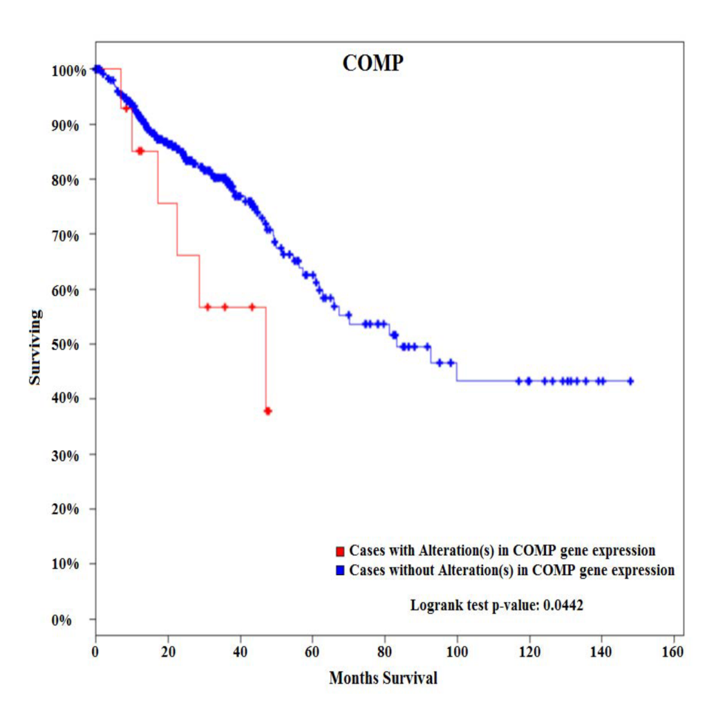
02.05 COMP Gene Is Over-expressed in Early-Onset Colon Cancer and Associated with Poor Survival.

V. N. Nfonsam1, J. Koblinski1, J. Jandova1  1University Of Arizona,Surgery,Tucson, AZ, USA

Introduction: Although the overall incidence of colon cancer (CC) has steadily declined over the last three decades, the incidence has increased in patients younger than 50. The etiology of early-onset (EO) CC is not fully understood. COMP gene has been shown to confer tumor aggressiveness in pancreatic cancer. The aim of this study was to elucidate gene expression patterns in EOCC and show its uniqueness compared to late-onset (LO) disease.

Methods: Two cohorts of patients with sporadic CC were identified. Tumors and matching noninvolved tissues from six EOCC patients (<50) and six late-onset colon cancers (LOCC) patients (>65) were obtained from pathology archives. De-paraffinized tissues were macro-dissected from FFPE sections, RNA isolated, and used for expression profiling of 770 cancer-related genes representing 13 canonical pathways. Survival analysis was performed using the cBioPortal for cancer genomics using 382 CRC patients from the TCGA Provisional database.

Results:Among 770 genes assayed, changes in expression levels of 93 genes were statistically significant between EOCC and matching noninvolved tissues. There were also significant differences in expression levels of 118 genes between LOCC and matching noninvolved tissues. Detailed comparative gene expression analysis between EOCC and LOCC normalized to their matching noninvolved tissues revealed that changes in expression of 88 genes were unique to EOCC using the cutoff criteria of expression levels difference >2 fold and P value <0.01. From these differentially expressed genes specific to EOCC, 28 genes were upregulated and 60 genes downregulated. At the pathway level, PI3K-AKT signaling was the most deregulated pathway in EOCC and cell cycle in LOCC. COMP glycoprotein was one of the genes that were uniquely over-expressed in EOCC. Survival analysis of 382 patients with CRC tumors using the cBioPortal for cancer genomics showed that CRC patients with alterations (all of them with up-regulated COMP) in COMP expression present with poorer overall survival compared to patients without alterations in COMP expression.

Conclusion:
These results suggest that sporadic EOCC is characterized by distinct molecular events compared to LOCC. In addition, COMP glycoprotein is associated with poor overall survival. COMP gene can potentially serve as a novel biomarker associated with EOCC as COMP glycoproteins could be detected in serum and urine.  
 

02.06 Obesity-induced Sphingosine-1-phosphate in Breast Cancer Interstitial Fluid Promotes Angiogenesis

J. Tsuchida1, M. Nakajima1, K. Moro1, A. Ohtani1, M. Endo1, T. Niwano1, K. Tatsuda1, C. Toshikawa1, M. Hasegawa1, M. Ikarashi1, Y. Koyama1, J. Sakata1, T. Kobayashi1, H. Kameyama1, T. Wakai1, K. Takabe2, M. Nagahashi1  1Niigata University Graduate School Of Medical And Dental Sciences,Division Of Digestive And General Surgery,Niigata, NIIGATA, Japan 2Roswell Park Cancer Institute,Breast Surgery, Department Of Surgical Oncology,Baffalo, NY, USA

Introduction: It is well established that obesity evokes chronic inflammation, which aggravates cancer progression by facilitating interactions between cancer cells and stromal cells including blood endothelial cells. Sphingosine-1-phosphate (S1P) is a bioactive lipid mediator produced by sphingosine kinases (SphKs) that plays critical roles in inflammation and cancer progression. We have recently discovered that the SphK1/S1P/S1PR1 axis play critical roles in inflammation-associated cancer progression, and that the S1PR1 functional antagonist, FTY720, suppresses cancer progression by inhibition of the S1P axis during chronic inflammation. We have recently developed a method to measure S1P levels in tumor interstitial fluid (IF), which is a component of the tumor microenvironment that bathes cancer cells in the tumor. Our findings suggested the possibility that S1P secreted from tumor cells to IF may be important for metastasis. We hypothesized that obesity and its related inflammation up-regulate SphK1, which produces more S1P, and the elevated levels of S1P in both tumor and tumor IF stimulate angiogenesis and progression of breast cancer. In this study, we test this hypothesis utilizing animal models with obesity.

Methods: Orthotopically-implanted E0771 syngeneic breast cancer mouse models were used. Mice were fed with normal or high-fat diet (HFD). FTY720 was administered orally (1 mg/kg/day). Angiogenesis in the primary tumor was determined by measurement of microvessel density. S1P levels in tumor and tumor IF were measured by mass spectrometry.

Results: HFD induced obesity significantly worsened the cancer progression. Obesity elevated inflammatory cytokines such as IL-6 and TNF-α in both circulation and in the tumor. Mass spectrometry analysis revealed that while S1P levels in the normal breast mammary fat pad were increased with HFD feeding, S1P levels were even higher in breast tumors. Consistent with increased SphK1 and S1P in tumors, S1P was also significantly increased in the tumor IF. Further, microvessel density analysis revealed that significantly more angiogenesis in peri-tumor area in mice fed with HFD than in mice fed with normal diet. Importantly, FTY720 dramatically reduced the levels of cytokines as well as SphK1, S1PR1 and S1P in primary tumor. Further, FTY720 suppressed not only primary tumor growth, but also angiogenesis in the primary tumor with suppression of aggregation of tumor-associated macrophages.

Conclusion: Our results indicate an important role of S1P in tumor microenvironment, which is affected by obesity, for obesity-related angiogenesis and breast cancer progression. S1P will be one of the promising targets for breast cancer patients with obesity.