02.07 Peri-tumor Sphingosine-1-phosphate Levels Are Affected by the Tumor

J. Tsuchida1, K. Moro1, A. Ohtani1, M. Endo1, T. Niwano1, K. Tatsuda1, C. Toshikawa1, M. Hasegawa1, M. Ikarashi1, M. Nakajima1, Y. Koyama1, J. Sakata1, T. Kobayashi1, H. Kameyama1, K. Takabe2, T. Wakai1, M. Nagahashi1  1Niigata University Graduate School Of Medical And Dental Sciences,Division Of Digestive And General Surgery,Niigata, NIIGATA, Japan 2Roswell Park Cancer Institute,Breast Surgery, Department Of Surgical Oncology,Buffalo, NEW YORK, USA

Introduction: Sphingosine-1-phosphate (S1P), a pleiotropic bioactive lipid mediator, has been implicated as a key regulatory molecule in cancer through its ability to promote cell proliferation, migration, angiogenesis and lymphangiogenesis. Although many researchers including us have reported important roles of S1P in breast cancer progression utilizing in vitro and in vivo models, there have been very few data on human patients due to the difficulty in accurate measurements of S1P levels in the clinical samples. Recently, we have reported that high S1P levels in the tumor determined by mass spectrometry are associated with lymph node metastasis in breast cancer patients. In this study, to reveal further roles of sphingolipids in the patients, we measured the levels of sphingolipids including S1P in plasma and breast tissue including peri-tumor tissue.

Methods: Forty-nine serum prior to operation and 20 tumors from invasive cancer larger than 1.5cm were collected from breast cancer patients who underwent operation from November 2015 to March 2016 in Niigata University Medical and Dental Hospital. The levels of sphingolipids were determined by mass spectrometry.

Results: The levels of sphingolipids including sphingosine, dihydro-sphingosine, S1P, dihydro-S1P were detected successfully in the plasma from 49 breast cancer patients and in the tissue samples from 20 patients. Despite our expectation, no significant differences in plasma S1P levels were identified by hormone receptors (ER, PgR) and HER2 status, Ki-67 index, nuclear grade, lymphatic and vascular invasion of the tumor, pT, pN, and pStage. The levels of sphingosine in patients with lymph node metastasis (pN1,2) were significantly higher than those in patients without metastasis (pN0) (P = 0.023). Interestingly, dihydro-S1P levels in patients with high nuclear grade (NG2,3) were significantly lower than those in patients with low nuclear grade (NG1) (P = 0.025). Moreover, there were significant differences among S1P levels in tumor, peri-tumor, and normal breast tissue (P = 0.0004). Significant differences were also seen among sphingosine (P = 0.0007) and dihydro-sphingosine levels (P = 0.0236) in the three different tissue types.

Conclusion: This is the first report to measure the S1P levels in peri-tumor tissue compared to the tumor and normal breast tissue. This is in agreement with our notion that S1P that is generated in cancer is secreted out to peri-tumoral microenvironment where it aggravates cancer progression, such as lymphangiogenesis. Further study will be needed to reveal the role of S1P in breast cancer patients.
02.08 Paradoxical Association of Postoperative Sphingosine-1 Phosphate and Breast Cancer Aggressiveness

R. Ramanathan1, A. Raza1, J. Young2, J. Sturgill1, D. Lyon1, K. Takabe2  1Virginia Commonwealth University,Richmond, VA, USA 2Roswell Park Cancer Institute,Breast Surgery,Buffalo, NY, USA

Introduction: Sphingosine-1 phosphate (S1P) is bioactive lipid mediator that has been shown to serve an important regulatory function in breast cancer progression. The dynamics of the circulating S1P levels during the postoperative period, however, have yet to be delineated. In this study, we analyze plasma S1P levels in breast cancer patients undergoing adjuvant therapy as compared to control healthy volunteers.

Methods: With institutional review board approval, plasma S1P was measured in 20 healthy women without breast cancer (control) as well as 158 patients with breast cancer who underwent adjuvant chemotherapy with or without irradiation after surgical resection. Among patients with breast cancer, postoperative plasma S1P was measured two weeks prior to adjuvant therapy (baseline), prior to the 4th cycle of chemotherapy (midpoint) and two weeks after completion of adjuvant therapy (completion). Correlations with demographics, treatments and inflammatory markers were analyzed with the use of chi-square tests, paired and non-paired t-tests and ANOVA analyses.

Results: 452 plasma S1P samples among 158 breast cancer patients were analyzed, along with 20 control healthy volunteers. Mean S1P levels did not differ between cancer patients and controls (1221.7 vs. 1139 pmol/mL, p=NS). S1P levels similarly did not associate with age or race in our cohort. Smoking was associated with higher S1P levels (1445.4 vs. 1163.3 pmol/mL, p<0.05). Midpoint S1P levels during adjuvant therapy were lower than baseline (1088.8 vs. 1221.8, p<0.05), with near return to baseline after completion (1121.5 vs. 1221.8, p=0.06), indicating a relationship between chemotherapy and circulating S1P. Furthermore, while stage of disease did not correlate with plasma S1P levels, they were lower among patients with Her2 enriched and triple negative breast cancer as compared to luminal type cancer (1119.2 and 1167.1 vs. 1280.8, p<0.05). Additionally, patients with intermediate and high grade tumors similarly had lower S1P than those with low grade tumors (1230.0 and 1176.5 vs. 1570.8 pmol/mL, p<0.05), further suggesting an inverse relationship between plasma S1P levels and tumor aggressiveness. There were no significant differences based on chemotherapy regimen and radiation.

Conclusions: We found that plasma S1P levels are paradoxically suppressed in aggressive breast cancer and during adjuvant chemotherapy, which raises the possibility that plasma S1P levels do not reflect S1P secretion from cancer cells.

02.09 cFLIPS is a Critical Regulator of EMT-Associated Resistance to Apoptosis

C. Padmanabhan1, E. J. Rellinger1, H. An1, A. G. Waterson2, C. W. Lindsley2, A. Means1, R. D. Beauchamp1  1Vanderbilt University Medical Center,Surgery,Nashville, TN, USA 2Vanderbilt University Medical Center,Chemistry,Nashville, TN, USA

Introduction:  

Epithelial cancers comprise the top four causes of cancer related deaths in the United States. Epithelial-to-mesenchymal transition (EMT) is a major cellular reprogramming of epithelial cancers associated with loss of E-cadherin expression and resistance to apoptosis. The mechanism that drives this phenotype, however, is unclear and this knowledge gap has hindered the development of apoptosis inducing therapeutics. It has previously been reported that E-cadherin expression is necessary for apoptosis induction by the tumor necrosis factor related apoptosis inducing ligand (TRAIL). Using a small molecule, ML327, that we previously showed to partially reverse EMT, we have identified the short variant of the cellular FLICE-like inhibitory protein (cFLIP) as a critical regulator of EMT-associated resistance to TRAIL that is independent of E-cadherin re-expression. 

Methods:

Trail Sensitization: Solid tumor cancer cell lines were treated with either ML327 (10 μ M) or vehicle control for 24 hours. TRAIL was added at 24 hours (50 – 500 ng/mL) for 4 hours. Cells were lysed and analyzed via western blot for cFLIP and markers of apoptosis. Cells were also fixed and stained with propidium iodide (PI) and analyzed via FACS cell cycle analysis.

cFLIP Knockdown: si-RNA knockdown of cFLIP was performed and cells were subsequently treated with TRAIL as above. Cells were lysed and analyzed via western blot for cFLIP and markers of apoptosis.

cFLIP Overexpression: cFLIPS was exogenously overexpressed using a CMV promoter based plasmid. Transfected cells were treated with ML327 and TRAIL as above. Cells were lysed and analyzed via western blot for cFLIP and markers of apoptosis.

E-Cadherin Independence: si-RNA knockdown of E-cadherin was performed in cancer cells and cells were subsequently treated with ML327 or vehicle and TRAIL as above. Cells were lysed and analyzed via western blot for e-cadherin, cFLIP, and markers of apoptosis. 

Results:

ML327 treatment caused a significant reduction of cFLIPS protein at 24 hours. This reduction in cFLIPS was associated with TRAIL sensitization as measured by cleaved PARP and sub G0 population on cell cycle analysis. si-RNA knockdown of cFLIP mimicked ML327 treatment and sensitized cancer cells to TRAIL-induced apoptosis. cFLIPS overexpression blunted apoptosis induction by ML327 and TRAIL. E-cadherin re-expression was not required for ML327-induced TRAIL sensitization.

Conclusion:

EMT-associated resistance to apoptosis in epithelial cancers is well described but the mechanism that drives this phenotype is unclear. Here, we demonstrate that cFLIPS is a critical regulator of this phenotype and that E-cadherin expression may not be necessary for apoptosis induction. Further study is required to determine the exact mechanism of cFLIP loss with EMT reversal and whether this finding can be exploited for therapeutic development.

02.11 Inhibitor of NF-κB Enhances the Antitumor Effect of Radiation Therapy in Colorectal Cancer.

H. Sugano1,2, Y. Shirai1, N. Saito1,2, T. Horiuchi1,2, H. Shiba1, K. Eto1, T. Uwagawa3, T. Ohashi2, K. Yanaga1  1The Jikei University School Of Medicine,Department Of Surgery,Minato-ku, TOKYO, Japan 2The Jikei University School Of Medicine,Department Of Gene Therapy, Research Center For Medical Science,Minato-ku, TOKYO, Japan 3The Jikei University School Of Medicine,Department Of Hematology And Medical Oncology,Minato-ku, TOKYO, Japan

Introduction:

Colorectal cancer is the third most commonly diagnosed malignancy and the fourth leading cause of cancer death in the world. Most patients with CRC are diagnosed at advanced stages, and the prognosis of such patients remains very poor. Neoadjuvant chemoradiothrapy (CRT) followed by radical surgery is currently the standard of care for patients with locally advanced low rectal cancer. CRT has the potential to downsize tumors before surgery and to decrease locoregional recurrence. However, previous findings indicate that irradiation promotes tumor migration, distant metastasis, and the invasive potential of cancer cells. This radiation-induced invasiveness is associated with an increased expression of matrix metalloproteinase (MMP) through nuclear factor kappa B (NF-κB) pathways. Irradiation also activates NF-κB that plays an important role in the regulation of cell apoptosis, inflammation, and oncogenesis including invasion and angiogenesis. We previously reported that nafamostat mesilate, a synthetic serine protease inhibitor, inhibited NF-κB activation and induced antitumor effects for pancreatic cancer. We hypothesized that nafamostat mesilate may inhibit radiation-induced NF-κB activation and tumor invasiveness, and improve therapeutic outcome of colorectal cancer. 

Methods:

We assessed NF-κB activity, cell viability, induction of caspase cascade,  quantification of apoptosis, and cell invasiveness of human colorectal cancer cell line (SW620) in the following four groups: 1) radiation alone, 2) nafamostat mesilate alone, 3) combination (radiation and nafamostat mesilate), or 4) vehicle as control. In combination therapy, we incubated the cells with nafamostat mesilate at 3 hour before radiation therapy.

Results:

NF-κB activity in radiation group was higher than that in vehicle group (p<0.001). NF-kB activity was significantly surpressed in nafamostat mesilate group and combination group as compared with radiation group (p<0.001). Cell viability in combination group was lower than that in radiation group (p<0.001). Cleaved caspase-3, 8, and 9 level in combination group was the greatest in the 4 groups. Apoptosis in combination group was significantly higher than that in radiation group. In the assessment of cell invasiveness, invasive ability in radiation group was higher than that in vehicle group (p<0.01). Invasive ability was significantly suppressed in nafamostat mesilate group and combination group as compared with radiation group (p<0.001). MMP expression in nafamostat mesilate and combination groups was lower than that in radiation group.

Conclusion:

Nafamostat mesilate enhanced the antitumor effect of radiation therapy,  degraded radiation-induced tumor invasiveness, and inhibited MMP through  downregulation of NF-κB in colorectal cancer cell.


02.12 Sphingosine Kinase 1 Contributes to Breast Cancer Cell Metabolism and Survival

M. Nagahashi1, M. Nakajima1, T. Saito2, M. Komatsu2, T. Soga3, R. Zhao4, H. Zhou4, S. Okuda5, K. Takabe6, T. Wakai1  1Niigata University Graduate School Of Medical And Dental Sciences,Division Of Digestive And General Surgery,Niigata, NIIGATA, Japan 2Niigata University Graduate School Of Medical And Dental Sciences,Department Of Biochemistry,Niigata, NIIGATA, Japan 3Keio University,Institute For Advanced Biosciences,Tsuruoka, YAMAGATA, Japan 4Virginia Commonwealth University School Of Medicine,Department Of Microbiology And Immunology, Medical College Of Virginia Campus,Richmond, VA, USA 5Niigata University Graduate School Of Medical And Dental Sciences,Bioinformatics,Niigata, NIIGATA, Japan 6Roswell Park Cancer Institute,Breast Surgery, Department Of Surgical Oncology,Buffalo, NY, USA

Introduction: Sphingosine-1-phosphate (S1P) is a pleiotropic bioactive lipid mediator that regulates many physiological and pathological processes. S1P exerts its function either intracellularly or extracellularly after produced by sphingosine kinases (SphK1 and SphK2) inside the cells. Previous studies suggested that S1P and SphKs play important roles in cancer cell survival. Indeed, expression of SphK1 has been reported to be associated with worse prognosis of breast cancer patients. We hypothesized that SphK1 affects cancer cell-specific metabolism, such as “Warburg effect” in which cancer cells predominantly produce energy by a high rate of glycolysis, and plays a role in cancer cell survival. The aim of this study is to test the hypothesis that SphK1 regulates breast cancer cell metabolism by metabolome analysis.

Methods: SphK1 was downregulated by SphK1-shRNA in 4T1 murine mammary adenocarcinoma cell line. Proliferation and migration assays were performed to examine the cell biology. Metabolic changes in 4T1 cells were analyzed using capillary electrophoresis time-of-flight mass spectrometry (CE-TOFMS) between sh-control (sh-Ct) or sh-SphK1 treated cells.

Results: Proliferation assays revealed significantly less cell proliferation of 4T1 sh-SphK1 cells compared to sh-Ct cells under serum free condition. Cell migration was also suppressed with down-regulation of SphK1. CE-TOFMS analysis revealed the metabolomics profiles of 4T1 sh-SphK1 cells, which were dramatically changed in the glycolysis pathway, amino acid synthesis, and tricarboxylic acid (TCA) cycle compared to the sh-Ct cells. The levels of most nucleotides such as ATP, ADP, GTP, and GDP in 4T1 sh-SphK1 cells were significantly lower than those in sh-Ct cells. Moreover, 4T1 sh-SphK1 cells contained lower amount of glutathione (GSH) than sh-Ct cells, which indicate an important role of SphK1 in not only cell survival, but also oxidative stress and drug resistance.

Conclusion: Our results indicate that SphK1 plays pivotal roles in cancer specific metabolism, which strengthen resistance to oxidative stress and cancer cell survival. SphK1 will be a promising target for patients with breast cancer.


02.13 Clinical Relevance of Annexin A1 in Triple-negative Breast Cancer Patients

M. Okano1,2, H. Okayama1, T. Ohtake1, T. Kawaguchi2, L. Yan3, Q. Qi3, S. Liu3, K. Takabe2  1Fukushima Medical University School Of Medicine,Department Of Organ Regulatory Surgery,Fukushima, FUKUSHIMA, Japan 2Roswell Park Cancer Institute,Department Of Surgical Oncology,Buffalo, NY, USA 3Roswell Park Cancer Institute,Department Of Biostatistics & Bioinformatics,Buffalo, NY, USA

Introduction: Annexin A1 (ANXA1) is a calcium-dependent phospholipid-linked protein, involved in anti-inflammatory effects, regulation of cellular differentiation, proliferation and apoptosis. Recently, we reported that ANXA1 is associated with triple-negative breast cancer (TNBC) and its poor prognosis. Inhibition of programmed death-1 (PD-1) receptor/PD-1 ligand (PD-L1) was reported to suppress progression of TNBC. I In the present study, we evaluated the association between ANXA1 and PD-L1 expression in TNBC.

Methods: The ANXA1 and PD-L1 protein expression in the breast cancer (BrCa) tissues was assessed by IHC staining using an anti-ANXA1 and PD-L1 antibody in 48 TNBC patients and compared the levels with clinicopathological factors. We also validated the result using The Cancer Genome Atlas (TCGA) including RNA-seq data and clinical data.

Results:ANXA1 was positively expressed in 17 of the 48 cases (35.4%). Overall survival was significantly shorter in patients with ANXA1 positive tumors compared with ANXA1 negative tumor (p = 0.0082). No other clinicopathological factor was associated with ANXA1 expression. Utilizing TCGA dataset (n = 1060) for a validation study, high expression of ANXA1 associated with worse overall survival in TNBC patients (n = 111, p = 0.0459), while high expression of ANXA1 demonstrated conversely better overall survival in non-TNBC patients (n = 785, p = 0.0287). There was no survival difference with PD1 expression in our cohort, while high expression of PD-L1 associated with better prognosis in all BrCa patients in TCGA cohort (p = 0.0246). There was no association between ANXA1 and PD-L1 expression in the primary cohort, while high expression levels of ANXA1 demonstrated significant association with high PD-L1 expression in TCGA cohort (p < 0.0001).

Conclusion:These results suggested that ANXA1 is associated with poor prognosis of TNBC .


01.06 Notch1 Signaling Is a Key Regulator of Fibroblasts in Angiogenesis and Wound Healing

Z. Liu1, P. P. Parikh1, H. Shao1, Y. Li1, R. Prokupets1, S. Liu1, O. C. Velazquez1  1University Of Miami,Surgery,Miami, FL, USA

Introduction:  Wounds heal in coordination of a myriad of types of cells, including keratinocytes, inflammatory cells, endothelial cells and fibroblasts. Fibroblasts play pivotal roles in supporting wound healing and tissue remodeling by producing extracellular matrix proteins (ECM) and regulatory soluble factors, providing structural scaffolding and contracting the wound. However, the molecular mechanisms that determine the function of fibroblasts in wound healing remain unknown. Here, we uncover the Notch1 signaling pathway as a molecular determinant that controls the function of fibroblasts in wound healing and angiogenesis. 

Methods:  To explore the role of Notch1 signaling in regulating function of fibroblasts in wound healing and neovascularization, we generated two new mouse lines in which Notch1 signaling is specifically activated (Gain-Of-Function, GOF) or inactivated (Loss-Of-Function, LOF) in fibroblasts, respectively. A 6-mm skin wound was created on mouse dorsal skin by punch biopsy (n=6/group). Wound healing rates were evaluated based on daily photography. Wound tissue angiogenesis was assessed by Dil-LDL perfusion and confocal microscopy.

Results: Using novel mouse models, we demonstrated that Notch1 pathway activity could determine the function of fibroblasts in wound healing and angiogenesis. Notch1 activation in fibroblasts significantly inhibited wound angiogenesis (~40%, P<0.05) and wound closure rate (~30%, P<0.05). However, inactivation of Notch1 signaling in fibroblasts did not promote angiogenesis and wound healing, likely because the basal level of Notch1 activity in fibroblasts is already very low, lower Notch1 activity by deletion of Notch1 would not be expected to add additional effect. 

Conclusion: These findings identify the Notch1 signaling pathway as a molecular determinant that regulates the function of fibroblasts in wound healing and angiogenesis, unveiling Notch1 pathway as a potential target for therapeutic intervention in delayed wound healing and/or vascular diseases. 


01.08 Nuclear Co-Repressor Complex Inhibition Reverses Benefit of HDAC2 Deletion in Renal Ischemia

D. D. Aufhauser Jr1, D. Murken1, Z. Wang1, G. Ge1, T. Bhatti3, W. Hancock2,3, M. Levine1,4  1University Of Pennsylvania,Surgery,Philadelphia, PA, USA 2University Of Pennsylvania,Pathology And Laboratory Medicine,Philadelphia, PA, USA 3Children’s Hospital Of Philadelphia,Pathology And Laboratory Medicine,Philadelphia, PA, USA 4Children’s Hospital Of Philadelphia,Surgery,Philadelphia, PA, USA

Introduction:  Class I histone/protein deacetylases (HDACs) 1 and 2 bear >85% protein sequence homology and conventionally are thought to have similar function via occupancy in nuclear co-repressor complexes.  Inhibition of the HDAC-axis has been shown to mitigate renal warm ischemia-reperfusion injury (IRI) in mice. 

Methods:  Wild type C57BL/6 (WT) and whole animal-inducible HDAC1- or 2- gene deleted mice (HDAC1-/- or HDAC2-/-) were used in this study.  Standardized warm renal IRI consisted of unilateral clamping of the renal pedicle and contralateral nephrectomy.  Primary renal tubular epithelial cell (RTEC) cultures were derived from each genetic strain for use in protein analysis.

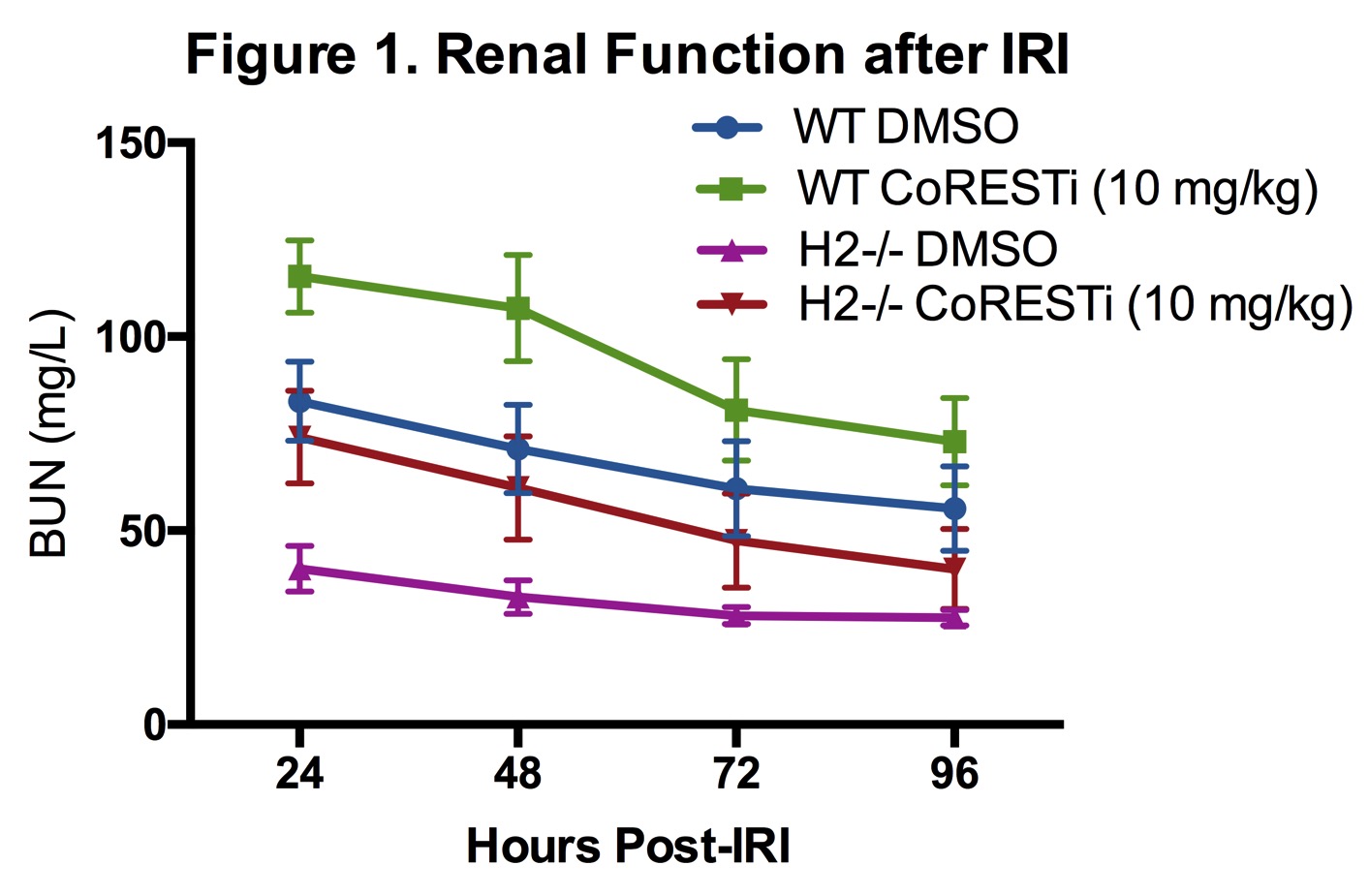
Results: HDAC1-/- mice had impaired IRI tolerance compared to B6 controls with higher daily BUN levels following IRI whereas HDAC2-/- mice demonstrated profound protection from renal IRI. Protein analysis of nuclear extracts from WT, HDAC1KO, and HDAC2KO RTEC showed that deletion of either HDAC1 or 2 was associated with compensatory enhanced protein expression of the other.  Pulldown experiments from RTEC extracts demonstrate that HDAC2KO leads to enhanced HDAC1 association with CoREST, NuRD and Sin3A nuclear co-repressor complexes and increased protein expression of these co-repressor components.  In vivo inhibition of the CoREST complex in both WT and HDAC2-/- mice increased vulnerability to renal IRI injury and returned HDAC2-/- mice to a WT renal IRI phenotype (Figure 1).

Conclusion: HDAC1 and 2 have reciprocal effects on murine renal IRI tolerance, with HDAC1 deletion increasing vulnerability and HDAC2 deletion providing substantial protection from warm and cold ischemia.  HDAC2 deletion leads to alteration in expression and possibly stability of nuclear co-repressor complexes, and its effect can be reversed with inhibition of the CoREST complex. 


01.09 The Circulating Plasma miRNA Profile In Response To Vascular Injury

T. Yoo1, M. Haurani1, C. Rink1  1Ohio State University,Vascular Surgery,Columbus, OH, USA

Introduction: miRNA are short non-coding nucleotides that post-transcriptionally regulate messenger RNA. Recently, they have been discovered to be stable and amplifiable components of circulating plasma that are implicated in inflammation and remodeling. Here, we seek to identify novel circulating miRNA targets that are differentially expressed in response to vascular injury. 

Methods:  Rats underwent no surgery, sham left external carotid ligation, or left carotid balloon injury. Plasma samples were obtained at day 3 and day 14 after surgery. Carotid arteries were collected, sectioned and H&E stained to measure intimal hyperplasia. High resolution in vivo ultrasound was performed and analyzed to assess wall variation and intimal thickness in response to balloon angioplasty. Plasma miRNA was purified, then quantified via nCounter. Differentially expressed targets were validated with qPCR. Target Scan and Ingenuity Pathway Analysis were used to predict possible in silico targets of validated miRNA.

Results: Of the 449 genes on array, 35 were detected over background in cell free plasma samples with no indication of cellular contamination by qPCR or cell housekeeping markers. At day 3 after balloon injury there was no significant change in the miRNA profile. However, at post-injury day 14, miRNA-16 and miRNA-223 which are known to regulate proliferative and inflammatory pathways, significantly decreased compared to sham and non-operated animals (miRNA-16, Relative Expression 0.16, p = 0.001, miRNA-223 Relative Expression 0.08, p< 0.001) This corresponded to an increase in intimal thickness as detected by high-resolution, non-invasive in vivo ultrasonography and histology.

Conclusion: We have identified key circulating plasma miRNA transcriptome components that correlate to the formation of intimal hyperplasia. Identification of these targets may further elucidate miRNA regulation of vascular inflammation as well as serve as predictive biomarkers of re-stenosis following balloon injury.

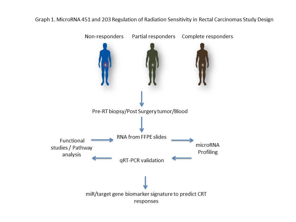
02.01 MicroRNA 451 and 203 Regulation of Radiation Sensitivity in Rectal Carcinomas

K. A. Kelley1, R. Ruhl3, S. Rana2, C. R. Thomas2, S. Anand2,3, V. L. Tsikitis1  1Oregon Health And Sciences University,Department Of Surgery,Portland, OREGON, USA 2Oregon Health And Sciences University,Department Of Radiation Medicine,Portland, OREGON, USA 3Oregon Health And Sciences University,Department Of Cell, Developmental, Cancer Biology,Portland, OREGON, USA

Introduction:
Chemoradiotherapy (CRT) response is an independent predictor of overall survival in locally advanced rectal cancer, highlighting the importance of improving CRT response rates. It is known that several tumor intrinsic factors govern responses to radiation therapy.  Emerging evidence suggests that microRNAs (miRs) modulate gene expression profiles in response to radiation.

Methods:
microRNA profiles were examined and analyzed in approximately forty rectal cancer patients that had either a pathological partial response (PR) or no response (NR).  Using Nanostring technology, a miR profiling platform, miRNAs significantly down and up regulated were identified in FFPE biopsy specimens.   mRNA differentially expressed was followed by in vitro radiation models and tumor sphere assays.  Graph 1.

Results:
miR-451a was among the most upregulated miRs in the PR group whereas miR-203 was among the most downregulated miR.  Inhibition of miR-451a in HCT-116 significantly increased survival in response to a 2 Gy dose of radiation; conversely, transfection of a miR-451 mimic significantly decreased cell survival, with more than a 12-fold decrease when combined with a 5 Gy dose fraction.  Consistent with these results, miR-451a inhibition increased proliferation in 2D as well as in a tumor sphere assay, whereas transfections of a miR-451a mimic robustly decreased proliferation.  In contrast to miR-451a, miR-203 was downregulated in patients with a partial response to CRT.  Inhibition of miR-203 increased apoptosis in combination with a 5 Gy dose fraction.

Conclusion:
A gain of miR-451a and an inhibition of miR-203 in tumor cell lines enhanced cell death and decreased survival in combination with radiation. In this context, we hypothesize that differential expression of miRs regulates rectal cancer radiation sensitivity and therefore can be used as a biomarker to predict therapeutic responses to radiation therapy.
 

02.02 LY2090314 suppresses cholangiocarcinoma cell growth via apoptosis and reduction in GSK-3β

K. M. Sokolowski1, S. Kunnimalaiyaan1, T. C. Gamblin1, M. Kunnimalaiyaan1  1Medical College Of Wisconsin,Surgical Oncology/Surgery,Milwaukee, WI, USA

Introduction:

Cholangiocarcinoma (CCA) is characterized by poor prognosis and therapeutic inefficacy. The current standard of practice for advanced CCA is systemic chemotherapy with gemcitabine and cisplatin. However, a limited survival benefit along with increasing resistance has ushered in a renaissance in alternative approaches. Increased understanding and application of new technologies has led to the identification of more pathway-focused treatment strategies. The glycogen synthase kinase-3 (GSK-3) pathway is a potential therapeutic target as it is overexpressed in various cancer types including CCA. However, the role of targeting GSK-3 in CCA progression remains elusive. We hypothesize that GSK-3 stabilizes cellular growth thereby promoting proliferation and with subsequent inhibition, leading to a reduction in cellular growth in vitro. For this, we have used a clinically well-characterized GSK-3 inhibitor, LY2090314. 

Methods:

The effects of LY2090314 on CCA cell lines (CCLP-1 and SG-231) were assessed by MTT and colonogenic assay. Cell lysates were analyzed via Western blotting for pro-apoptotic, anti-apoptotic and cell cycle proteins. Apoptotic induction was further evaluated through caspase-glo 3/7 assay. Mechanism of LY2090314 administration on CCA cellular proliferation was also examined. 

Results:

Increasing LY2090314 treatment (0μM – 20μM) had a significant dose and time-dependent growth reduction (p<0.01) compared to control. The concentration at which 50%growth inhibition (IC50) was observed in vitro following 96hr treatment for CCLP-1 and SG-231 is 13.7µM and 9µM respectively. Similar concentrations inhibited colony formation in both CCA cell lines.  Western analysis indicated that growth suppression was due to apoptosis as well as  cell cycle arrest as evidenced by increased expression of pro-apoptotic (cleaved PARP and cleaved caspase-3), reduced anti-apoptotic (survivin) proteins and mitigation of cell cycle arrest proteins (p21 and cyclin D1). Functionally, this was confirmed by an increase in caspase activity. Importantly, LY2090314 treatment reduced the expression levels of only GSK-3β in CCA cell lines. 

Conclusion:

LY2090314, a GSK-3 inhibitor, effectively reduces CCA growth in vitro and appears through reduction of GSK-3β. To our knowledge, this is the first study on the effect of LY2090314 in cholangiocarcinoma cell lines in vitro and provides rationale for further preclinical analysis.

02.03 The Tumor Microenvironment Stroma Promotes Cancer By Using Cell-Type Specific Stress Kinase Pathways.

S. G. Patel1, L. Li1, A. Maas1, R. Ghukasyan1, J. Williams1, P. Toste1, A. Nguyen1, I. Elliott1, N. Wu1, T. Donahue1  1University Of California Los Angeles,Surgery,Los Angeles, CA, USA

Introduction:
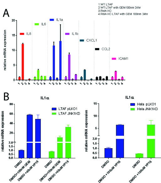
We have demonstrated that pancreatic cancer (PDAC) tumor associated fibroblasts (TAFs) support pancreatic cell growth and invasion from a pro-inflammatory, DNA damage (DDR) mediated pathway.  This pathway referred to as the senescence associated secretory phenotype (SASP) requires both stress kinases and Nfkb signaling for maximum signal production after stimulus.  Within the tumor microenvironment, genotoxic chemotherapies can stimulate the stromal cells to produce factors that promote tumor cell fitness.  Contrary to the prevailing model, we have identified that JNK isoforms are cell-type specific and that both JNK1 and JNK3 can transduce this inflammatory pathway in the tumor microenvironment.  We additionally have found that not all of the SASP is regulated by Nfkb, but rather a key mediator, IL-1 alpha, can be expressed in its absence.

Methods:
Immortalized pancreatic cancer tumor associated fibroblasts (Logsden Lab) as well as Hela cells were used to create both gene knockouts as well as stable knockdowns for JNK1, JNK3 and p65 (Nfkb signaling).  SASP induction was verified after a DNA damage response using RTPCR.  Small molecules specific to JNK isoforms were used to verify genetically manipulated cell lines in WT cells.

Results:

Gemcitabine treatment (100 nm) of PDAC TAFs induced a pro-inflammatory gene expression program including statistically significant (p < 0.05) upregulation of a number of cytokines: including IL-6, IL-8, IL-1 alpha and beta, CXCL1, CCL2 and ICAM1.   Homozygous knockout TAFs for p65 (TAF p65 -/-) stops the induction of these all genes tested (p<0.05) except IL-1 alpha (p = 1) [Figure 1A].   Nfkb (p65) is required for upregulation of IL-1 alpha in the presence of recombinant (10 ug/mL) TNF-alpha or IL-1 alpha (p <0.05).  We found that TAFs do not express JNK3, but rather JNK1 and 2.  We identified using a combination of shRNAs to JNK1 and JNK3 as well as small molecule inhibitors to JNK1/2 that TAFs require JNK1 for upregulation of IL-1 alpha after a DDR (p<0.05) [Fig 1B].   This implies that in the tumor microenvironment, the SASP is propagated by specific stress kinases expressed.

Conclusion:
Previous studies have attributed the SASP to be critically dependent on Nfkb signaling as well as amplified in culture with JNK3.  We identify that some elements of the SASP are not dependent on Nfkb signaling.   Furthermore, we find that JNK3 is not a ubiquitous stress kinase in this response, but rather JNK isoforms are cell-type restricted in expression.  These findings highlight the complexity of the pro-inflammatory signaling within the tumor microenvironment and suggest a more careful analysis of cell and tissue specific gene expression is needed for developing optimal treatment strategies.

01.03 Urinary N-acetyl-beta-D-glucosaminidase Activity is Increased in Renal Transplant Rejection

A. MacDonald1, K. P. Samy1, B. S. Parker1, T. Truong3, M. Kuchibatla3, J. Espinosa1, M. McRae1, J. Cheeseman1, A. D. Kirk1,2, T. V. Brennan1  1Duke University,Department Of Surgery,Durham, NC, USA 2Emory University,Department Of Surgery,Atlanta, GA, USA 3Duke University Medical Center,Biostatistics And Bioinformatics,Durham, NC, USA

Introduction:
Serum creatinine (sCr) remains the primary screening tool for renal transplant rejection, increasing only after significant damage has been done to the allograft. Acute injury-specific markers may provide an earlier and more sensitive measure of rejection. Urinary N-acetyl-beta-D-glucosaminidase (NAG) is produced in the nephron proximal tubules, which are injured during renal allograft rejection. We retrospectively analyzed urine samples from renal transplant patients to investigate its use as a biomarker for allograft rejection. 

Methods:
We retrospectively analyzed 126 urine samples from renal transplant patients collected at two institutions for urinary NAG activity and Cr levels. Raw urinary NAG activity and NAG activity corrected for urinary Cr were correlated to clinical outcome. Analyses were run both with the raw NAG values and also corrected for urine concentration by dividing by the urine creatinine (corrected-NAG).

Results:
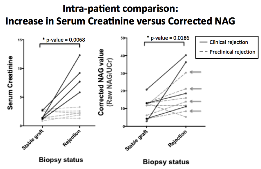
There was a statistically significant elevation in urinary NAG between healthy controls and transplant patients when samples were tested independently (raw NAG p=0.028, corrected-NAG p=0.003). There was a statistically significant increase from a patient’s baseline (during normal graft function) to periods of rejection within single patients for both raw NAG (p=0.002) and corrected-NAG (p=0.02) (Figure 1). Many patients with confirmed histological rejection on protocol biopsy had significant elevations in corrected NAG activity without elevation in sCr (Arrows, Figure 1). There was no significant difference between stable allograft and rejection NAG activity when testing independent samples (raw NAG p=0.143, corrected-NAG p=0.488). Elevations in NAG also occurred in the setting of BK nephropathy.

Conclusion:
Urinary NAG activity was associated with renal injury in transplant patients, with an acute elevation in NAG activity observed in the setting of biopsies showing rejection or BK nephropathy. However, it was not specific for acute rejection versus acute kidney injury from sources such as BK virus. Importantly, urinary NAG was elevated in patients without changes in sCr, indicating that it may serve as an earlier and more sensitive marker of kidney injury than sCr in the workup of a transplant patient.  
 

01.16 Alterations in Mitochondrial DNA Density in Right Ventricular Failure and Recovery

J. Winward1, M. E. Bowen1, H. Li1, S. H. McKellar1  1University Of Utah,Cardiothoracic Surgery,Salt Lake City, UT, USA

Introduction:
Increasing evidence indicates that abnormal energy metabolism plays a leading role in right ventricular failure (RVF), but the specific role in RVF and RV recovery (RVR) is unknown. Our analyses showed failed fatty acid oxidation (FAO) and changes in hypoxia markers in RVF and RVR. These observations led us to hypothesize that the buildup of FAO precursors was the result of insufficient mitochondrial (MT) biogenesis. Our objective was to discover how MT biogenesis is altered in RVF and RVR cardiomyoctes compared to healthy controls in a rabbit model.

Methods:
Fifteen rabbits were assigned to one of three groups—control, RVF, and RVR—and RV tissue samples were taken from each rabbit’s RV. MT gene expression and transcriptional variance were measured via RT-PCR and RNA sequencing. Immunoblotting of MT biogenesis activators was also performed.

Results:
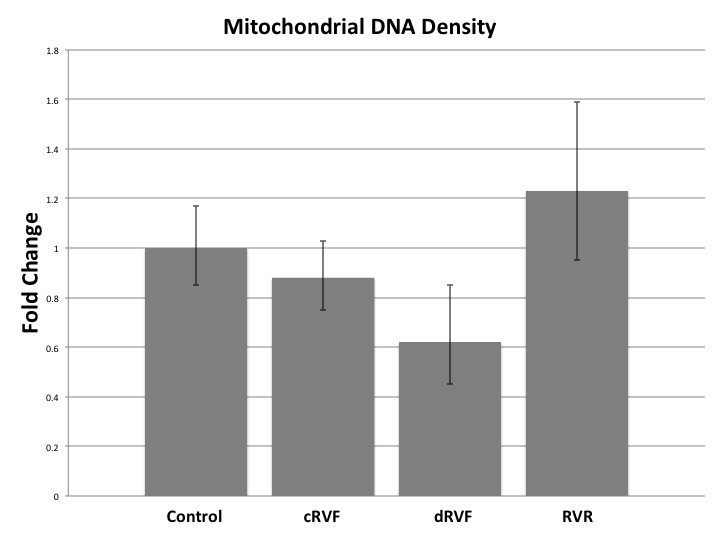
MT DNA density decreased during RVF with fold changes of RVF=0.75 (p=0.22) returning to RVR=1.06 (p=0.84). Further division of the RVF group into compensated RVF (cRVF) and decompensated RVF (dRVF) revealed that MT density was qualitatively greater in cRVF (FC=0.88, p=0.58) than in dRVF (FC=0.62, p=0.26). RNA sequencing revealed significantly increased transcription of HIF in RVF when compared to the control (FC=0.93; p<0.04) and RVR (FC=0.90; p<0.03). However, the rate of transcription of downstream activators of MT biogenesis (i.e. PGC-1-α, NRF-1, NRF-2, Akt3, Perm-1, TFAM, VEGF, and AMPK) was not significantly altered between the groups. Despite its unaltered transcription, immunoblotting of activated AMPK revealed a significant fold increase in RVF (1.50) and RVR (1.61) compared to the control (1.06) (p=0.01). 

Conclusion:
Our data qualitatively suggest that MT DNA density decreases in a manner directly proportion to the severity of RVF, and that it increases in RVR.  Other studies show similar findings in human subjects. Additionally, the increased transcription of HIF in the RVF samples leads us to hypothesize that hypoxia plays a significant role in the MT pathogenesis of RVF. The discrepancy between the rate of transcription and the enzyme activation of AMPK, in conjunction with the altered MT DNA density in RVF and RVR despite the unchanged rate of transcription of upstream MT biogenesis regulatory enzymes, leads us to hypothesize that the functionality rather than the quantity of MT biogenesis regulators is leading to the variance in MT DNA density, as seen with the increased levels of the activated AMPK protein in RVF and RVR, despite its unaltered rates of transcription. Further research investigating the alterations of MT biogenesis markers at the transcriptional and translational level, and hypoxia’s role therein, is warranted.