15.20 Level 1 Trauma Centers: More is Not Necessarily Better

J. C. He2, L. A. Kreiner2, N. Sajankila1, D. L. Allen3, J. A. Claridge2 1Case Western Reserve University School Of Medicine,Cleveland, OH, USA 2MetroHealth Medical Center,Department Of Surgery,Cleveland, OH, USA 3Northern Ohio Trauma System,Cleveland, OH, USA

Introduction:
The optimal number of level I trauma centers (L1TCs) in a region has not been elucidated. To begin addressing this, we compared mortalities for patients injured in counties or regions with 1 L1TC to those with more than 1 L1TC.

Methods:
Recent state trauma registry data from 2010-2012 were analyzed. Trauma patients with age ≥15 from counties or regions with L1TC were included in this study. Region was defined as a L1TC containing county and its neighboring counties. Counties boarding more than one L1TC containing county were excluded from analysis. Two analyses were performed. For the county-level analysis, counties containing only 1 L1TC were compared to counties with more than 1 L1TC. For the regional analysis, regions covered by only 1 L1TC were compared to regions covered by greater than 1 L1TC. The following patient subgroups were included a priori for both analyses: Injury Severity Score (ISS) ≥ 15, age ≥ 65, and trauma mechanisms.

Results:
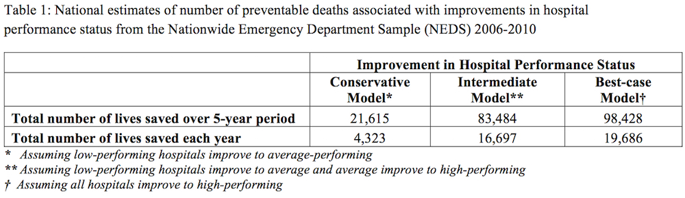
A total of 54,471 patients were analyzed. Their mean age was 59; 90% had blunt injuries. Their median ISS was 5, and the overall mortality was 4.8%. Both the county-level and regional analyses showed that patients in counties or regions with only 1 L1TC were older (60 vs. 57, p<0.001), but had similar ISS as compared to patients in counties or regions with more than 1 L1TC. Counties and regions with only one L1TC also had less total number of trauma centers (3.5 vs. 3.8 and 2.4 vs. 2.9, respectively; all p <0.001) Mortalities for the county-level and regional analyses for all patients and patient subgroups are compared in Table 1. Multivariable logistic regression adjusting for age, ISS and trauma mechanism demonstrated that having more than 1 L1TC in a region was an independent predictor for death (Odds Ratio=1.2; 95% CI: 1.1-1.3, p<0.001).

Conclusion:
Despite older patients and fewer total number of trauma centers, counties and regions with only one L1TC had lower mortality. This suggests that having multiple L1TCs in a county or region may not lead to increased patient survival.

15.15 Pre-hospital Tourniquets for Severe Extremity Injury: Decreased Mortality from Hemorrhage

M. Scerbo1, J. B. Holcomb1, K. Gates1, B. A. Cotton1 1University Of Texas Health Science Center At Houston,Houston, TX, USA

Introduction: Field use of tourniquets (TQ) in military medicine is regarded as an effective adjunct for preventing hemorrhage-related deaths from extremity trauma. Application in the civilian setting, however, has been accepted sporadically due to the paucity of evidence supporting efficacy. Subsequently, there have been no randomized, controlled trials assessing the efficacy of pre-hospital (PH) TQ application. Our urban EMS system has been training pre-hospital personnel to use TQ since 2009. The purpose of this study was to assess whether PH TQ use in the civilian setting decreases death from hemorrhagic extremity trauma.

Methods: Following IRB approval, patients arriving to a level-1 trauma center between 01/2009 and 05/2013 with an extremity injury and a PH TQ applied were reviewed. Control patients were obtained from the trauma registry prior to PH TQ implementation (01/2003 to 12/2008) and were eligible for matching if they had (1) Extremity Abbreviated Injury Severity (AIS) ≥ 2 and (2) OR exploration /control of extremity vascular injury or (3) death with extremity injury likely to have a vascular injury. Patients were propensity-score matched by age, gender, body mass index, mechanism of injury, method of transport and race. Matching occurred via a 1:1 ratio, with caliper and no replacement. Continuous data are presented as medians with 25th and 75th interquartile ranges. Categorical data are reported as proportions. Univariate and multivariate analyses were performed.

Results:

110 patients had PH TQ placement. Of 6961 control patients with AIS Extremity ≥ 2, 380 fulfilled all criteria for matching. Propensity matching yielded a sample size of 61 patients per group. Cases and controls displayed predominate isolated extremity injury (ISS 9 (4, 16) vs. 9 (8,18); AIS Extremity 3 (2, 3) vs 3 (2, 3)). The rate of vascular repair was similar between the two groups (89% vs 80%, p=0.212). Patients without a PH TQ had lower systolic blood pressure (109 mmHg (83, 135) vs 124 mmHg (93, 154) P <0.05) and hemoglobin 11 g/dL (9, 13) vs 13 g/dL (11,14) p<0.001) upon arrival. Failure to apply a PH TQ was associated with an increase mortality (21% vs 5%, OR 5.2 95% CI 1.31-29.9, p < 0.01). Similarly, there was a lower percentage of expected survivors in the patients that did not receive a PH TQ (31% vs 67%, p=0.001). Patients that did not receive a PH TQ had a higher resuscitation intensity (4.1 (1, 11.25) vs. 7.5 (3.4, 14.2) p<0.05).

Conclusion: Pre-hospital TQ application is associated with improved mortality from severe extremity injury in the civilian setting.

15.16 Rural Risk: Geographic Disparities in Trauma Mortality

M. P. Jarman1, R. C. Castillo1, A. R. Carlini1, A. H. Haider2 1Johns Hopkins University School Of Public Health,Health Policy,Baltimore, MD, USA 2Brigham And Women’s Hospital,Surgery,Boston, MA, USA

Introduction: Treatment at a designated trauma center is proven to reduce mortality from traumatic injuries, but the majority of US residents in rural areas do not have timely access to Level I or II trauma centers. To date, no studies have used nationally representative date to quantify the impact of barriers to trauma care on injury mortality in rural populations.

Methods: We performed a retrospective analysis of 2006-2011 National Emergency Department Sample data to determine if mortality following traumatic injury differs across urban/rural classifications. Emergency department (ED) visits with ICD-9-CM codes for injuries (ICD-9-CM 800-959.9) as the primary or secondary diagnosis were included in these analyses, excluding superficial and foreign body injuries, late effects of injury, as well as records with missing urban/rural status or disposition from the ED (N=8,887,575). Mortality was defined as dying in the ED or in the hospital during the admission associated with the ED visit. Overall, 0.12% of ED encounters in the sample resulted in death (N=10,665) Odds of death were calculated using multiple logistic regression with patient residential urban/rural status, Injury Severity Score, comorbidities, trauma center designation, patient age, and patient gender as covariates. All analyses were performed using Stata 12.1.

Results: Residents from rural communities were 25% (p = 0.002) more likely to die of traumatic injury than non-rural residents, when controlling for severity, comorbidities, trauma center designation, age, and gender. Rural residents treated at Level I trauma centers were 3.06 times (p < 0.001) more likely to die of their injuries, compared to non-rural residents. Rural residents at Level II centers were 73% more likely to die (p = 0.002), and rural residents at Level IV centers were 13% more likely to die (p = 0.016), compared to non-rural residents. There was no statistically significant difference in mortality between rural and non-rural residents with treated at Level III (p = 0.151).

Conclusion: People living in rural communities are significantly more likely than non-rural residents to die following traumatic injury. This disparity is largest at Level I trauma centers decreases at lower level trauma centers. Distance and travel time to treatment likely play a significant role in injury outcomes for rural residents, but measures of distance and time generally not available for nationally representative data that also include measures or rural/urban residence. Future analyses should explore the interaction between time to treatment, level of care, and outcomes for rural residents.

15.17 Trends and Outcomes in the Management of Vascular Injuries: Open vs. Endovascular Approaches

B. K. Richmond1, A. F. AbuRahma1 1West Virginia University/Charleston Division,Department Of Surgery,Charleston, WV, USA

Introduction:

Controversy exists in vascular trauma regarding the best method of treatment – open vs. endovascular techniques. Little has been published on this complex topic.

Methods:

Patients from 2005-2013 at a Level 1 trauma center with vascular injury/repair were identified via a prospectively maintained registry. Patient data, injury type/severity, treatment and 30 day outcomes were obtained from the trauma registry and chart review. Adverse events (limb loss, major disability, death) were outcomes of interest. Univariate analysis and multivariate logistic regression were used to identify predictors of adverse events.

Results:

In all, 346 patients were included (median age 34, range 1- 93 years) Median injury severity score (ISS) was 10(1- 59). Endovascular repairs (n=52)increased from 0%(2005) to 32%(2013), and demonstrated equivalent outcomes to open approaches(p = 0.24). On multivariate analysis, higher ISS(p =0.001), increasing age (p=0.01) and lower extremity injuries (p=0.001) were associated with adverse outcomes across the entire series. Endovascular approaches were most commonly utilized in vascular injuries of the chest/abdomen (39 of 52, 75% of all endovascular procedures in the series,p<0.001), older patients (p=0.003), blunt injury mechanism(p<0.001), and patients with a higher ISS at presentation(p<0.001).

Conclusions:

In this large series, the use of endovascular procedures increased over time, and was associated with equivalent outcomes to open approaches, despite their higher usage in older patients, those with chest/abdominal injuries, and those with a higher ISS at presentation. These results are encouraging and stress the need for further prospective study into the role of endovascular therapies in the treatment of vascular injuries.

15.10 Assessing Clot Strength Using Thromboelastography: Are There Coagulopathies After Burn Injury

J. N. Luker1,2, J. D. Karalis2, S. Tejiram1,2, J. Zhang2, K. M. Johnson2, L. T. Moffatt2, M. M. McLawhorn2, J. W. Shupp1,2 1MedStar Washington Hospital Center,Burn Center, Department Of Surgery,Washington, DC, USA 2MedStar Health Research Institute,Firefighters’ Burn And Surgical Research Laboratory,Washington, DC, USA

Introduction:

Acute coagulopathy of trauma has been studied extensively in the setting of blunt and penetrating injury. Conversely, little is known about how thermal injury augments the homeostasis of coagulation. To understand how clot strength is augmented after burn injury Rapid thromboelastography was performed post-injury in a cohort of thermally injured patients. This study aimed to assess whether burn patients exhibit changes in clot strength that could be associated with occult or gross coagulopathy.

Methods:

A prospective study of patients with burn injury who presented to a regional urban burn center was conducted. Patient demographics and injury characteristics were collected and RapidTEG™ was performed on blood samples on admission and at regular time points over a 21-day period while hospitalized. Parameters analyzed were R (time to initial clot formation), α angle (rate of clot amplification), and MA (maximum clot strength). Coagulopathy was defined as either hypocoagulable or hypercoagulable with at least one parameter outside of the normal range.

Results:

The TEG profile of 88 burn patients with a mean age of 41.7 and a mean TBSA of injury of 22.2% were studied. Of these patients, 82% demonstrated abnormal metrics, with an observed trend from a normal or hypocoagulable state, to a more hypercoagulable state occurring between 48 and 72 hours. To further analyze this transition, patients without TEG analysis for three time points beyond 72 hours post-injury (due to death, discharge or decline to further participate) were excluded and the remaining subset of 33 patients were examined. This subset of patients had a mean age of 43.6 and a slightly higher mean TBSA of 33%. Of these patients, 15% (n=5) had R metrics within the hypocoagulable range on admission while no patients had hypercoagulable metrics. When comparing early time points versus late time points, greater than 72 hours, 79% had a statistically significant change (p<0.05) in parameters indicating a hypercoagulable state. This included all but two of the initially hypocoagulable subset of patients, one of which remained hypocoaguable throughout all time points. When stratified by injury severity 80% of the less than 10%TBSA (n=10), 86% of the 10-30%TBSA (n=14), 57% of the 31-50%TBSA (n=7), 100% of the 57-70%TBSA (n=1), and 100% of the greater than 70%TBSA (n=2) patients had significant changes in TEG parameters indicative of a hypercoagulable state. Initial fluid resuscitation and transfusions were explored for potential associations with the observed coagulopathy, but no significant correlations were identified.

Conclusions:

RapidTEG™ analysis demonstrates hypercoagulability in a time dependent fashion post burn injury. The clinical significance of this coagulopathy needs to be further explored and on going reseach efforts will be aimed at correlating these results with other functional and plasma assays to better understand coagulation after thermal injury.

15.11 Rib Fracture Number Thresholds Independently Predict Worse Outcomes in Older Adults

N. O. Shulzhenko1, H. Jung1, M. V. Beems1, T. J. Zens1, A. P. O’Rourke1, A. E. Liepert1, J. E. Scarborough1, S. K. Agarwal1 1University Of Wisconsin,Department Of Surgery,Madison, WI, USA

Introduction:

Rib fractures have been previously associated with significant morbidity and mortality in injured older adults. Contemporary studies have reported that when adjusted for age, comorbidities and trauma burden, the number of rib fractures is not associated with worse outcomes. Our study sought to investigate this risk-adjusted relationship in older adults. We hypothesized that, in an older adult population, the number of rib fractures was a predictor of worse outcomes independent of patient comorbidities and trauma burden.

Methods:

A retrospective review of the prospectively collected American College of Surgeons’ National Trauma Data Bank registry was performed for all patients 65 years of age and older who had sustained rib fractures between 2009 and 2012. Patients with accompanying sternal fractures, penetrating or burn mechanisms, and those with missing data on the number of rib fractures were excluded for a total study cohort of 67,695 patients. Data were collected for age, gender, injury severity score, mechanism of injury, comorbidities, number of rib fractures, hospital mortality, hospital and ICU lengths of stay (LOS), need for ICU care, need for ventilator support, and ventilator duration. Data were also collected for the occurrence of any complication, the occurrence of any pulmonary complication; as well as the occurrence individually of pneumonia, acute respiratory distress syndrome (ARDS), and unplanned intubation. To account for International Classification of Diseases defined database coding, rib fracture data was modeled both as an ordinal variable with groups (1-2, 3-5, 6-7, 8+ fractures) and as an interval variable (one to seven). Bivariate analysis was performed with all candidate predictor and outcome pairs to identify significant factors (α<0.1) to include in multivariate models. Multivariate logistic regression analysis was then performed for all dichotomous outcomes and log-transformed multiple linear regression analysis was performed for all continuous outcomes.

Results:

Eight or more rib fractures were independently associated with hospital mortality, ICU LOS, need for mechanical ventilation, ventilator duration, pulmonary complications, ARDS, and unplanned intubation (p<0.001). Six or more rib fractures were independently associated with LOS, need for ICU care, overall complications, and pneumonia (p<0.001). In a subset excluding patients with serious injuries (AIS>2) in body regions other than the chest, six or more rib fractures were independently associated with all outcomes except for ventilator duration (p<0.03). In this subset, every additional rib fracture increased LOS and the need for ICU care (p<0.03).

Conclusion:

In older adults, the number of rib fractures is a significant predictor of trauma outcomes independent of comorbidities and trauma burden. It is unclear whether rib fractures in isolation cause the outcomes measured or are an independent variable for injury severity.

15.12 The Potential for Trauma Quality Improvement: One Hundred Thousand Lives in Five Years

Z. G. Hashmi1, S. Zafar2, T. Genuit1, E. R. Haut4, D. T. Efron4, J. Havens3, Z. Cooper3, A. Salim3, E. E. Cornwell III2, A. H. Haider3 1Sinai Hospital Of Baltimore,Department Of Surgery,Baltimore, MD, USA 2Howard University Hospital,Department Of Surgery,Washington, DC, USA 3Brigham And Women’s Hospital,Department Of Surgery,Boston, MA, USA 4Johns Hopkins University School Of Medicine,Department Of Surgery,Baltimore, MD, USA

Introduction: Nationwide efforts at trauma quality improvement aim to reduce in-hospital trauma mortality. However, the magnitude of this mortality reduction at the national level remains largely unknown. Our objective was to determine a nationwide estimate of number of lives that could potentially be saved if high-mortality trauma centers improved their performance.

Methods: Adults with blunt/penetrating injuries included in the Nationwide Emergency Department Sample 2006-2010 were analyzed. Hospitals were classified as high, average or low-performers based on risk-adjusted in-hospital mortality using the standardized Trauma Quality Improvement Program (TQIP) benchmarking methodology. Generalized linear modeling, adjusting for demographics and injury severity characteristics, was then used to estimate the relative-risk of death for patients treated at high/average performing hospitals versus low-performing centers. Subsequently, weighted national estimates of preventable mortality were determined for each of the following; 1)Conservative model: low-performing hospitals improve to average-performing, 2)Intermediate model: low-performing hospitals improve to average and average improve to high-performing and 3)Best-case model: all hospitals improve to high-performing.

Results: A total of 9,992,202 trauma patients from 1771 hospitals were included. 151 (8.5%) hospitals were classified as high-performing, 1,506 (85.0%) as average and 114 (6.4%) as low-performing. For conservative and intermediate models, an estimated 4,323 and 16,697 trauma deaths, respectively, could be prevented annually. Additionally, if all hospitals were to deliver the highest quality of care, an estimated 19,686 lives could potentially be saved each year.

Conclusion: If all trauma centers achieved outcomes similar to those at the highest-performing centers, nearly 100,000 lives could be saved over 5 years. These national estimates demonstrate the tremendous societal benefits associated with provisioning high quality of trauma care. Concerted efforts aimed at the standardization and implementation of high quality trauma care should therefore be a priority.

15.13 10-Year Analysis of Crystalloid Resuscitation after Traumatic Brain Injury

A. Ko1, G. Barmparas1, B. J. Sun1, E. Smith1, M. Y. Harada1, E. Chen1, D. Mehrzadi1, E. J. Ley1 1Cedars-Sinai Medical Center,Division Of Trauma And Critical Care,Los Angeles, CA, USA

Introduction: Patients with traumatic brain injury (TBI) are often resuscitated with crystalloids in the emergency department (ED) to maintain cerebral perfusion. The purpose of this study was to evaluate whether crystalloid resuscitation volume impacts the mortality of TBI patients.

Methods: This was a retrospective study of trauma patients with head AIS ≥ 2, who received crystalloids during ED resuscitation between January 1, 2004 and December 31, 2013. Demographics, clinical data, and volume of crystalloid received in the ED were collected. Patients who received < 2L of crystalloids were categorized as low volume (LOW) while those who received ≥ 2L were considered high volume (HIGH). Mortality and outcomes were compared and multivariate regression analysis was used to determine independent risk factors for mortality.

Results: Over the 10-year study period, 879 patients met inclusion criteria. Overall mortality was 12.9%. 743 (85%) were in the LOW cohort and 136 (15%) in the HIGH cohort. Gender and mean age were similar between the two groups. The HIGH cohort had lower mean admission SBP (125 vs. 138, p<0.001), lower admission GCS (10 vs. 12, p<0.001), higher head AIS (3.8 vs. 3.3, p<0.001) and higher ISS (26 vs. 18, p<0.001). The LOW group had shorter ICU LOS (5 vs. 7 days, p=0.01), hospital LOS (9 vs. 13 days, p=0.02), ventilator days (1 vs. 3 days, p=0.02) and lower mortality (10% vs. 28%, p<0.001). Multivariate analysis demonstrated that ED resuscitation with ≥ 2L of crystalloid independently predicted higher mortality (AOR 1.85, p=0.035).

Conclusion: Higher volume crystalloid resuscitation after TBI is independently associated with increased mortality. When possible, resuscitation with crystalloids less than two liters for TBI patients is recommended.

15.07 Shark Attack Related Injuries: Implications for Surgeons

J. A. Ricci1, C. R. Vargas1, O. A. Ho1, D. Singhal2, B. T. Lee1 1Beth Israel Deaconess Medical Center,Division Of Plastic Surgery,Boston, MA, USA 2University Of Florida Health System,Division Of Plastic Surgery,Gainesville, FL, USA

Introduction: Society’s fear of sharks is caused by the media hype surrounding attacks and by movies. Although few sharks are considered dangerous, attacks on humans can result in large soft tissue defects; necessitating the early intervention of reconstructive surgeons. This study aims to determine the characteristics of shark related injuries so that they may be better treated.

Methods: The Global Shark Accident File, maintained by the Shark Research Institute (Princeton, NJ) is a compilation of all known worldwide shark attacks. Database records since 1900 were evaluated to identify differences between fatal and non-fatal attacks. Characteristics evaluated for both fatal and non-fatal attacks included: victim age, victim gender, geographic location of attack, anatomic injury pattern, shark species, and the victim’s activity at the time of attack. The T-test and Chi-Squared tests were used to analyze the data for significant features between the groups.

Results: Since 1900 there have been 5034 shark attacks, with 1205 (24.0%) fatal attacks and 3829 (76.0%) non-fatal attacks documented. While the number of reported attacks per decade has increased, the percentage of attacks that were fatal has decreased. Bites to the legs (41.8%) or arms (18.4%) were most common, with limb loss occurring in 7% of attacks and no injuries in 14.2% of incidents. Characteristics of fatal attacks included swimming (p = 0.001), boating (p = 0.001), three or more anatomical bite sites (p = 0.03), limb loss (p = 0.001), bites to the torso (p = 0.05) or Tiger shark attack (p = 0.002). Non-fatal attacks tended to be associated with fishing (p = 0.001), surfing (p = 0.001), only one anatomical bite site (p = 0.001), bites to the hands or arms (p = 0.001) and bites to the legs (p = 0.001). Geographically, the majority of attacks occurred in North America (1847; 36.7%), followed by Australia (1335; 26.5%) and Africa (676; 13.4%). Within the United States, the individual states with the highest number of attacks were Florida (896; 49.1%), California (248; 13.6%) and Hawaii (240, 13.2%).

Conclusion: Although rare, shark attacks cause devastating injuries to patients. These injuries often involve multiple bite sites and limb loss which can create significant challenges for surgeons by limiting the available reconstructive options. A proper evaluation of the characteristics of the attack can lead to the development of optimal, well-coordinated care plans and improved patient outcomes.

15.08 VTE Chemical Prophylaxis For Traumatic Neurosurgical Injuries: Finding The Balance

B. M. Tracy1, C. O’Neal1, J. Dunne1 1Memorial University Medical Center,Surgery,Savannah, GA, USA

Introduction:

Trauma patients are at an increased risk of venous thromboembolism (VTE) for a multitude of reasons. The purpose of this study is to determine if delay in VTE prophylaxis initiation in neurosurgical trauma patients is associated with an increased incidence of VTE. Furthermore, we seek to identify the appropriate timing of initiation in various subgroups of VTEs as to prevent VTE development and not worsen the initial injury.

Methods:

With permission from our institutional review board, we performed a retrospective review of patients enrolled in Memorial’s TQIP database from 2010 to 2014. We included patients who sustained any traumatic brain or spinal cord injury (SCI), who were placed on chemical VTE prophylaxis. Our data points included date of arrival, date of initiation of chemical VTE prophylaxis, and presence or absence of VTE development. We stratified the patients into injury groups: subarachnoid hemorrhage (SAH), subdural hemorrhage (SDH), a combination of SAH/SDH together, any other brain hemorrhage (BH), concussions, and SCI. We compared all injury groups, then subdivided them into VTE and non-VTE development groups.

Results:

Of all the 2,062 neurosurgical trauma patients, 142 had SAH, 155 had SDH, 160 had SAH/SDH combinations, 91 had BHs, 1,497 had some form of isolated SCI, and 17 had only concussions. When all injuries were stratified, we found that the SAH/SDH group was started on prophylaxis significantly later at 8.3 days (±6.2) than SAH (p<0.01), SDH (p<0.01), BH (p<0.01), and SCI (p<0.01). Furthermore, isolated SCIs were all started on prophylaxis significantly earlier at 3.2 days (±4.3) compared to SAH (p<0.01), SDH (p<0.01) and BHs (p<0.01).

The overall time to initiation of chemical prophylaxis in the non-VTE group was 4.0 days (±4.8) compared to 6.2 days (±4.7) in the group that did develop VTEs (p<0.01). When further broken down by type of neurosurgical injury, there was also a significant delay in initiation of prophylaxis in the VTE subgroups SAH/SDH (p<0.01) and BHs (p<0.01) with and without SCI.

In the subgroups that developed VTEs, the SCI subgroup was started on chemical prophylaxis at 3.82 days (±2.74), which was significantly earlier than the SAH (p<0.01), SDH (p<0.01), SAH/SDH (p<0.01), and BH subgroups.

Conclusion: This study suggests that VTEs in patients with traumatic brain injuries correlate with an increase in time to initation of chemical prophylaxis. Specifically, SDH/SAH combinations are started significantly later than any other injury and carried the highest rate of VTE events. Future studies will investigate the relationship between the time to initation of chemical prophyalxis and worsening of the neurosurgical injury to devise a formalized protocol for management.

15.09 Surgery for Patients In Extremis: Reasonable Care or Surgical Futility?

N. D. Martin1, S. P. Patel1, K. Chreiman1, J. Pascual1, D. N. Holena1, B. Braslow1, P. M. Reilly1, L. Kaplan1 1University Of Pennsylvania,Department Of Surgery,Philadelphia, PA, USA

Introduction: Critically ill patients in extremis are often evaluated for an intra-abdominal catastrophe. With or without a pre-operative diagnosis, abdominal exploration is often performed despite the subjectively high incidence of morbidity and mortality. We hypothesis that: 1) abdominal exploration for patients in extremis leads to an inordinately high mortality and thus may be inefficacious, and 2) common physiologic measures at laparotomy predict mortality.

Methods: All patients undergoing laparotomy while in extremis by the Acute Care Surgery (ACS) service at a mature academic medical center during a 4-year period were reviewed. Surgery in extremis was defined by American Society of Anesthesiologists (ASA) score of 4 or 5 or if surgery was a bedside laparotomy (BSL) in an ICU. Outcomes were stratified by patient demographics, primary service, surgical findings, physiology at operation, and mortality. Comparisons were by made using the Chi Squared and Students t-test as appropriate.

Results: 144 patients had surgery performed in extremis (45 BSL and 99 in the operating room (OR)). Overall mortality was 55.6% (77.8% for BSL and 45.5% for OR, p<0.001). Primary services and mortality rates included cardiac [71.4% (n=42)], medicine [70.0% (n=30)], ACS [42% (n=50)], and other [36.4% (n=22)]. At laparotomy, significant differences in survival were noted for Lactate level (2.7 vs. 8.5mmol/L, p<0.001), vasopressor use (62.5% vs. 97.5%, p< 0.001), acute kidney injury (51.6% vs. 72.5%, p<0.01), leukocytosis (53.1% vs. 71.3%, p<0.04), and anemia (45.3% vs. 71.3%, p<0.01). Operative findings at BSL revealed a mortality rate of 53.3% without identified abdominal pathology and 90% with any pathology (p<0.01).

Conclusion: Therapeutic BSL has an extremely high mortality rate and thus likely represents futile care. OR procedures for patients in extremis also carries significant mortality that may be predicted by physiology at operation. This data suggests that surgical consultation for patients in extremis should be scrutinized for efficacy prior to offering surgical intervention.

15.04 Does Universal Insurance Attenuate Racial Disparities in Trauma Outcomes?

L. M. Kodadek1, W. Jiang2, C. K. Zogg2, S. R. Lipsitz2, J. S. Weissman2, Z. Cooper2, A. Salim2, S. L. Nitzschke2, L. L. Nguyen2, L. A. Helmchen3, L. Kimsey4, S. T. Olaiya4, P. A. Learn4, A. H. Haider2 3George Mason University,Department Of Health Administration And Policy,Fairfax, VA, USA 4Uniformed Services University Of The Health Sciences,Bethesda, MD, USA 1Johns Hopkins University School Of Medicine,Surgery,Baltimore, MD, USA 2Brigham And Women’s Hospital,Center For Surgery And Public Health,Boston, MA, USA

Introduction: Race and insurance status are both independent predictors of outcome disparities after traumatic injury, but it remains unclear whether universal insurance may attenuate racial disparities. We investigated for the presence of racial disparities in a cohort of adult trauma patients with TRICARE coverage (military health system insurance).

Methods: We identified patients (age ≥18), including uniformed service personnel, dependents and retirees, who were treated in the United States for non-combat index injuries between 2006 and 2010. Included patients had a primary diagnosis of traumatic injury (ICD-9 800-959.9) and an Injury Severity Score (ISS) ≥9. Patients with superficial injuries, foreign body injuries and late effects were excluded. Patient demographics as well as clinical and hospital characteristics were compared by race. Multilevel logistic regression, adjusting for potential confounding and accounting for clustering of patients within hospitals, determined whether race is an independent predictor of mortality, major morbidity or readmission following traumatic injury among patients with universal insurance coverage. Interaction between trauma outcomes by race and hospital type (civilian or military) was tested.

Results: Identified trauma patients (N=19,024) were young (58% of patients age <35), predominantly male (76%) and healthy (89% of patients had Charlson Comorbidity Index = 0); 77% were White, 13% Black and 5% Asian/Pacific Islander. The remaining 5% identified with other races. The largest proportion of patients was active duty or guard (64%) and received care at a civilian hospital (63%). Compared to White patients, minority patients admitted for primary trauma did not experience worse outcomes with respect to morbidity, mortality or readmission. Some groups experienced better outcomes than White patients: Asians/Pacific Islanders had significantly lower odds ratios of 90+ day morbidity and 30+ day readmission, while patients of minority races other than Black and Asian/Pacific Islander experienced lower mortality at 90 and 180 days. There was no significant interaction between race and hospital type (civilian versus military). Risk-adjusted regression results are presented in Table 1.

Conclusion: Universal military insurance coverage was associated with resolution of racial disparities in morbidity, mortality and readmission after traumatic injury. While unmeasured confounders, including socioeconomic status, may limit direct comparison with an injured civilian population, these findings highlight a role for universal insurance coverage for traumatic injury to mitigate known racial disparities in outcomes.

15.05 Trauma Resuscitation Teams Add Little to the Initial Treatment of Ground Level Falls

D. Kim1, A. Lai1, S. Lorch1, C. Kapsalis1, D. Ciesla1 1University Of South Florida College Of Medicine,Tampa, FL, USA

Introduction:
The trauma resuscitation team (TRT) adds to the care of the injured by performing immediately life saving interventions, completing the diagnostic workup, and planning definitive care. Triage guidelines target TRT activation to those patients who are at high risk of severe injury. Ground level fall (GLF) is the most common mechanism associated with injury related hospital discharges. Although GLF is a low energy transfer mechanism, patients often present with physiologic findings that trigger TRT activation. The purpose of this study was to measure the need for life saving interventions in patients presenting after ground level falls.

Methods:
We queried our institutional trauma registry for all patients presenting after ground level fall from 2012-2014. Records were reviewed to determine the number of life saving interventions that occurred during the initial treatment phase. Life saving interventions were defined as CPR, intubation, chest tube placement, central line placement, packed red blood cell transfusion and transfer directly to the OR. Patients were grouped according to TRT Activation (full), Alert (partial), consult or none.

Results:

We identified 1398 patients who suffered ground level falls. Only 0.1% of patients underwent CPR in the ER. Intubation was required in 4.2% of all patients. Of the 59 patients that required intubation, 36 were intubated in the pre-hospital setting. Other life saving interventions included chest tube placement in 0.5% of patients, central line placement in 0.8% of patients, packed red blood cell transfusion in 1.1% of patients and transfer directly to the OR in 5.4% of patients.

Of those who were transferred directly to the OR, 12 required decompressive craniotomies. Operative fixation of fractures occurred in 55 patients. Laminectomy with fusion was required in 2 patients. Ophthalmic injuries necessitating operative intervention occurred in 7 patients. No patients required a trauma laparotomy.

Conclusion:
The main value of a trauma team is to perform life saving interventions and to approach the injured patient in an organized fashion. Patients who suffer from ground level falls rarely require life saving interventions. Trauma triage is a dynamic process. In a setting where patients can be rapidly evaluated and their triage upgraded at any time, the best use of resources may be to forgo pre-hospital trauma team activation for the ground level falls.

15.06 Disparities in Failure to Rescue for Injured Patients

D. Metcalfe1, C. K. Zogg1, J. W. Scott1, O. A. Olufajo1, A. H. Haider1, J. M. Havens1, A. J. Rios Diaz1, B. Yorkgitis1, A. Salim1 1Harvard Medical School,Center For Surgery And Public Health,Boston, MA, USA

Introduction:

Uninsured patients and those from historically disadvantaged racial/ethnic groups have higher odds of death following severe injury. However, the reasons for these disparities are not fully understood. ‘Failure to rescue’ (FTR) is an indirect measure of the effectiveness with which providers respond to patients that develop a major adverse event (MAE) while in hospital. This study explored whether there are racial and/or insurance disparities in FTR following injury.

Methods:

Patients admitted to hospital in California (2007-2011) with a primary injury diagnosis (ICD-9-CM 800-957) were identified from the California State Inpatient Database (SID). The SID provides a comprehensive population cohort inclusive of 98% of hospital admissions. Patients who developed a MAE (myocardial infarction, deep vein thrombosis, pulmonary embolism, acute renal failure, respiratory failure, pneumonia, or post-operative hemorrhage) were identified and used to define an ‘at-risk’ denominator group. Patients with a MAE who subsequently died comprised the numerator and were used to calculate FTR rates for categories based on race/ethnicity (White, Black, Hispanic) and primary payer status (privately insured, public insurance, uninsured). Multivariable logistic regression compared potential risk-adjusted differences in the odds of MAE and FTR, accounting for potential confounding associated with differences in patient- and hospital-level factors (Table 1).

Results:

A total of 744,584 trauma admissions were identified; 73,885 (9.9%) developed a MAE and 4,860 died, giving an overall FTR rate of 6.6%. Multivariable logistic regression found a lack of significant racial/ethnic differences in MAE and FTR (Table 1). The only significant difference was a protective effect reported in Hispanic relative to White patients (OR 0.88, 95% CI 0.83-0.93). Differences in MAE by payer status were only significant for publicly- (including Medicare) versus privately-insured patients. However, among patients with a MAE, differences in FTR were profound. Uninsured patients with a MAE had 59% higher odds of death (95% CI 1.25-2.02) relative to privately-insured patients – 28% for publically-insured patients (95% CI 1.14-1.44). These findings persisted for sub-group analyses using Injury Severity Score (ISS) thresholds of >9 and >15.

Conclusion:

FTR is a mechanism that could partially explain worse outcomes for trauma patients without private insurance. This study did not find any evidence of disparities in rates of FTR for historically disadvantaged racial/ethnic groups.

15.01 Trauma Patients Who Present in a Delayed Fashion: a Unique and Challenging Population

M. J. Kao1, H. Nunez1, A. H. Stephen1, S. F. Monaghan1, S. N. Lueckel1, D. J. Heffernan1, C. A. Adams1, W. G. Cioffi1 1Alpert Medical School Of Brown University, Rhode Island Hospital,Division Of Trauma And Surgical Critical Care, Department Of Surgery,Providence, RI, USA

Introduction: Although trauma is an illness that occurs in an acute fashion, often demanding immediate attention, significant numbers of trauma patients present to the hospital hours or days after their initial injury. Delays in presentation occur for a variety of reasons such as lack of awareness of the consequences of injury or limited access to healthcare. Such delays are often associated with increasingly complex disease patterns and management options. There has been little investigation into whether there are differences in trauma patients who present delayed compared to patients that present immediately after injury. We hypothesize that trauma patients who present delayed have different demographic characteristics, comorbidities, and may be at increased risk for worse outcomes.

Methods: This is a retrospective review of the trauma registry from 2010-2014 at a Level I trauma center. Patients admitted to the trauma service were categorized as delayed if they presented greater than 24 hours after their initial injury. All patients admitted to the trauma service within 24 hours of their initial injury served as the control group. Charts were reviewed for demographics, mechanism of injury, comorbidities, hospital course and complications and outcomes. Continuous data was analyzed with Student’s t test and categorical data with Chi squared technique.

Results: During the five-year period, 11,705 patients were admitted of whom 588 patients (5%) presented greater than 24 hours after their initial injury. Patients in the delayed presentation group were older (65 vs. 55 years, p<0.001) and more likely to have psychiatric (33% vs. 24%, p = 0.0001) or endocrine comorbidities (27% vs. 23%, p = 0.04) than the control group. ISS in the delayed group was slightly lower (9 vs 10, p=0.003). In the delayed group, the average presenting time was 4.9 days after injury and there were a higher percentage of burns (7.1% vs. 2.8%, p<0.001) and falls (76.4% vs. 49.3%, p<0.001). Patients in the delayed group had shorter ICU length of stay (1.29 vs. 1.79 days, p = 0.03) but were more likely to suffer from alcohol withdrawal during their hospital stay (8.9% vs. 4.1%, p<0.001).

Conclusion: Trauma patients who presented to the hospital in a delayed fashion after their initial injury were older and had different patterns of injury with more burns and falls. They also had more psychiatric comorbidities and were more likely to suffer negative outcomes such as alcohol withdrawal. It is important to identify these patients so they can receive more timely and focused care as it pertains to prevention of alcohol withdrawal. Future goals will include exploring strategies for early intervention, such as automatic alcohol withdrawal monitoring and social work referral for all patients who present in a delayed fashion.

15.03 Thromboelastography is Superior to the Platelet Function Assay-100 in Detecting Clopidogrel

A. Bartels1, C. Jones1, A. Scott1, J. Coberly1, S. L. Barnes1, R. D. Hammer1, S. Ahmad1 1University Of Missouri,Columbia, MO, USA

Introduction:

Antiplatelet therapy is prevalent due to the important role platelets play in preventing and treating coronary artery disease and cerebrovascular accidents. These medications can potentiate bleeding complications associated with trauma; identification of chemical coagulopathy in trauma patients is important to guide initial resuscitation.

We previously conducted a retrospective review demonstrating the failure of the Platelet Function Assay (PFA)-100 to reliably detect aspirin and clopidogrel in traumatic brain injuries and cerebrovascular accidents. The overall sensitivity of the PFA-100 was 48.6% with a specificity of 74.8%.

The purpose of this study was to evaluate the effectiveness of thromboelastography with platelet mapping (TEG-PM) as an alternative to the PFA-100 in identifying antiplatelet medications in trauma patients.

Methods:

All TEG-PM studies from September 2013-2014 were collected. Admission diagnoses and home antiplatelet medications were reviewed. Trauma patients were selected and evaluated for concurrent PFA-100 tests to allow direct comparison of the two studies.

Results:

A total of 256 TEG-PM studies were performed. 106 were trauma patients and constituted our research group. Both TEG-PM and PFA-100 studies were performed on 21 patients.

TEG-PM identified aspirin with an overall sensitivity of 73.1% and specificity of 37%; clopidogrel was detected with 100% sensitivity and 12.7% specificity.

In the 21 patients who had both a TEG-PM and PFA-100, the overall sensitivity and specificity of the PFA-100 was 50% and 61.9%, respectively. Individually, the sensitivity of the PFA for ADP inhibition was 33.3%. There was 100% sensitivity and 8.3% specificity of the TEG-PM for ADP inhibition. Overall sensitivity and specificity for the TEG-PM was 94.4% and 25%, respectively.

Conclusion:

The overall sensitivity and specificity of the PFA-100 was similar to our previous study. TEG-PM was more sensitive for detecting clopidogrel than the PFA-100 in the same set of patients.

The high sensitivity of TEG-PM for detecting clopidogrel was also seen in the larger trauma group. A 100% negative predictive value of the TEG-PM for ADP inhibition is key and indicates its possible role as a screening tool for antiplatelet medications in the initial evaluation of trauma patients.

Interestingly, there is a low specificity of the TEG-PM for ADP inhibition – high levels of ADP inhibition are seen in trauma patients despite no known antiplatelet medication use. Acute traumatic coagulopathy (ATC) is a known phenomenon. The etiology of this inhibition is unclear; however, coagulopathy in the trauma patient remains an independent predictor of death and warrants further investigation.

14.19 Technique of Ablation of Primary Liver Tumors Influences Peri-Operative Outcomes

N. G. Berger1, R. Rajeev1, S. Tsai1, K. K. Christians1, F. M. Johnston1, T. C. Gamblin1, K. K. Turaga1 1Medical College Of Wisconsin,Department Of Surgery, Division Of Surgical Oncology,Milwaukee, WI, USA

Introduction: Oncologic equivalence for laparoscopic (LA) vs. open ablation (OA) of malignant liver tumors has been previously demonstrated in a large multi-institutional study. The aim of this project was to compare peri-procedural outcomes of LA vs. OA for malignant primary liver tumors.

Methods: Patients with primary hepatic malignancies undergoing ablation of their primary tumors were queried from the American College of Surgeons National Surgical Quality Improvement Program (NSQIP) Database (2008-2013) using CPT codes. Multivariable logistic regression analyses were used to determine the association of technique of ablation with 30-day morbidity and mortality.

Results: Of 5,747 cases with primary hepatic malignancies, 11.4% (n=656) underwent ablation, 3.1% (n=177) had OA and 8.3% (n=479) had LA. Patients undergoing LA had a lower mortality (1.9% vs. 5.1%, p=0.03), lower minor morbidity (2.3% vs. 5.7%, p=0.03), and lower major morbidity (4.2% vs. 17.0%, p<0.001). Patients undergoing LA had marginally lower median platelet count (115 vs. 140, p=0.001), and higher INR (1.16 vs. 1.13, p=0.03). After adjusting for demographic characteristics (including BMI), and sequelae of liver failure (ascites, albumin, INR, platelet count), the mortality and minor morbidity were similar in both groups, while major morbidity was lower in LA (OR 1.13 95% CI (1.05-1.22) p=0.001). OA was associated with increased length of stay (5 vs. 2 days, p<0.001), and longer operative time (152 vs. 112 minutes, p<0.001).

Conclusion: Laparoscopic and open ablations have similar peri-procedural mortality and minor morbidity, while laparoscopic technique shows improved major morbidity and reduced peri-operative resource utilization. When technically feasible, LA should be preferred to OA.

14.20 External Beam Radiation Does Not Improve Survival in Locally Advanced, Differentiated Thyroid Cancer

J. M. Lee1, R. Cress1, Y. Chen1, W. T. Shen2, M. J. Campbell1 1University Of California – Davis,Sacramento, CA, USA 2University Of California – San Francisco,San Francisco, CA, USA

Introduction: The role of external beam radiation therapy (EBRT) in patients with differentiated thyroid cancer remains ill defined. Previous studies have suggested that some patients with locally advanced cancers and those > 60 years of age may have improved outcomes with EBRT.

Methods: We used the California Cancer Registry (CCR) to evaluate the influence of EBRT on survival in patients with T4 differentiated thyroid cancer. The overall and cause-specific survival were estimated using a Kaplan-Meier method and the unadjusted differences compared using a log-rank test. Cox proportional-hazard models were used to evaluate the effect of EBRT in predefined subgroups.

Results: We identified 1664 patients who underwent a thyroidectomy for a T4 differentiated thyroid cancer. 134 patients received EBRT. Patients who received EBRT had a worse disease specific survival (61% vs 93%, p<0.001) and overall survival (47% vs 79%, p<0.001) when compared to patients who did not receive EBRT. In a subgroup analysis, receiving EBRT increased the risk of death regardless of age (<60 years old [HR = 8.7, CI = 4.5 – 16.9], >60 years old [HR = 6.0, CI = 3.8 – 9.5]), gender (women [HR = 6.7, CI = 4.0 – 11.1], men [HR = 7.0, CI = 3.9 – 12.5]), tumor size (<4cm [HR = 6.4, CI = 3.5 – 11.7], >4cm (HR = 6.4, CI 3.9 – 10.5]), or M-stage (M0 [HR = 7.6, CI = 4.7 – 12.3], M1 [HR = 3.6, CI = 1.8 – 6.9]). No histologic subtype showed an improved survival after receiving EBRT (follicular cancer HR = 11.3, CI = 3.5 – 36.9, papillary cancer HR = 6.6, CI = 4.6– 10.7, mixed follicular and papillary HR = 6.7, CI = 2.4 – 18.6).

Conclusion: Although often reserved for select patients with a poor prognosis, EBRT does not appear to improve survival in patients with locally advanced thyroid cancer regardless of their age, gender, tumor size, M-stage, or histologic subtype.

14.22 Identification of actionable genes for nine cancer types using microarray and a large-scale WGCNA

J. Yu1, G. Zhou1, S. Liu1, R. Sanchez1, R. Damoiseaux2, F. C. Brunicardi1 1University Of California At Los Angeles,Department Of Surgery, David Geffen School Of Medicine,Los Angeles, CA, USA 2University Of California At Los Angeles,Department Of Physiology, David Geffen School Of Medicine,Los Angeles, CA, USA

Introduction:

While each type of malignancy has its own genotype and phenotype, there are commonalities among all cancers as a disease. In the era of precision medicine, it is essential to determine the genomic signature of each type of cancer, which could reveal commonalities among cancer. Microarray and next-generation sequencing techniques have revealed a series of somatic mutations and differentially expressed genes (DEG) associated with multiple types of cancers. Our objective was to identify a set of potentially actionable genes for nine cancers using a novel combination of systematic genomic analysis and published cancer microarray databases and to determine whether there exist overlapping actionable genes among these cancers.

Methods:

A total of 12 gene expression microarray datasets containing 9 different solid cancer types (n=1016) were downloaded from the Gene Expression Omnibus, including 104 breast cancer, 117 brain tumor, 36 colon cancer, 108 gastric cancer, 155 liver cancer, 72 pancreatic cancer, 72 renal cancer, 6 prostate cancer and 346 matching non-tumor control tissue samples. A weighted gene co-expression network analysis (WGCNA) was used to compute gene expression network and to determine the connectivity and significance of genes for each cancer type and whether there were common genes among the nine cancer types.

Results:

WGCNA of a total of 1016 gene expression data revealed specific gene modules for each cancer type. Gene co-expression networks were constructed and actionable genes for the nine cancer types were identified. Importantly, one particular module contained differentially overexpressed genes across all nine cancer types versus their matching non-tumor controls. Of these, the actionable genes BIRC5, TPX2, CDK1, and MKI67, well-known cancer genes, were significantly enriched in cell cycle and cell proliferation pathways. In addition, a DEG-survival correlation matrix was constructed and resulted in a list of actionable genes that were significantly associated with the overall patient survival across these cancer types. Strikingly, the solute carrier family proteins SLC6A13, SLC13A1 and SLC5A12 mediating ion/glucose transport led the gene list, suggesting a strong correlation between ion/glucose transport and cancer patient survival.

Conclusion:

The systematic genomic analysis utilizing a large collection of cancer gene expression microarray datasets and WGCNA reveals potentially actionable cancer genes unique for each cancer type. A shared gene module containing actionable genes common to the nine cancer types involving cell cycle and cell proliferation pathways was identified in support that cancer types may have a shared core molecular pathway. Specific gene modules for each type of the cancers may provide better understanding of molecular mechanisms for these cancers, and provide potential therapeutic targets for precision medicine.

14.14 Defining the Impact of Malnutrition on the GI Surgical Patient with a Standardized Evaluation

C. Mosquera1, N. J. Koutlas1, K. Chandra1, N. A. Vohra1, E. E. Zervos1, A. Strickland1, F. L. Timothy1 1East Carolina University Brody School Of Medicine,Division Of Surgical Oncology,Greenville, NC, USA

Introduction: The impact of malnutrition on surgical outcomes is difficult to define as studies often use indirect measures of nutrition such as albumin. In order to better understand the effects of malnutrition in the surgical patient, we reviewed a cohort of patients from a single high-volume surgical oncology unit.

Methods: Patients undergoing abdominal surgical procedures on a single surgical oncology unit from June 2013 to March 2015 were reviewed. A dietitian evaluation based on the most recent American Society of Parenteral and Enteral Nutrition/Academy of Nutrition and Dietetics (ASPEN/AND) criteria was used to diagnose malnutrition.

Results: A total of 490 patients were included. Median age was 64 years, a majority were female (50.6%), white (60.2%), well-nourished (81.0%), underwent elective procedures (77.6%), had a Charlson comorbidity score of 3-5 (40.0%), and a Clavien complication grade of 0-II (81.2%). Surgical interventions included colectomy (18.6%), intestinal resection (18.0%), pancreatectomy (18.0%), hepatic resection (10.6%), gastrectomy (5.9%), and other abdominal procedures (29.0%). A total of 93 (19.0%) patients were diagnosed with moderate/severe malnutrition. On univariate analysis, malnourished patients were more likely to be older (66.8 vs. 62.6 years; p= 0.0066) and undergo emergent/urgent procedures (58.1% vs. 14.1%; p= <0.0001). Malnutrition was also associated with a longer length of stay (LOS) (13.3 vs. 7.4 days; p< 0.0001), higher cost ($45,433 vs. $24,658; p< 0.0001), greater mortality (7.5% vs. 2.3%; p= 0.021), more severe complications (30.1% vs. 16.1% Grades III-V; p= 0.0030), and higher readmission rate (22.6% vs. 16.1%; p= 0.045). Gender (p= 0.10) and race (p= 0.60) were similar. On multivariate analysis malnutrition continued to be associated with LOS (1.73; p= 0.045), and cost (2.56; p= 0.0012) while difference in complications (1.52; p= 0.18), mortality (2.21; p= 0.20), and readmission (1.32; p= 0.39) failed to reach significance.

Conclusion: In order to understand the implications of malnutrition, proper assessment is of paramount importance. In this study, malnutrition was associated with prolonged LOS and increased cost. Nutritional interventions to mitigate risk in this at-risk population are warranted.