14.16 Increasing Extent Of Treatment For Differentiated Thyroid Cancers

J. A. Davies1, G. Leverson1, C. J. Balentine1, S. C. Pitt1, R. S. Sippel1, D. F. Schneider1 1University Of Wisconsin Madison School Of Medicine And Public Health,Section Of Endocrine Surgery, Department Of Surgery,Madison, WI, USA

Introduction: The incidence of differentiated thyroid cancer (DTC) has increased exponentially over the last 25 years. Small, low risk tumors account for most of this increase. During this same period, the U.S. population continues to age with the average life expectancy of 79 years. In this study, we explored trends in the extent of treatment for DTC over the last 25 years, with a focus on the elderly.

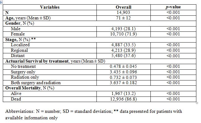
Methods: This study is a retrospective analysis using the Survey Epidemiology and End Results (SEER) database. Patients 20 years and older with thyroid cancer who underwent surgery from 1988 to 2012 were included. Cases were considered low risk if they were either classical papillary or follicular carcinoma, T1 (a or b) N0 M0 by TNM classification, and without extrathyroidal extension. Larger tumors, with unfavorable histology, nodal or distant metastases or extrathyroidal extension were considered high risk. We defined elderly as ≥ 70 years old. Trends in rates of total thyroidectomy (TT), radioactive iodine treatment (RAI) and lymph node dissection (LND) (removal of at least three lymph nodes) were analyzed and compared using chi square test, student's t-test, weighted least square linear regression and multivariate logistic regression where appropriate.

Results: 131,590 cases of DTC met our inclusion criteria and 11.3% were elderly. Overall, rates of TT have increased since 1988 by an average of 0.4% annually. The rate of increase was greater in the elderly compared to the younger cohort (0.58 vs. 0.38%/year, p = 0.02). This disparity between the elderly and younger was most pronounced within the low risk group alone (0.66 vs 0.3%/year, p = 0.03). Through the entire study period, the elderly remained less likely to receive a TT than those under 70 when controlling for all patient and tumor features (OR = 0.66, p < .001).

Rates of RAI increased between 1988 and 2012 by 0.19%/year. Annual rates of RAI treatment averaged 12.4 % lower in the elderly than the younger cohort (p = 0.063). The elderly and younger patients experienced similar rates of increasing RAI treatment (p = 0.99).

Annual rates of LND rose by 0.88%/year. Considering year of diagnosis alone, patients were much more likely to receive a LND in the most recent five years (2008 – 2012) compared to early years (OR 2.33, p < 0.01). Overall, elderly patients were half as likely to receive LND as younger patients in our model (OR 0.52, p < 0.01).

Conclusion: Treatment for DTC has become more aggressive over the last 20 years with increasing use of TT and LND. The elderly experienced this trend, but older age alone was not independently associated with more aggressive treatments. The overall increase in more extensive treatment for low risk tumors warrants improved risk adjusted treatment decisions.

14.17 Utilization of Preoperative Endoscopic Ultrasound for Pancreatic Adenocarcinoma

R. K. Schmocker1, D. J. Vanness2, C. C. Greenberg1, H. B. Neuman1, E. R. Winslow1 1University Of Wisconsin,Surgery,Madison, WI, USA 2University Of Wisconsin,Population Health,Madison, WI, USA

Introduction:
Technologies, such as endoscopic ultrasound (EUS), have increased the available staging modalities for patients with resectable pancreatic cancer. However, there currently is not a clear understanding about EUS utilization patterns and impact on management decisions, especially given that tissue diagnosis is not required prior to resection. Therefore, we set out to determine the extent of preoperative EUS use and the factors that increase the likelihood of EUS receipt for patient with resectable pancreatic cancer.

Methods:
We used the Surveillance, Epidemiology, and End Results (SEER) Medicare-linked database to identify patients with pancreatic adenocarcinoma. We included patients ≥66yo with the diagnosis of pancreatic adenocarcinoma, who underwent major pancreatic resection. The date of diagnosis was defined as the first staging procedure within the 6 months prior to surgery, with the staging period extending until the time of surgery. Univariate analysis compared demographic and clinical variables for those with EUS and those without. Logistic regression was used to determine factors most associated with preoperative EUS. The main outcome was EUS use in the staging period. Patient and disease factors associated with receipt of EUS were also identified. Secondary outcomes were number of staging tests, and time to surgery.

Results:
2,782 patients were included, 55% female with an average age of 74.7±5.5 years. 72% had regional disease (n=2002), and 56% were treated at an academic hospital (n=1563). Of all operations, 83.4% (2321/2782) had a proximal, total, or other pancreatectomy. 1204 patients underwent EUS (43.3%). On logistic regression the factors most strongly associated with receipt of EUS were: date of diagnosis, SEER area, a NCI or academic hospital, and a gastroenterologist as the first consultant (Table). Additionally, on multivariate regression, EUS was associated with mean increased time to surgery (17.79 days; p<0.0001), and independently associated with an increased number of staging tests (40 tests per 100 patients; p<0.0001).

Conclusion:
EUS is commonly used in the preoperative period, despite guidelines stating that tissue diagnosis is not required prior to surgery. Factors most associated with receipt of EUS are most strongly associated with geographic, temporal, and institutional factors, not clinical/disease factors. Additionally, patients with EUS had a longer time to surgery and an increase in the number of staging tests. This suggests that EUS may be overused in patients with resectable disease, with an increased cost and treatment delay without clearly adding clinical value.

14.18 Assessing Outcomes In Patients With Colon Cancer: The Effect Of Increasing Age

J. Harkins1, V. Pandit1, V. Nfonsam1 1The University Of Arizona,Surgery,Tucson, ARIZONA, USA

Introduction: Colon cancer (CC) continues to burden the health care system in the United States. In recent years, there has been a demographic shift in the incidence of CC among patients however; the factors associated with patient outcomes in patients with CC remains unknown. The aim of the study was to assess the factors associated with mortality and complications in patients with CC. We hypothesized that increasing age is associated with worse outcomes

Methods: We abstracted the national estimates for colon cancer diagnosis from the National Inpatient Sample database 2011 (representing 20% of all in-patient admissions). Patients undergoing surgical intervention were included. Patients were divided into groups based on age (Age 0-25yrs, 26-50yrs, 51-75yrs, and ≥76yrs). Outcome measures were: in-hospital complications (cardiac, sepsis, renal, hematological) and mortality. Regression analysis was performed.

Results:A total of 16,815 patients were included with mean age 69±13.5 years, median Charlson Comorbidity Index 2 [0-6] and 52% (n=8,751) females. 22.5% (n=3,782) patients had complications and mortality rate was 2.8% (n=465). After controlling for all factors, increasing age was independently associated with development of in-hospital complications (OR: 7.6 [1.9-16.2], p=0.001). Patient with increasing age were 1.87 times more likely to die compared to younger patients (OR: 1.87 [1.4-2.8], p=0.001).

Conclusion: Increasing age is associated with adverse outcomes in patients with colon cancer undergoing surgical intervention with worse outcomes in oldest patient (age≥76yrs). Further research is required to understand the reasons for the differences in these outcomes among patients independent of the cancer burden.

14.11 Features of Eligible Nipple-Sparing Mastectomy Patients Declining Nipple Preservation

L. A. Dossett1,2, J. Lowe1, W. Sun1, C. Lee1, P. D. Smith1,2, C. Laronga1 1Moffitt Cancer Center And Research Institute,Comprehensive Breast Program,Tampa, FL, USA 2University Of South Florida College Of Medicine,Tampa, FL, USA

Introduction: Nipple-sparing mastectomy (NSM) has rapidly grown in popularity as an approach to treating select patients with breast cancer and for risk reduction in patients with genetic mutations. Mounting data supports the oncologic safety of preservation of the nipple areola complex (NAC), and many presume quality of life (QOL) and other patient-centered outcomes are improved compared to skin-sparing mastectomy (SSM). The predictive factors for selection of NSM by eligible women are largely unknown. We sought to describe demographic, clinical and QOL characteristics of patients declining nipple preservation despite eligibility for NSM.

Methods: Women electing mastectomy with immediate reconstruction and eligible for NSM were prospectively offered inclusion in an IRB-approved nipple sensation and body image/QOL cohort study. Eligibility criteria for NSM: unifocal, small (<3cm) or no tumor (prophylaxis), tumor >2cm from the nipple-areolar complex (NAC), clinically node negative, body mass index (BMI) < 30, estimated breast weight <700 grams, and no or grade 1 ptosis. After signing informed consent, the women self-selected the control arm (SSM) or investigational arm (NSM). Baseline nipple/skin sensation testing and body image/QOL questionnaires were obtained pre-operatively. Demographic, clinical, and QOL data were collected prospectively. Descriptive and comparative statistics were performed.

Results:53 patients were enrolled; 38 (72%) patients selected NSM while 15 (28%) patients selected SSM without NAC preservation. Patients declining NSM despite eligibility were younger (43 ± 10 versus 49 ± 10 years, p=0.05), had a higher BMI (26 ± 4 versus 23 ± 3, p=0.02) and were more likely to have an indication of prophylaxis (strong family history or confirmed genetic carrier) (53% versus 21%, p<0.01). Corresponding to increased BMI, patients declining NSM also had larger breasts by weight (564 v 364 grams, p<0.001) as measured by gross pathology. On baseline QOL evaluation, patients declining NAC preservation were less satisfied with preoperative nipple sensation, as well as the size and appearance of their breast and nipple; these differences were not statistically significant.

Conclusion:Not all women eligible for NSM select this approach. Patient characteristics including age, BMI, satisfaction with the natural breast, and indication for mastectomy may predict patient preference and motivation with regards to preservation of the NAC during mastectomy. Patient motivation and acceptance of future cancer risk may explain these differences. Ongoing research regarding patient preferences may clarify decisions for NAC preservation and improve overall patient outcomes.

14.12 Gallbladder Carcinoma in the United States: Epidemiology and Outcomes Involving 14,903 Patients

K. Mahendraraj1, C. S. Lau1,3, R. S. Chamberlain1,2,3 1Saint Barnabas Medical Center,Surgery,Livingston, NJ, USA 2New Jersey Medical School,Surgery,Newark, NJ, USA 3Saint George’s University,Grenada, Grenada, Grenada

Introduction: Gallbladder carcinoma (GBC) is the most common malignancy of the biliary tract and third most common gastrointestinal tract malignancy. Although GBC is much less prevalent in North America compared to Asia, it is associated with extremely poor prognosis. This study examines a large cohort of GBC patients in the United States in an effort to define demographics, clinical and pathologic features impacting clinical outcomes in GBC patients.

Methods: Demographic and clinical data on 14,903 GBC patients was abstracted from the SEER database (1973–2012). Statistical analysis was performed with SPSS©v20.0 software using Chi-square test, paired t-test, multivariate analysis, and Kaplan-Meier functions.

Results: 14,903 cases of GBC were abstracted with a mean age of 71 years. GBC was significant more common among Caucasian (65.3%) females (71.9%), p<0.001. GBC was most often poorly or moderately differentiated (40.9% and 40.1%), p<0.001). 37.6% of GBC cases presented with distant metastasis, p<0.01. Surgery was the most common treatment modality for GBC patients (60.7%), p<0.001. Combination surgery and radiation, which was utilized in 11.5% of GBC patients, achieved similar survival rates compared to surgery alone (3.637 vs. 3.435 years, p<0.001). There was a significant increase in GBC incidence since 2000, with a rise in mean incidence of 1.8% between 1973 and 1999 period to 4.6% between 2000 and 2010, accompanying a rise in the mean incidence of surgical resection from 2.5% from1973 to 1999 to 5.0% from 2000 to 2010. Overall mortality was 86.8%, with 1- and 5-year survival rates of 35% and 13%, respectively, p<0.001. Multivariate analysis identified regional (OR 2.8) or distant disease (OR 2.1), lymph node positivity (OR 1.6), undifferentiated grade (OR 1.3), Caucasian race (OR 2.0), and male gender (OR 1.2) as independently associated with increased mortality for GBC, p<0.001.

Conclusions: GBC is an uncommon malignancy that presents most often among Caucasians females in their 8th decade of life, with over a third of cases presenting with distant metastasis. The incidence of GBC has doubled in the last decade concurrent with increases in cholecystectomy rates attributable in part to improved histopathological detection, as well as laparoscopic advances and enhanced endoscopic techniques. Surgical resection confers significant survival benefit in GBC patients, although a significant portion of GBC patients with regional disease remain untreated or undertreated. Given its rarity, all GBC patients should be enrolled in clinical trials or registries to optimize treatment and clinical outcomes for these patients.

14.13 Laparoscopic Colectomy for Cancer: Improved Compliance with Guidelines for Chemotherapy and Survival

R. H. Kim1, Q. D. Chu1, G. C. Caldito2 1Louisiana State University Health Sciences Center – Shreveport,Surgical Oncology,Shreveport, LA, USA 2Louisiana State University Health Sciences Center – Shreveport,Neurology,Shreveport, LA, USA

Introduction: Laparoscopic surgery for colon cancer has been demonstrated in clinical trials to have short term benefits, including shorter hospital stay and recovery period, when compared to the open surgical approach. NCCN guidelines recommend that patients with stage III or high risk stage II colon cancer undergo adjuvant chemotherapy. We hypothesized that laparoscopic colectomy is associated with increased compliance to recommendations for chemotherapy, shorter time to start of chemotherapy, and increased overall survival.

Methods: The National Cancer Data Base was queried to identify patients with stage III or high risk stage II (T4, positive margins, LN<12, or high tumor grade) colon adenocarcinoma diagnosed between 2010 and 2012. Patients were divided into laparoscopic colectomy (LC) and open colectomy (OC) groups based on the surgical approach used for their definitive surgery. Intent-to-treat analysis was used, with converted cases included in the LC group. Rates of receiving adjuvant chemotherapy, time from diagnosis and surgery to start of chemotherapy, and overall survival were compared. Chi-square test, two-sample t-test and Kaplan-Meier method were used for statistical analysis.

Results: A total of 48,257 patients were included for analysis. 18,801 patients underwent LC and 29,456 underwent OC. LC patients received adjuvant chemotherapy at a higher rate than OC (66.2% vs 59.4%, p<0.01). Among patients who received chemotherapy, time to start of chemotherapy after definitive surgery was shorter for LC than OC (p<0.01). Two-year overall survival was higher for LC than OC (81.9% vs 73.2%, p<0.01).

Conclusion: Laparoscopic colectomy is associated with higher rates of compliance with NCCN guidelines for adjuvant chemotherapy for stage III and high risk stage II colon cancer, as well as a shorter time to start of chemotherapy and improved overall survival, compared to open colectomy.

14.07 Hand-Assisted Laparoscopic Vs Open Colectomy Improves Outcomes Without Increasing Operative Time

C. T. Ong1, Z. Sun1, M. A. Adam1, J. Kim1, B. F. Gilmore1, B. Ezekian1, U. P. Nag1, C. R. Mantyh1, J. Migaly1 1Duke University Medical Center,Durham, NC, USA

Introduction:
Hand-assisted laparoscopic surgery (HALS) is gaining wider application in colorectal procedures as it bridges the divide between open and laparoscopic technique. However, there are concerns for whether this hybrid approach carries the complications of the open approach while sacrificing the perioperative benefits of completely minimally invasive technique. Our aim was to compare outcomes between HALS and open colectomy and delineate predictive factors for selecting the HALS technique.

Methods:
Adult patients who underwent elective open or HALS colectomies from the 2012-2013 combined National Surgical Quality Improvement Program dataset were selected. Short-term perioperative outcomes were compared between 1:1 propensity-matched groups. A subset analysis was performed in those who received segmental colectomies only. Multivariable logistic regression modeling was used to determine predictors of utilizing either operative approach.

Results:
In total, 8,791 patients were included. 2,707 (30.8%) patients received planned open colectomies and 6,084 (69.2%) received HALS. Independent predictors of utilizing HALS include male sex (OR 1.17, p=0.006), increasing BMI (OR 1.01, p=0.02), a benign indication for surgery (OR 1.48, p<0.001), and total abdominal colectomy compared to segmental (OR 10.39, p<0.001), while younger age, black race, ASA class >3, inflammatory bowel disease, and surgery requiring low pelvic anastomosis were predictive of open surgery (all p<0.05). After matching for clinical, disease, and treatment factors, HALS was associated with lower rates of overall complications (13.6% vs 21.5%, p<0.001), wound complications (8.8 vs 13.8%, p<0.001), anastomotic leak (3.1% vs 4.7%, p=0.014), transfusion requirement (5.0% vs 10.7%, p<0.001), postoperative ileus (8.8% vs 18.0%, p<0.001), length of stay (median 4 vs 6 days, p<0.001), and readmissions (6.9% vs 11.4%, p<0.001), without requiring longer operative time (median 148 vs 150 minutes, p=0.111). When examining those undergoing segmental resection only, HALS was still associated with improved rates of overall complications, wound complications, respiratory complications, postoperative ileus, anastomotic leak, transfusion requirement, length of stay, and readmissions (all p<0.05).

Conclusion:
Compared to open colectomy, hand-assisted laparoscopic surgery is associated with improved perioperative outcomes without requiring longer total operative time. In cases considered too difficult for a totally minimally invasive approach, HALS provides a more favorable alterative than the traditional open approach.

14.08 Predictors of Unplanned 30-Day Reoperation after Pancreatic Resection

H. G. Lyu2, G. Sharma1,2, E. Brovman2, R. Urman2, J. S. Gold2,3, E. E. Whang2,3 1Dana Farber Cancer Insititute,Surgical Oncology,Boston, MA, USA 2Brigham And Women’s Hospital,Surgery,Boston, MA, USA 3VA Boston Healthcare System,Surgery,West Roxbury, MA, USA

Introduction: Pancreatic resections are associated with a significant rate of postoperative complications, some of which may require reoperation. However, to date, studies reporting the frequency, indications, and risk factors for reoperation have been limited. We hypothesized that specific demographic, clinical, operative, and postoperative characteristics would predict the need for reoperation after pancreatectomy.

Methods: We examined clinically abstracted information of patients undergoing pancreatic resections at one of the 435 U.S. hospitals participating in the American College of Surgeons National Surgical Quality Improvement Program (ACS NSQIP) between January 1, 2011 and December 31, 2013. Debridements for pancreatitis and patients with missing reoperation data were excluded. Baseline, preoperative patient characteristics, index procedure characteristics, and postoperative course were retrospectively analyzed. Comparison of the cohort requiring unplanned 30-day reoperation with the cohort which did not require reoperation was performed utilizing Student’s t-test for continuous variables and Pearson’s Chi-Squared test with Yates’ continuity correction for categorical variables. All analyses were conducted using R Project for Statistical Computing, v3.1.2.

Results: Of the 15,658 patients undergoing pancreatectomy in our sample, 784 (5.0%) patients required unplanned reoperation. Reoperative patients were more likely to be male, have higher preoperative body mass index, cardiorespiratory comorbidities, lower preoperative baseline functional status and serum albumin, and higher American Society of Anesthesiologists class. Administration of neoadjuvant therapy was not significantly different between the two groups. Patients undergoing index conventional pancreaticoduodenectomy carried the highest reoperation rate (6.2%) whereas enucleation carried the lowest (2.4%) (Table.) Longer mean operative duration also predicted need for reoperation. Patients who ultimately underwent reoperation were more likely to have experienced postoperative cardiac, respiratory, thromboembolic, and infectious events. The rate of transfusion-requirement was more than 2-fold among patients who subsequently required reoperation. 100% of reoperations occurred during the index hospitalization, and total length of stay was more than double in the reoperative group (Table.)

Conclusion: Demographic characteristics, BMI, comorbid conditions, procedure type and duration, and postoperative events predict unplanned 30-day reoperation after pancreatectomy in the largest and most diverse sample to date. These findings identify at-risk patients; further study is required to identify preoperative measures to mitigate the risk of reoperation.

14.09 Survival is Related to Neutrophil/Lymphocyte Ratio after Neoadjuvant Therapy in Pancreatic Cancer

E. S. Glazer1, O. Rashid1, J. Pimiento1, P. J. Hodul1, M. P. Malafa1 1Moffitt Cancer Center,Gastrointestinal Oncology,Tampa, FL, USA

Introduction: The neutrophil to lymphocyte ratio (NLR, neutrophil count divided by lymphocyte count) is a marker for systemic inflammation and has been studied as a prognostic biomarker in numerous cancers. An NLR value greater than ~3-5 is associated with systemic inflammation. We hypothesized that changes to this ratio in patients undergoing neoadjuvant therapy for borderline pancreatic carcinoma would be predictive of long term survival after resection.

Methods: All patients in our borderline resectable pancreatic carcinoma database (2006-13) were included if they completed neoadjuvant chemoradiotherapy, underwent resection, and complete data was available. Neoadjuvant chemotherapy usually consisted of gemcitabine, docetaxel, & capecitabine while radiotherapy was usually 40 Gy with SBRT. The difference in NLR was calculated as the NLR before surgery (after neoadjuvant therapy) minus the NLR before neoadjuvant therapy. A clinically significant increase in NLR was defined as an increase by 2.5 or more units while any differences less than 2.5 units (including negative differences) were defined as stable NLR. Statistical significance was α = 0.05, survival was investigated with the Kaplan-Meier method, and uncertainties are standard deviations.

Results: 58 patients were identified; all patients completed neoadjuvant therapy and 97% had R0 margins. The mean age was 65 ± 9 years; 60% were male. The mean pre-neoadjuvant NLR was 3.1 ± 2.4 while the mean post-neoadjuvant/pre-surgical NLR was 4.4 ± 3.5. Overall survival was significantly worse in the increased NLR group compared to the stable NLR group (see figure, P = 0.009) with a Cox hazard ratio of 2.9 (P = 0.02). There were no significant differences in age, gender, tumor location, or Ca19-9 levels between the 2 groups. Survival was not associated with adjuvant chemotherapy administration (P = 0.4) or AJCC tumor stage (P = 0.9), but N0 disease conferred a survival advantage over N1 disease (Cox hazard ratio = 3.0, P = 0.01). There was no association between changes in NLR and nodal stage (P = 0.8). On multivariate Cox regression analysis, both increased NLR and N1 disease remained independent predictors of worse survival (P < 0.008 for both).

Conclusion: This is the first investigation demonstrating an independent, inverse association between survival and increase in NLR after neoadjuvant therapy followed by resection for patients with borderline resectable pancreatic carcinoma. We theorize that a pro-inflammatory state is related to worse survival despite a high rate of R0 resections. It is unclear if modulation of inflammation will improve survival in patients with pancreatic carcinoma, but we are actively investigating this in the laboratory.

14.04 Preoperative Radiation Compared with Surgery Alone in the Treatment of Retroperitoneal Sarcomas.

A. Porpiglia1, j. M. Farma1, M. Von Mehren1, S. Movva1, P. R. Anderson1, A. S. Patchefsky1, S. Reddy1 1Fox Chase Cancer Center,Surgical Oncology,Philadelphia, PA, USA

Introduction: Retroperitoneal sarcomas account for 15% of all soft tissue sarcomas and they have a worse prognosis than extremity soft tissue sarcomas. The reason for this is multifactorial, including delay in diagnosis, surgically unresectable disease, and high recurrence rates. Recurrence rates for retroperitoneal sarcomas have been reported as high as 70%. Complete surgical resection is the most important prognostic factor for prolonged survival. Radiation therapy has been utilized to help achieve negative surgical margins and to potentially decrease rates of local recurrence. The study was to review the experience at a tertiary referral center specializing in the multidisciplinary care of treating patients with retroperitoneal sarcomas. The goal of the study was to determine if there was a difference in local recurrence rates or postoperative morbidity for patients treated with preoperative radiation therapy and surgery or for those treated with surgery alone.

Methods: Retrospective review of patients treated for retroperitoneal sarcomas at Fox Chase Cancer Center from 1997-2015, with IRB approval. Chi-square and Student t-tests were used to assess primary endpoints of recurrence and postoperative morbidity.

Results: There were 73 patients identified with retroperitoneal sarcomas treated with preoperative radiation therapy and surgery (18patients) or surgery alone (55 patients). The median age was 60 years and there were more females than males, 55% versus 45% respectively. The predominant histologies were: liposarcoma, leiomyosarcoma, and malignant fibrous histiocytoma. Sixty-eight percent of patients had tumors greater than 10cm. There were more low-grade tumors in the surgery alone group and there were more high-grade tumors in the XRT/surgery group. However this was not statistically significant, p=0.17. Fifty-one percent of the patients developed recurrence. The surgery alone group had more local recurrences compared to the surgery/XRT group (36% versus 17%, p=0.014). Complication rate for the entire group was 35.6%. However, there was no difference in postoperative morbidity between the two groups.

Conclusion: The mainstay of treatment of patients with retroperitoneal sarcomas is surgery. However, with the high rates of local recurrence rates, the large size of the tumors, and involvement of adjacent organs radiation therapy is a viable adjunct to surgery. Our study demonstrated improved local recurrence rates with radiation therapy with no increase in morbidity.

14.05 Utility Of The ‘Mini-Shave’ Technique In Reducing Positive Margin Rates After Partial Mastectomy

J. Hanna1, D. Lannin1, M. Butler1, A. B. Chagpar1 1Yale University,Surgery,New Haven, CT, USA

Introduction: Recent randomized controlled trials have demonstrated that taking cavity shave margins in women undergoing partial mastectomy (PM) results in a halving of positive margin and re-excision rates. We sought to determine whether taking a ‘mini-shave’, or a tiny sample of each margin, would similarly reduce the positive margin rate, while removing less tissue.

Methods: A retrospective cohort study was performed of patients undergoing PM with mini-shave excision of margins between October 21, 2011 and November 22, 2013. Patient demographics and clinicopathologic tumor features were collected, along with margin status and volume of tissue excised from the primary PM specimen as well as the mini-shaves. A positive margin was considered to be no tumor at ink for invasive cancer and tumor within < 1 mm for DCIS. Statistical analyses were performed using SPSS Version 21 software.

Results: There were 121 patients in the cohort of interest. The median patient age was 62. 21 (17.4%) had invasive cancer, 31 (25.6%) had DCIS, and 69 (57.0%) had both. The median size of invasive cancer was 1.3 cm, and the median size of DCIS was 1.2 cm. 8 (6.6%) patients had invasive lobular cancer, and 3 (2.5%) had neoadjuvant chemotherapy. The median volume of the initial PM specimen was 85.8 cm3, and 27 (22.3%) had initially positive margins. While the same number of patients (27, 22.3%) also had additional tissue removed intraoperatively prior to mini-shave excision, only 7 (25.9%) of these had positive margins on their PM specimen. Selective excision of additional tissue did not significantly reduce the positive margin rate (22.3% to 19.8%, p=0.250 by McNemar test). All but one patient had mini-shaves taken from all six faces; the median total volume of mini-shaves excised per patient was 1.3 cm3. The median ratio of mini-shave:PM surface area was 0.03. The mini-shaves were negative in all but 4 patients, resulting in a reduction of the positive margin rate from 19.8% to 3.3%, p<0.001. Six patients underwent re-excision; three of whom had a mastectomy. 5 (83.3%), including the two who had negative mini-shaves, were found to have residual cancer. All five had DCIS and one had invasive cancer; the median size of the residual disease was 1.6 cm.

Conclusion: Taking mini-shaves at the time of PM results in an 83% reduction in positive margin rates. However, mini-shaves sample only 3% of the surface area of the initial cavity, and therefore may not accurately reflect the presence or absence of residual disease. It is unclear whether this potential sampling error will affect recurrence rates, particularly in the current era of nearly ubiquitous adjuvant systemic and radiation therapy.

14.06 Using Administrative Claims to Understand Care Coordination and Treatment in Stage III Colon Cancer

R. L. Hoffman1, K. D. Simmons1, C. B. Aarons1, R. R. Kelz1 1University Of Pennsylvania,Philadelphia, PA, USA

Introduction: The ability to link cancer patient records across encounters could provide important information regarding the patterns of care associated with best practice. Using colon cancer as a model, the aim of this study was to investigate the role of physician type and order on the receipt of guideline-based therapy(GBT).

Methods: Patients aged 65-84yrs who underwent resection for AJCC stage III colon cancer were identified within the SEER-Medicare database(2005-2009). The administration of chemotherapy and/or radiation was determined using CPT codes across all encounters within 365 days of the colectomy date. Guideline adherence was assigned using stage-specific NCCN guidelines. Provider specialty(surgery, oncology, internal medicine, gastroenterology) and dates of service were identified from the first 4 claims which contained an ICD-9 diagnosis of colon cancer. Univariate analysis was performed. Forward stepwise multivariate logistic regression controlling for patient demographics and stratified by age group was performed to determine the association between physician type/order with inappropriate care.

Results:A total of 6139 stage III colon cancer patients were identified. The cohort was 56% female, 83% white, 73% <80 years of age. 69% of patients had ≥3 comorbidities. 57% received chemotherapy and 5% underwent radiation. 47% did not receive appropriate GBT(2880), 90%(2596) of whom were undertreated. A surgeon was seen within the first 4 claims following the diagnosis for 64%(3924) of cases and an oncologist in 15%(947). Patients saw multiple physicians on the same date 18%(1100) of the time. On univariate analysis, seeing a surgeon(OR 1.19; 95% CI 1.07-1.32), oncologist(OR 1.22; 95% CI 1.06-1.40) or gastroenterologist(OR 1.11; 95% CI 1.00-1.24) was associated with an increased odds of appropriate GBT, however physician order did not make a difference. On multivariate analysis, seeing a surgeon was associated with a significantly increased odds of appropriate GBT at all ages except >80 years(65-69 yrs OR 1.30, 95% CI 1.00-1.70) (70-74 yrs OR 1.35, 95% CI 1.07-1.69) (75-79 yrs OR 1.33, 95% CI 1.08-1.63) (≥80 yrs OR 1.06, 95% CI 0.85-1.33) and seeing an oncologist was significant for those age 75-79(OR 1.39, 95% CI 1.05-1.85). Other physician types were not significant. The physician of first contact was not significant except for those age 80-84yrs, where seeing an internist first was associated with an decreased odds of appropriate treatment(OR 0.60; 95% CI 0.38-0.95).

Conclusion:Rates of receipt of GBT in stage III colon cancer are low in the Medicare population, however seeing a surgeon within the first 4 physician visits has a significant impact on improving the rates of GBT in patients less than 80 years of age. Understanding the influence of provider type/order on the receipt of appropriate cancer care as a proxy for care coordination may allow a more robust understanding of where deficits occur in the patient experience.

14.02 Impact of Minimally Invasive Distal Pancreatectomy on Use of Chemotherapy for Cancer and Survival

K. L. Anderson1, M. A. Adam2, S. Thomas3, S. A. Roman2, J. A. Sosa2,4 1Duke University School Of Medicine,Durham, NC, USA 2Duke University Medical Center,Department Of Surgery,Durham, NC, USA 3Duke University,Department Of Biostatistics,Durham, NC, USA 4Duke Clinical Research Institute,Durham, NC, USA

Introduction: Interest in minimally invasive distal pancreatectomy (MIDP) has increased. Published data examining impact of MIDP on survival are limited to experiences from high-volume institutions. Our aims were to compare adjuvant chemotherapy use and overall survival between MIDP and open surgery (ODP).

Methods: Adult patients undergoing distal pancreatectomy for Stage I and II pancreatic adenocarcinoma were identified from the National Cancer Data Base, 2010-2012. Univariate and multivariate regression and survival analysis were employed to examine the association between MIDP (laparoscopic or robotic) vs. ODP and use of adjuvant chemotherapy and oncologic outcomes.

Results: A total of 1,807 patients underwent distal pancreatectomy for adenocarcinoma at 506 institutions: 505 (28%) MIDP, and 1,302 (72%) ODP. Use of MIDP from 2010 to 2012 increased from 131 (26%) to 209 (41%) cases. The majority of MIDP cases were performed at academic institutions (68%) compared to comprehensive community (30%) and community hospitals (2%). After adjustment, compared to patients who underwent ODP, those who underwent MIDP were more likely to have complete tumor resections [OR 1.53 (CI 1.09-2.17), p=0.02] and a shorter hospital length of stay (LOS) [-11%, (CI -5% to -16%), p=0.0002]. MIDP is associated on average with an 11% shorter LOS, which would be a reduction of about 1 day from the average ODP LOS of 10 days. The rates of 30-day readmission [OR 1.11 (0.72-1.72), p=0.62] and 30-day mortality [OR 0.83 (0.65-1.06), p=0.13] were similar between groups. Patients undergoing MIDP vs. ODP were more likely to receive adjuvant chemotherapy [OR 1.26 (1.00-1.60), p=0.05]; time to initiation of adjuvant chemotherapy was not different between groups [-2%, (CI -8% to 4%) p=0.45). Median follow-up was 17 months (IQR 10-26 months). In unadjusted analysis, 3-year survival was similar between MIDP and ODP (40% vs. 37%, p=0.42). After adjustment, overall survival for MIDP vs. ODP remained similar (HR 0.85, (CI 0.67-1.10) p=0.21).

Conclusion: Over the years, utilization of MIDP has increased, with the majority of procedures performed at academic institutions. It is associated with increased use of adjuvant chemotherapy; further study is needed to understand the etiology and impact of this association.

14.03 Comparative Outcomes Among Surgical and Ablative Treatments for Small Localized Renal Cell Carcinoma

T. Wittmann1, S. Best1, S. Wells1, T. Ziemlewicz1, J. L. Hinshaw1, M. Lubner1, F. Shi1, F. T. Lee1, S. Nakada1, E. J. Abel1 1University Of Wisconsin School Of Medicine And Public Health,Madison, WI, USA

Introduction: While surgery is the gold standard treatment for small (≤4cm) renal cell carcinoma (RCC), percutaneous cryoablation provides a nephron-sparing alternative that may also result in curative outcomes. In addition, the recent development of high-powered microwave ablation (MWA) may offer potential benefits over cryoablation. The objective of this study was to evaluate disease recurrence after treatment for patients with small RCC treated with ablation or surgery.

Methods: A total of 489 patients were treated with percutaneous ablation or surgery for localized RCC (confirmed with biopsy or surgical pathology) at our institution from 2001-2015. Differences in patient and disease characteristics among patient cohorts were evaluated using t-test or chi squared analysis.

Results: Of the 489 patients, 314 (64.2%) were treated surgically, 100 (20.4%) were treated using MWA and 75 (15.3%) had cryoablation. There was no difference in gender, race, body mass index, smoking history, R.E.N.A.L. nephrometry score, laterality, tumor diameter, or histologic RCC subtype among ablative treatments or surgery (p=0.13, 0.08, 0.08, 0.18, 0.16, 0.29, 0.80, 0.3).

Patients treated surgically were younger (p<0.0001) with median age 56.9 (IQR 48-66) compared to MWA (median age 67.0 IQR 59-72) or cryoablation (median age 65, IQR 60-74). Patients treated surgically were also more likely to have symptoms at presentation (p=0.01) compared to ablative treatments and more likely to have higher nuclear grade compared to ablative treatments (0.0002). Median Charlson Comorbidity Index was 2 (IQR 1-3) in surgically treated patients, compared to 3 (IQR 2-4) and 3 (IQR 2-4) in MWA and cryoablation respectively (p<0.0001).

Median follow-up was 9 months (IQR 6-18) for patients treated with MWA compared to 40 months (IQR 24-60) for cryoablation and 41 months (IQR 15-80) for surgery (p<0.0001).

Local recurrence was identified in 11(14.7%) patients following cryoablation compared to 1(1%) patients following MWA or 3(1%) after surgery, (p<0.0001). Development of metastatic RCC was observed in 2 (2.7%), 0 (0%), and 12 (3.8%) of patients following cryoablation, MWA or surgery respectively, (p=0.13).

Conclusion: Local recurrence is rare after surgery or percutaneous microwave ablation for small localized RCC. Cryoablation is associated with higher rates of local recurrence and retreatment compared to surgery. Future studies with longer follow-up are necessary to determine whether local recurrence rates are superior after treatment with MWA compared to cryoablation.

09.20 A State-wide Assessment of Cholecystectomy Outcomes in the Elderly

D. J. Li1, M. Terjimanian1, L. M. Napolitano1, M. Englesbe2, D. A. Campbell2, K. B. To1 1University Of Michigan,Acute Care Surgery / Dept Of Surgery,Ann Arbor, MI, USA 2University Of Michigan,Dept Of Surgery,Ann Arbor, MI, USA

Introduction:
Laparoscopic cholecystectomy (LC) is the procedure of choice for treatment of cholelithiasis/cholecystitis, and carries a lower risk for morbidity and mortality when compared to open cholecystectomy (OC). Likewise, elective/semi-elective cholecystectomy (EL) carries a lower complication rate than emergency cholecystectomy (EM). When assessing risk across age groups, there is limited data regarding complication and mortality risk for older patients. We hypothesize that while there will be an expected difference in older patients, the difference will be greater for emergency operations in older patients, and especially so for those undergoing open cholecystectomy.

Methods:
Prospective data were obtained from a state-wide Surgical Quality Collaborative (SQC) database with a random sample of 20-30% of all surgeries performed 1/1/2005-12/31/2010. Patient characteristics, preoperative comorbidities, complication and mortality rates were compared for emergent and elective cases. Patient outcomes were stratified according to age groups: (1) age <45years; (2) age 45-64 years; (3) age 65-79 years; and (4) age >80 years

Results:
Over the 5-year time period, 18106 cholecystectomies were logged into the SQC database. A total of 1409 cases were classified as emergency operations, of which 1080 were completed laparoscopically and 329 were completed open. A total of 16697 patients in the database underwent elective operations, of which 15185 were laparoscopic and 1512 were open. Overall, 71.5% of patients were female, with a mean age of 49.9 years. Overall morbidity was 2.2% for elective LC, 14.6% for elective OC, 3.7% for emergent LC and and 24% for emergent OC cases. Patient data as stratified by age groups are listed in Table 1. The difference in outcome between age groups 3&4 in the emergent setting is much greater than the difference between the same age groups in the elective setting (P<0.001).

Conclusion:
Patient who are >80 years of age have significant risk for morbidity and mortality in both elective and emergency cholecystectomies. This increase in risk overall, the difference is much higher in the emergent population, regardless of surgical approach. This demonstrates a need for closer analysis for improvement in the care of elderly patients undergoing cholecystectomy, especially in the emergent setting.

13.20 SPECT/CT Improves SLN Yield and Nodal Positivity Rate in Head and Neck Melanoma

B. C. Chapman1, A. Paniccia1, J. Merkow1, J. J. Kwak2, P. Koo2, B. Bagrosky2, N. Pearlman1, C. Gajdos1, M. McCarter1, N. Kounalakis1 1University Of Colorado School Of Medicine,Department Of Surgery,Aurora, CO, USA 2University Of Colorado School Of Medicine,Department Of Radiology,Aurora, CO, USA

Introduction: Sentinel node positivity is the single most important prognostic factor in predicting survival in cutaneous

melanoma. Traditionally, sentinel lymph nodes (SLN) are identified preoperatively using 2-D planar lymphoscintigraphy;

however, a new technique utilizing SPECT/CT may improve nodal detection rate in head and neck melanoma. The

purpose of this study is to compare lymph node yield and nodal positivity rates utilizing SPECT/CT versus conventional

lymphoscintigraphy.

Methods: Retrospective review of a prospectively maintained database of patients undergoing SLN biopsy for cutaneous

melanoma of the head and neck between February 1998 and June 2015. Patient demographics, melanoma pathologic

features, number of SLN, and nodal positivity rates were compared in patients utilizing SPECT/CT versus conventional

lymphoscintigraphy. A multivariable logistic regression analysis was utilized to identify factors associated with the

identification of a positive sentinel lymph node.

Results: Two hundred seventy-eight patients underwent SLN biopsy: 201 underwent traditional lymphoscintigraphy and

77 patients underwent SPECT/CT. There was no difference in gender (75% vs. 75% males; p=0.95), however the

SPECT/CT group trended towards being older (57 vs. 53 years, p=0.05). The depth of primary lesion was similar in the

two groups (2.1 vs. 2.1 mm; p=0.76) and incomplete data in the lymphoscitigraphy group limited the analysis on

ulceration and mitotic rate. The total number of SLN identified was greater in the SPECT/CT group (2.7 vs. 2.4;

p=0.0292) and a positive SLN was identified more frequently in the SPECT/CT group (n=16, 20.8% vs. n=24, 8.6%;

p=0.060). Age , gender, location of primary lesion, presence of ulceration, total number of lymph node harvested, and

intraoperative technique utilized to identify SLN (radiocolloid with or without blue dye injection), were not associated with

SLN positivity; however, depth of primary lesion (OR 1.40; p=0.002) and use of SPECT/CT (OR 2.75; p=0.023) were

significantly associated with a positive SLN. The multivariable logistic regression model c-statistic was 0.72, indicating a

moderate predictive value.

Conclusion: Patients with head and neck melanoma who undergo SPECT/CT have higher SLN yields. After controlling

for common factors associated with the presence of positive SLN, the use of SPECT/CT has 3-fold higher likelyhood of

identifying a positive SLN compared to traditional lymphoscintigraphy. Long-term follow-up is needed to further define the

impact of SPECT/CT on recurrence and survival.

13.21 Weighing the Oncogenic Role of the A-Kinase Anchor Protein-12 in Pancreatic Cancer

M. Beveridge1, D. Delitto1, J. Trevino1, G. A. Sarosi1,2, S. J. Hughes1, R. M. Thomas1,2 1University Of Florida,College Of Medicine, Department Of Surgery,Gainesville, FL, USA 2NF/SG Veterans Health System,Department Of Surgery,Gainesville, FL, USA

Introduction: A large majority of patients with pancreatic ductal adenocarcinoma (PDAC) have metastatic disease at the time of diagnosis. The mechanisms whereby PDAC is able to metastasize are still poorly understood. A-kinase anchor proteins (AKAP) are a family of scaffolding proteins that mediate signal transduction and cellular localization of protein kinase A, an inducer of cell motility. AKAP12 is one member that has shown a role in a variety of highly-metastatic cancers including colon, melanoma, and prostate but its role in PDAC is undefined.

Methods: AKAP12 expression was determined by immunoblot in both commercially available PDAC cell lines as well as from patient-derived PDAC tumors. The effect of AKAP12 knockdown with siRNA on cell proliferation and migration was determined by measurement of Alamar Blue reduction and transwell migration assay to culture media supplemented with 5% fetal bovine serum, respectively. The AKT, ERK, and MEK pathways were interrogated by immunoblot to ascertain changes in signaling associated with abrogation of AKAP12 expression.

Results: A differential expression was noted between PDAC cell lines with a 19, 27, and 328-fold increase in expression of AKAP12 in BxPC3, L3.6pl, and Panc-1 cell lines compared to MiaPaCa. Additionally, a 17.6-fold increased mean expression of AKAP12 was noted in patient-derived PDAC samples compared to normal (p=0.05). While no statistical difference was seen in proliferation after AKAP12 knockdown, migration at 48hrs was significantly reduced by 95.5% (p=0.05) and 86.5% (p=0.02) in BxPC3 and Panc-1, respectively after AKAP12 siRNA knockdown compared to control (Figure 1). Finally, AKAP12 knockdown resulted in the 23-fold, 6-fold, and 4.5-fold increase in phosphorylation of AKT, ERK, and MEK proteins in Panc-1 cells, respectively and 34-fold, 56-fold, and 1.6-fold increase in BxPC-3, respectively.

Conclusions: AKAP12 is differentially expressed in human PDAC tissue samples and cell lines. Inhibition of AKAP12 results in decreased migration in vitro but no effect on proliferation. This may result in compensatory increases in the activation of the AKT and MEK-ERK pathways. AKAP12 may play a role in pancreatic carcinogenesis but further studies are needed to clarify its role.

14.01 A New Index ‘Air–Bubble Sign’ for Early Detection of Anastomotic Leakage After Esophagectomy.

Y. Shoji1, H. Takeuchi1, H. Kawakubo1, K. Fukuda1, R. Nakamura1, T. Takahashi1, N. Wada1, Y. Kitagawa1 1Keio University, School Of Medicine,Department Of Surgery,Tokyo, TOKYO, Japan

Introduction: Operation for esophageal cancer is one of the most invasive operation in digestive surgery. Incidence of postoperative complication is relatively high than that of other gastrointestinal tract surgery. Anastomotic leakage is one of the critical complications after esophagectomy. Early diagnosis and initiation of treatment is essential. In our institute, we developed a new diagnostic procedure for postoperative anastomotic leakage using computed tomography examination (CT) at the 6th postoperative day after esophagectomy for esophagial cancer.?

Methods: From January 2012 to April 2015, 156 patients with esophageal cancer underwent curative resection under thoracolaparotomy in our institute. We made a comparative review of patient characteristics, surgical outcomes, and findings from the postoperative CT and the videofluorographic examination for swallowing (VF) of the 138 patients, which were reconstructed primary by the gastric tube (including salvage operation). Anastomotic leakage was defined as discharge of the digestive fluid from wounds or drains, which required medical and/or operative treatment (above grade 2 in Clavien – Dindo classification). Gastric tube stump leakage and anastomotic leakage were undifferentiated.

Results: Twenty-four cases, 17% suffered Anastomotic leakage (AL (+) group), and 114 cases, 83% did not (AL (-) group). There were no significant differences in patient characteristics such as age, sex, stage of the disease, location of the tumor, and preoperative treatment such as endoscopic extraction and chemotherapy and/or radiation therapy. Surgical outcomes such as usage of laparoscope / thoracoscope, field of lymph node dissection, reconstruction route, anastomotic site, operative duration, and intraoperative blood loss were equivalent in both groups. Mean number of air-bubbles (larger than 2 mm in diameter) in the cervical division and the mediastinal space by the CT image was significantly higher in the AL (+) group (AL (+) group, 4.8; AL (-) group, 0.7; P<0.001). When we decide 3 air-bubbles as a cutoff value ("air – bubble sign"), sensitivity and specificity of "air – bubble sign" against anastomotic leakage were 92% and 96%, respectively. In contrast, sensitivity and specificity of the postoperative VF against anastomotic leakage were 63% and 100%, respectively.

Conclusion: "Air-bubble sign" in the postoperative CT has higher sensitivity?than VF for diagnosis of anastomotic leakage after esophagectomy. "Air – bubble sign" may be a substitute for VF.

13.17 Location and Nodal Status Predict Survival for High-grade Gastrointestinal Neuroendocrine Tumors

C. Mosquera1, N. J. Koutlas1, N. A. Vohra1, T. L. Fitzgerald1 1East Carolina University Brody School Of Medicine,Surgical Oncology,Greenville, NC, USA

Introduction: Because of limited sample size in existing case series, the natural history and optimal management for patients with high-grade gastroenteropancreatic neuroendocrine tumors (GEP-NET) is poorly understood. In order to better grasp the implications of this diagnosis, a large cohort study was undertaken.

Methods: Patients diagnosed with high-grade GEP-NET from 1988-2010 were identified in the SEER registry. Primary sites with 5 or fewer patients, unknown surgery, or stage IV (n=171) were excluded.

Results:A total of 335 patients met inclusion criteria. Median age was 65 years, a majority were female (52.2%), white (84.5%), and had surgery (89.0%). The most common primary site was colorectal (38.2%) followed by appendix (19.7%), small bowel (15.8%), pancreas (14.9%), and gastric (11.3%). Median tumor size was 3.5 cm. The most frequent T classification was T3 (33.7%) followed by T4 (21.5%), T1 (16.4%), and T2 (9.0%). Surgical resection was performed in 89% of patients and varied by site, more commonly for appendix (97.0%), colorectal (94.5%), and small bowel (88.7%) and less commonly for pancreatic (78.0%) and gastric (71.1%) (p< 0.0001) primaries. Nodal involvement was frequent (41.2%) and varied by site; pancreas (57.8%), colorectal (55.1%), small bowel (44.0%), appendix (27.4%), and gastric (24.2%) (p= 0.0002). Pancreatic (57.5%) and colorectal (52.4%) neoplasms were more likely to be > 4 cm in size (p< 0.0001). The 5-year disease-specific survival (5-y DSS) was 63.3%, longest for small bowel (81.1%), followed by gastric (76.9%), appendix (75.5%), colorectal (54.2%), and pancreas (45.4%) (p= 0.0003). 5-y DSS was also associated with age (< 65 years 70.7% vs. ≥ 65 years 56.1%; p= 0.0034), node status (N0 73.6% vs. N1 54.2%; p= 0.0012) and surgery (Yes 64.6% vs. No 51.5%; p= 0.058). On multivariate analysis, node status (OR 1.91; p= 0.0091), surgery (5.25; p= 0.0010), and primary site continued to be associated with survival while age was not (1.50; p= 0.089). More specifically, small bowel (5.41; p= 0.0003) and gastric (3.90; p=0.0075) primaries had better survival compared to appendix (1.89; p= 0.16), colorectal (1.16; p= 0.79), and pancreas (referent). The incidence of high-grade GEP-NET increased from 0.03 to 0.19 per 100,000 over the study period, (R2= 0.79, p< 0.0001).

Conclusion: High-grade GEP-NET is a rare neoplasm that is increasing in incidence. Primary site and nodal status are useful predictors of survival. In contrast to current literature for this disease, a benefit was demonstrated for patients with localized disease undergoing surgical resection.

13.18 Circulating Tumor Cells and Relapse in Stage IV Cutaneous Melanoma Patients

C. Hall1, M. Ross1, M. Karhade1, J. B. Bauldry1, J. Upshaw1, R. Royal1, L. Valad1, A. Lucci1 1University Of Texas MD Anderson Cancer Center,Department Of Surgical Oncology,Houston, TX, USA

Introduction: Our group (and others) have demonstrated that circulating melanoma cells (CMCs) can be detected in stage IV melanoma patients, yet there is limited data regarding the prognostic significance of CMCs. The aim of this study was to determine if CMCs detected with a semi-automated platform predict early relapse in stage IV melanoma patients.

Methods: Serial CMC assessments (7.5mL blood) were performed in patients with stage IV cutaneous melanoma (n=58) using the CellSearch® system (Janssen Diagnostics). CD146+ cells were immunomagnetically enriched; CD146+, HMW-MAA+/, CD45-/, and CD34- nucleated cells were considered CMCs. We correlated the identification of CMCs with tumor characteristics using chi-square or Fisher exact tests. Log-rank test and Cox regression analysis was applied to determine the association of circulating tumor cells with relapse-free survival. Relapse-free survival (RFS) was compared between patients with ≥1 CMC detected at baseline (first blood draw) or at second blood draw (6 months after baseline) versus those with no CMCs at baseline and at the second blood draw.

Results: CMCs were identified in 28 of 58 (48%) patients at either baseline or at second blood draw. We observed no significant association between CMC presence at baseline draw and primary tumor factors such as presenting Breslow thickness, number of mitotic figures, or ulceration (P=NS for all factors). Relapse occurred in 22 of 28 (79%) stage IV patients with ≥1 CMC versus 12 of 30 (40%) with no CMCs at baseline and at second draw (log-rank P =0.02, HR 2.24, 95% CI 1.10 to 4.55; P=0.02). No other primary tumor characteristic, including presenting Breslow thickness, number of mitotic figures, ulceration, or Braf status, predicted shortened relapse-free survival.

Conclusion: Using a semi-automated platform, CMCs detected at baseline or at a subsequent blood draw, predicted relapse in stage IV melanoma patients. These data strongly support further study with long follow-up and serial blood draw to validate the prognostic significance and utility of CMC measurement in melanoma patients.