06.12 Non-Platelet Length Polyphosphates Inhibits tPA Mediated Fibrinolysis

P. J. Lawson1,2, H. B. Moore1,2, A. W. Bacon1,2, E. Gonzalez1,2, A. P. Morton1,2, A. Banerjee1, J. H. Morrissey3, E. E. Moore1,2 1University Of Colorado Denver,Surgery,Aurora, CO, USA 2Denver Health Medical Center,Surgery,Denver, CO, USA 3University Of Illinois,Surgery,Urbana, IL, USA

Introduction:
Fibrinolysis shutdown is the dominant phenotype in severely injured patients, but the mechanisms driving post-injury tPA resistance remain unknown. Polyphosphates are ubiquitous inorganic compounds that have recently been linked to coagulation. Polyphosphates are stored in platelets, and platelets are known to inhibit fibrinolysis. We hypothesize that polyphosphate polymers that are similar in size to the length of chains stored in platelets will inhibit tPA mediated fibrinolysis.

Methods:
Polyphosphates of four different polymer lengths were evaluated. Monophosphate, platelet length polyphosphates, medium size polyphosphates found in tissue, and large polyphosphates of the size produced by microbes. Polymers of different sizes were added at a concentration of 20 micromolar in whole blood from eight healthy volunteers and assessed with thrombelastography (TEG) for changes in clotting parameters with and without tissue plasminogen activator (tPA) to quantify inhibition in fibrinolytic activity.

Results:
Large polyphosphates reduced R time and increased angle in the presence of tPA compared control blood (p<0.001, p=0.008) and trended towards an increase in clot strength (MA p=0.052). Large, medium, and mono polyphosphates reduced LY30 compared to control (p=0.034, Figure 1). The other polymer lengths in the presence of tPA did not significantly alter other clotting parameters. Without tPA, the same relationship of shortened clotting time and increased angle were appreciated with the large polyphosphates (p<0.001, p=0.015). None of the other polymer lengths affected clot parameters.

Conclusion:
Large chain polyphosphates are associated with a rapid forming strong clot. Polyphosphates polymers in the length stored in platelets, however, did not impact clotting parameters. But all other polymer lengths inhibited tPA mediated fibrinolysis. These experiments indicate that platelet derived polyphosphates are either modified after release from stored granules, or the polyphosphates regulating coagulation are from another source.

06.13 High Glucose Does Not Inhibit Aortic Endothelial Cell Sprouting and Stimulates Proliferation

A. D. Morris1, H. Li1, K. Kuo1, S. Dalal1, L. P. Brewster1,2 1Emory University School Of Medicine,Division Of Vascular Surgery,Atlanta, GA, USA 2Atlanta VA Medical Center,Division of Vascular Surgery,Atlanta, GA, USA

Introduction:
Diabetes increases the risk of cardiovascular disease and major amputation, but the aggressive control of blood sugar levels in recent clinical trials did not correspond to better clinical outcomes. Endothelial cell regeneration is critical to preventing thrombotic and myointimal complications after vascular injuries and interventions, but the impact of hyperglycemia on this process is not well understood. Since hyperglycemia directly inhibits cellular homeostasis, we propose that it negatively impacts endothelial cell regeneration.

Methods:

Proliferation Protocol:

Human aortic endothelial cells(HAEC) were grown in standard culture conditions, and then plated in a 96-well plate at 5000 cells per well. After reaching ~70% confluence, the cells were quiesced with serum starvation for 24 hours. The cells were restimulated with growth media (positive control) or quiescent media (negative control) with 40mM glucose or 40mM mannose (oncotic control) added to both groups. After 72 hours, HAEC proliferation was collected by commercially available cell viability assay using absorbance.

3D Angiogenesis Assay:

HAECs were formed into pellets (40,000 cells) using mechanical rotation. The pellets were suspended in a fibrin hydrogel. The cell pellets were exposed to either 40mM glucose in growth media, 40mM mannose in growth media, or growth media for 3 days. HAEC invasion was captured by daily microscopy and analyzed as average sprout length at 10-degree intervals through 360 degrees by Matlab programming.

Results:

Contrary to our hypothesis, HAECs had greater proliferation in a 72-hour high glucose and high mannose environment compared to normal growth media (p<0.0001, p=0.030). Glucose media had additional proliferation potential compared to mannose media (p=0.012). High glucose and mannose conditions did not inhibit HAEC sprout length at any time point in the 3D angiogenesis assay.

Conclusion:

While there is literature supporting an inhibitory effect of high glucose on endothelial cells, it is related to a combination of concentration and time of exposure to high glucose conditions. In this work, we are surprised to find not only a lack of inhibition of EC sprout formation, but also an increase in EC proliferation with both high glucose and high mannose conditions. Perhaps, the 24 hours of quiescence positively affects proliferation in functionally starving cells. We are actively testing this by including the high glucose levels from the beginning of the experiment. The lack of inhibition on EC sprouting suggests that acute hyperglycemia may not inhibit EC angiogenic activity in a meaningful manner. The duration and severity of hyperglycemia that must be avoided remains a clinically important idea that must be further pursued.

06.09 Leukadherins: A Novel Agonist Approach To Treat Atherosclerosis Via Mac-1 Activation

A. Mesa1, S. Rahimpour2, N. Fernandez1, L. Song1, S. M. Pham2, K. A. Webster1, V. Gupta3, R. I. Vazquez-Padron1 1University Of Miami,Miami, FL, USA 2University Of Maryland,Baltimore, MD, USA 3Rush University Medical Center,Chicago, IL, USA

Introduction:
Atherosclerosis is a chronic systemic disease characterized by the accumulation of lipids and leukocytes in the arterial intima which eventually lead to myocardial infarction and other detrimental vascular events. New investigations revealed the role of integrin Mac-1 in leukocyte adhesion, migration and recruitment during the development of vascular proliferative diseases.

Methods:
In this study, a novel Mac-1 agonist drug named leukadherins (LA-1) is used as an innovative therapeutic tool to control inflammation with the ultimate goal to diminish atherogenesis. To test our hypothesis, daily intraperitoneal LA-1 or vehicle (control) injections (10 mg/ml) were administered to 20 hypercholesterolemic mice (ApoE null mice) kept in high fat diet for 16 weeks.

Results:
Mice treated with LA-1 exhibited a significantly reduction of monocytes and pro-inflammatory molecules in blood (p ≤ 0.001). Assessment of aortic atherosclerosis burden of treated and control mice indicated that LA-1 treatment significantly decreased atherosclerotic development (p = 0.03). Additionally, the results suggested that LA-1 reduces inflammation by retaining monocytes in bone marrow and spleen given that treated mice showed a significant reclusion of monocytes in these primary hematopoietic organs (p ≤ 0.001).

Conclusion:
These results suggest LA-1 treatment via Mac-1 activation represents a novel agonist approach for treating vascular inflammation and atherosclerosis.

06.10 Radiographic Kidney Volume Perfusion Correlates with Aortic Anatomic Severity Grade

C. I. Aghaie1, L. E. Trakimas1, D. S. Mix1, K. Rasheed1, M. Seaman1, J. L. Ellis1, R. J. Glocker1, A. J. Doyle1, M. C. Stoner1 1University Of Rochester,Vascular Surgery,Rochester, NY, USA

Introduction:
Anatomic Severity Grade (ASG) represents a quantitative mechanism for assessing anatomical suitability for endovascular aortic repair. ASG has been correlated with perioperative and mid-term outcomes, and resource utilization. The purpose of this study was to identify the correlation of a novel renal perfusion metric and ASG.

Methods:
Elective infrarenal aortic repair cases were identified in a retrospective database, describing patient operative and anatomical factors. Anatomic grading was undertaken by independent reviewers. Using volumetric software, kidney volume and a novel measure of kidney functional volume – radiographic kidney volume perfusion (RKVP) – was recorded. Systematic evaluation of the relationship of kidney volume and RKVP to CKD (glomerular filtration rate (GFR) < 60 ml/min) and ASG was undertaken using linear regression and receiver-operator statistical tools.

Results:
A total of 386 cases with patient and anatomic data were identified and graded. Mean age was 72.9 ± 0.4 years. Renal volume < 281 mL was correlated with CKD (AUC = 0.708, p = <0.0001). RKVP < 22.5 HU*L was correlated with CKD (AUC = 0.764, p = <0.0001). High (≥15) ASG scores correlated with both renal volume (AUC = 0.628, p = < 0.0001) and RKVP (AUC = 0.628, p = < 0.0001). Regression analysis demonstrated a strong, inverse relationship between ASG and RKVP (Figure).

Conclusion:
These data demonstrate that the novel metric of RKVP correlates with both CKD and ASG in a large database of patients undergoing elective aneurysm repair. The inverse relationship between renal function (renal volume, RKVP or GFR) and ASG has not been described in the literature before. Outcomes in patients with poor renal function may be related to anatomical issues in addition to well-described systemic ramifications.

06.06 Novel Link Between Cholesterol & Hydrogen Sulfide Production Potential in Vascular Disease Patients

W. W. King1, A. Longchamp1, M. Tao1, R. Kulkarni1, J. R. Mitchell2, C. K. Ozaki1, G. Sharma1 1Brigham And Women’s Hospital,Vascular And Endovascular Surgery,Boston, MA, USA 2Harvard School Of Public Health,Department Of Genetics And Complex Diseases,Boston, MA, USA

Introduction: Hydrogen sulfide (H2S) is a vasoactive gasotransmitter that is endogenously produced in the vasculature. In animal models, H2S is required for the maintenance of normal vascular function and protects from surgical stress including visceral and muscular ischemia-reperfusion injury, cardiac overload, and stroke. However, data regarding H2S biology in humans remains sparse. We hypothesized that there are specific clinical factors and circulating biomarkers that link with H2S generative capacity.

Methods: After obtaining institutional review board approval, we prospectively collected demographic information, clinical history, peripheral blood, and subcutaneous and perivascular adipose tissues from consented patients undergoing carotid endarterectomy (CEA), major leg amputation (AMP), or open lower extremity (arterial) revascularization (LER). Standard laboratory data were extracted from the medical record. Nine adipose-associated biologic mediators (adiponectin, IL-1β , IL-6, IL-8, leptin, MCP-1, PAI-1, resistin, and TNF) were assayed in the adipose tissues and plasma. In addition, serum H2S production capacity was determined utilizing the lead sulfide method. For categorical clinical and demographic characteristics, the continuous H2S and mediator data were analyzed using either Wilcoxon rank-sum or Student’s t-test, while for continuous clinical and demographic variables, adipose mediators, and H2S Pearson or Spearman correlation testing was utilized depending upon the normality of distribution.

Results: The cohort included 78 patients (49 CEA, 12 AMP, 17 LER). While several weak associations were identified between adipose related mediators and H2S production potential, the most powerful association among the variety of factors examined was total serum cholesterol: regression analysis revealed a negative correlation between total cholesterol and H2S (r=-0.53, p=<0.001) (Figure 1).

Conclusions: In human patients with vascular disease, there is a strong negative correlation between total serum cholesterol and H2S generative capacity. This unexpected relationship likely holds important mechanistic implications for the genesis and management of arterial vascular disease.

06.07 Model of Hemodynamic Changes after Arteriovenous Fistula Creation

L. Laquian1, Y. He1, S. Berceli1,2 1University Of Florida,Division Of Vascular And Endovascular Surgery,Gainesville, FL, USA 2Malcom Randall Veterans Affairs Medical Center,Gainesville, FL, USA

Introduction: Chronic inflammation and hemodynamic changes are major components of the altered physiology in hemodialysis (HD) patients. Parallel studies in our laboratory have identified significant changes in leukocyte biology and their genomic profile in association with arteriovenous fistula (AVF) creation. The purpose of this study is to characterize the changes in blood flow and wall shear stress (WSS) associated with AVFs. We hypothesize that increased WSS and turbulence associated with AVFs act as a driving force for the altered biology in HD patients.

Methods: Utilizing vessel dimension and blood flow data compiled from the literature, normal systemic circulation was modeled and then adapted for left brachiocephalic AVF creation using postoperative values. For each vascular segment, transit time, peak WSS, and peak Reynolds number (as an indicator of turbulent flow) were calculated. Fraction of circulation time spent at high WSS (>40 dyn/cm2) and turbulent flow (Reynolds number >4000) was computed in normal and AVF models.

Results: Increased flow after AVF creation results in elevated peak WSS and Reynolds number in the subclavian and brachial arteries. Peak WSS in the left subclavian artery increases by more than seven fold from 32 dyn/cm2 preoperatively to 232 dyn/cm2 after AVF creation. Left brachial artery peak WSS increases from 46.0 dyn/cm2 to 209.5 dyn/cm2 after AVF placement. Overall, high peak WSS in normal upper extremity circulation occurs for 9.5% of circulation time but does not exceed 50 dyn/cm2 (Fig 1A). After AVF creation, 13.5% of circulation time is spent at high WSS, with 7.4% of circulation time spent at over 200 dyn/cm2. With an AVF, subclavian artery flow turns turbulent as Reynolds number increases from 1309 in normal circulation to 9366. Peak Reynolds number in the brachial artery increases from 660 to 3008 after AVF, reflecting more disordered and chaotic flow. In normal circulation, 2.5% circulation time is spent at turbulent flow, while an AVF increases circulation time at turbulent flow to 13.9% (Fig 1B).

Conclusion: AVF creation leads to elevated WSS and more turbulent flow compared to normal circulation. Furthermore, the time spent at high WSS and turbulent flow increases after AVF placement. Ongoing studies have been initiated to investigate the influence of this increased exposure time to higher WSS and turbulent flow on circulating immune cell phenotype and their role in the chronic inflammatory state that characterizes the typical HD patient.

06.08 Development of a Novel Murine Carotid Artery Neointimal Hyperplasia Model

A. M. Steely1,2, T. Mann-Gow2, B. S. Childs2, M. Kida2,3, P. Zvara1,2 1The University Of Vermont Medical Center,Department Of Surgery,Burlington, VT, USA 2The University Of Vermont,College Of Medicine,Burlington, VT, USA 3The University Of Vermont Medical Center,Department Of Pathology And Laboratory Medicine,Burlington, VT, USA

Introduction: Neointimal hyperplasia remains a common problem following vascular surgery. Current murine mechanical injury models, which denude the endothelium in a retrograde fashion and require suture ligation of some portion of the arterial tree, have been criticized for the small proliferative response and high inter-animal variability. Our aim was to develop a simple, reproducible, mechanical, antegrade murine common carotid artery (CCA) neointimal hyperplasia model that does not require suture ligation of any portion of the cervical arterial tree.

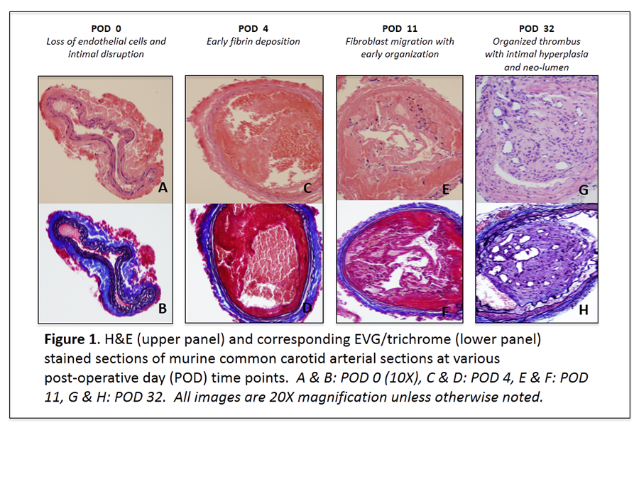
Methods: After creating an arteriotomy in the CCA with a 30G needle, a 0.010-inch hydrophilic wire was used to cannulate the CCA via the arteriotomy and denude the endothelium in an antegrade fashion. Hemostasis at the arteriotomy site was achieved by placing a topical cellulose matrix (Surgicel, Ethicon, Inc. Somerville, New Jersey) over the arteriotomy site. Mice were euthanized 0, 4, 11, and 32 days following CCA injury after systemic formaldehyde perfusion fixation. CCA sections were stained with hematoxylin and eosin (H&E), trichrome, and elastin van Gieson (EVG).

Results: Loss of endothelial cells and intimal disruption was observed at POD 0. Early fibrin deposition was seen in the injured arterial segment at POD 4, however, fibroblast migration with early organization was not observed until POD 11. Organized thrombus with intimal hyperplasia and neo-lumen formation was evident on POD 32.

Conclusion: Antegrade mechanical injury with a hydrophilic wire effectively and reproducibly denudes the endothelium of the murine CCA with subsequent neointima formation throughout the injured arterial segment. This novel, physiologic, and reproducible CCA injury model may aid in the development of innovative pharmaceuticals to prevent and treat neointimal hyperplasia.

06.03 The Effect of Autophagy on Wound Healing in the Murine Model

H. Albadawi1,2, M. W. Koulopoulos1, L. M. Crowley1, H. Yoo1, M. T. Watkins1,2 1Massachusetts General Hospital,Division Of Vascular And Endovascular Surgery, Department Of Surgery,Boston, MA, USA 2Harvard School Of Medicine,Brookline, MA, USA

Introduction: Delayed wound healing is a significant comorbidity associated with peripheral vascular disease. Autophagy is an important cellular process for homeostasis during tissue repair. The role of autophagy in the wound healing process has not been investigated. This study evaluated the effect of the autophagy inhibitor, chloroquine, on cutaneous wound healing in mice.

Methods: Two groups of mice were subjected to excisional full-thickness splinted wounds created on the upper dorsum using a 5mm biopsy punch. The first group of mice were treated daily with an intraperitoneal injection of 50mg/kg chloroquine (CQ, n=8) starting 24hrs before wounding. The second group received saline as an untreated control (CON, n=10). Wound closure was assessed using serial photography and image analysis at days 0, 3, 7, 11 and 14, and expressed as a percentage of the original wound size. On day 14, the mice were euthanized and the wounds were harvested and analyzed by western blotting for markers of autophagy, the ratio of the lipidated autophagosome localized isoform of the microtubule-associated protein Light Chain-3 (LC3-II) to its non-lipidated cytosolic localized isoform (LC3-I) and the expression of Sequestosome-1 (p62). Western blot data was expressed as arbitrary units (AU). Statistical analysis was performed using student-t test and Pearson correlation test.

Results: Wound healing was significantly delayed at days 11 (CQ: 78.0±4.9, CON: 55.7±6.4 percent, p<0.0001) and 14 (CQ: 37±6.8; CON: 18.1±4.8 percent, p=0.028) in the CQ group. Western blot analysis showed significant increase in p62 protein levels (CQ: 0.61±0.06; CON: 0.44±0.04 AU, p=0.02) and LC3-II/I ratio (CQ: 0.61±0.04; CON: 0.37±0.08, p=0.024) in the CQ group. There was a positive correlation between the wound size at day 14 and the p62 protein levels in the same wound (Pearson r=0.83, p=0.02).

Conclusion: These data suggest that autophagy plays a significant role in the wound healing process. The significant increase in the LC3-II/I and p62 protein accumulation demonstrated chloroquine’s inhibitory effect on autophagy. This finding was supported by the positive correlation between wound size at day 14 and the corresponding p62 levels.

06.04 Thrombin and Laminar Flow have Synergistic Effects on YAP Activation in Endothelial Cells

J. Kurita1,2, G. Chitragari1,2, B. E. Sumpio1,2 1Yale University School Of Medicine,Vascular Surgery,New Haven, CT, USA 2VA Connecticut Healthcare System,Vascular Surgery,West Haven, CT, USA

Introduction: The endothelium can be activated not only by mechanical forces of the circulation but also by circulating chemicals such as thrombin (Th). However, the effect of combined mechanical and chemical stimulation of endothelial cells (EC) is poorly understood. The purpose of this study was to investigate the effect of steady laminar flow (CLF), as a mechanical stimuli, and Th, as a chemical stimuli, on yes-associated protein (YAP) and extracellular signal-regulated kinase 5 (ERK5) activities in EC. YAP is a mechano-signaling protein that controls growth and proliferation depending on cell shape. ERK5 is an intracellular protein that maintains EC integrity.

Methods: Bovine aortic ECs seeded on fibronectin coated glass slides were grown to confluence in culture medium containing 10% fetal bovine serum. Thereafter, they were serum starved for 16 hours and were subjected to 0.5, 1, 2, or 4 hours of CLF utilizing a parallel plate flow chamber system controlled by a computerized pump for 4 hours at 37? in the presence or absence of Th (4U/ml). Western blot analysis of total and phospho-YAP and ERK5 was performed. The activities of YAP and ERK5 were determined based on the ratio of immunoblot intensity of phosphorylated to total protein. Fold change compared to static control (Ctrl) at 0 hour was calculated and compared for significant difference using t-test. P value of <0.05 was considered statistically significant.

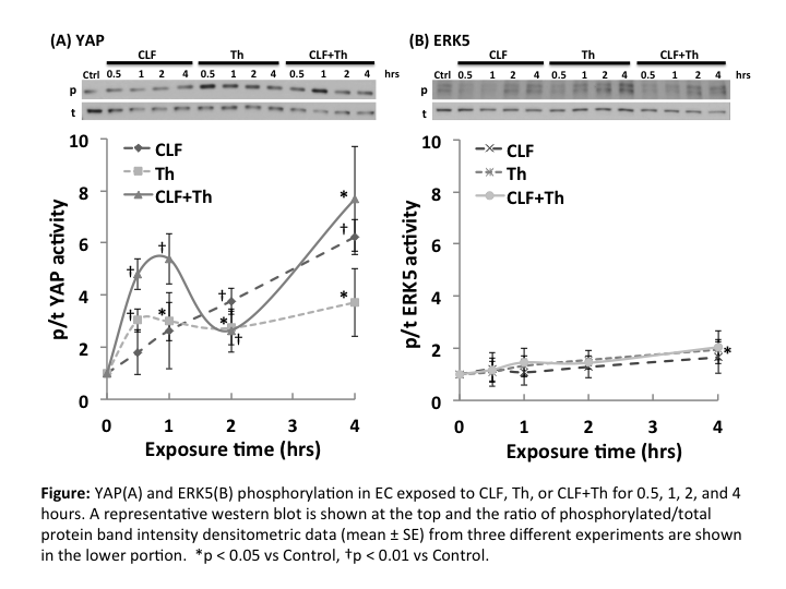
Results: ERK5 activity rose slightly through 4 h for all experimental groups but was only significantly increased in the Th group at 4 hours (1.95±0.39; p<0.03 vs Ctrl) (Figure B). YAP was phosphorylated by Th and the combination of CLF+Th (2.96±0.73; p<0.01 and 2.44±1.16; p<0.05 vs Ctrl, respectively). YAP phosph/total ratio was significantly increased with time through 4 h in the CLF group (p<0.05 vs Ctrl), while the ratio in the CLF+Th group showed the similar time course pattern as in the Th group within 2 hours. However, at 4 h it rose to the level of the CLF groups (Figure A).

Conclusion: Thrombin and flow did not activate ERK5. YAP was phosphorylated in EC exposed to both thrombin and flow although flow stimulation was stronger. The combination of flow and thrombin had an additive effect on YAP phosphorylation. These results indicate selective activation of intracellular proteins by mechano-chemical signals. Further studies have to be done to elucidate the interactions of these signaling pathways and to understand their roles in endothelial response to stimuli.

06.05 Baseline Adipose Phenotype Predicts Vascular Surgery Wound Complications

R. Kulkarni1, W. W. King1, S. Shah1, A. Longchamp1, M. Tao1, K. Ding1, C. K. Ozaki1, G. Sharma1 1Brigham And Women’s Hospital,Vascular And Endovascular Surgery,Boston, MA, USA

Introduction: Wound complication rates after vascular surgery may be as high as 30%, and represent a major cause of morbidity, mortality, and cost. The authors have previously demonstrated that local adipose tissue can exhibit an exacerbated inflammatory response to local surgical trauma. To date, however, links between human adipose phenotype and procedural outcomes such as wound complications have not been reported. We hypothesized that specific adipose-related biomarkers uniquely link to 30-day wound complication rates in patients undergoing open vascular surgical procedures.

Methods: Clinical history, peripheral blood, and subcutaneous and perivascular adipose tissue were prospectively collected from patients undergoing carotid endarterectomy (CEA), lower extremity revascularization (LER), and lower extremity amputations (AMP) at the time of surgery. Nine adipose-associated biologic mediators (adiponectin, IL-1β, IL-6, IL-8, leptin, MCP-1, PAI-1, resistin, and TNF) were assayed in the adipose tissues and plasma. Wound complications were classified according to a previously published grading system. Logarithmic transformation of mediator levels was performed based on positively skewed, non-Gaussian distribution and data were compared using the Student’s t-test. All statistical analyses were conducted using SAS software,v9.3 (SAS Institute, Inc., Cary, NC.)

Results: The cohort included 115 patients (49 CEA, 44 LER, 22 AMP). Median follow-up was 14 months (SD 9.97 months) and 30-day follow-up was 94.8%. At 30 days, 22 (19.1%) patients had wound complications—namely, superficial surgical site infections (14/22), hematoma (4/22), dehiscence (4/22), seroma or lymph leak (3/22), and deep surgical site infection (1/22). There were several statistically significant, plasma/perivascular/subcutaneous compartment-specific relationships between logarithmically transformed mediator levels and wound complications. Most notably, mean plasma TNF levels were higher in patients with superficial surgical site infections (figure, panel A), whereas plasma (figure, panel A), and perivascular (figure, panel B) TNF levels were lower in patients with wound dehiscence. There was also a trend relating elevated subcutaneous TNF levels and any wound complication (p=0.05.)

Conclusions: Adipose-associated mediator levels at the time of operation demonstrate a compartment-specific relationship to wound outcomes in patients undergoing vascular surgical procedures. These associations likely have implications for mechanisms underlying the pathogenesis of wound complications, and suggest novel interventional strategies to reduce wound complications based on the plasticity of the adipose organ’s phenotype.

06.01 Development of a Co-Culture Injury Model for Studying Vascular Smooth Muscle Cell Migration In Vitro

F. Galambo1, H. Bass1,2, R. Beard2, B. J. Cha2, P. R. Nelson1,2 2University Of South Florida College Of Medicine,Department Of Molecular Pharmacology And Physiology,Tampa, FL, USA 1University Of South Florida College Of Medicine,Division Of Vascular And Endovascular Surgery/Department Of Surgery/Morsani College Of Medicine,Tampa, FL, USA

Introduction: Vascular injury disrupts normal vessel wall architecture causing de-endothelialization, and SMC dedifferentiation. Unregulated SMC migration leads to neointimal hyperplasia and eventually restenosis. We sought to develop an in vitro co-culture model to study SMC physiology under conditions of injury. We hypothesized that disrupting quiescent co-cultures would lead to stimulation of SMC chemotaxis.

Methods: SMC/EC co-cultures were established by growing human aortic EC to confluence onto the bottom surface of 0.45µm porous polycarbonate membranes in 6-well Transwell® inserts. Human aortic SMC were then grown to confluence on the top/inner side of the membranes and the co-cultures were then incubated for 48-72 hours to reach equilibrium. The porous membrane allows both chemical and physical communication between the cells, but maintains the layered architecture found in the vessel wall in vivo. To confirm this, the model was characterized using multiphoton fluorescent microscopy. Migrating SMCs were grown to confluence, serum starved for 48 to 72 hours, and seeded onto a second 24-well Transwell® insert with 8.0 µm pores. These inserts were then suspended into SMC and EC monocultures, as well as our co-culture model, both uninjured and injured. Injury of co-culture was created using a cell scraper. Migration was measured using a 4 hour modified Boyden chemotaxis assay. Comparisons were performed using a Student’s T-test.

Results: Using a Z-stacked technique, 3-Dimensional renderings and transverse sections of co-culture membranes demonstrated not only the establishment of healthy co-culture, but also the presence of cell-cell contact through the pores. Under serum-free stimulant-free conditions, SMC demonstrated a low baseline level of migration. SMC exposed to either EC or SMC monoculture alone demonstrated significantly increased migration (P< .0001). SMC exposed to uninjured SMC/EC co-culture demonstrated migration that returned to unstimulated control levels (* P< .0001). SMC exposed to injured SMC/EC co-culture exhibited significantly increased migration levels compared to uninjured conditions (** P< .0001). Migrations results are summarized in Figure 1.

Conclusion: Confocal microscopy demonstrated the viability and utility of our co-culture model in studying vascular injury physiology in vitro. SMC and EC grown in co-culture induce a quiescence compared to either cell type alone, and as such had no influence on SMC chemotaxis. In contrast, disrupting this quiescence, by injuring the co-culture lead to a significant stimulation of SMC migration. This model holds promise to more accurately study the mechanism of restenosis.

06.02 Urotensin’s Mechanistic Role for Development of Intimal Hyperplasia in Androgen Deficient Males

J. Univers1, D. J. Mountain1, B. M. Freeman1, R. T. Fisher1, S. S. Kirkpatrick1, F. A. Klein1, M. B. Freeman1, O. H. Grandas1 1University Of Tennessee Graduate School Of Medicine,Department Of Surgery,Knoxville, TN, USA

Introduction: Androgen deficiency (AD) is associated with increased risk of vascular disease, though the underlying mechanisms remain unclear. We have previously demonstrated testosterone (TST) and dihydrotestosterone (DHT) inhibit vascular smooth muscle cell (VSMC) migration and proliferation in a dose dependent manner in vitro. Furthermore, we have shown that AD increases intimal hyperplasia (IH) in vivo in a rodent model of vascular injury, while physiological TST replacement attenuated this effect. Urotensin II (UTS) is a potent vasoconstrictor and can stimulate cellular proliferation. Activation of the UTS/UTS receptor mechanism has been shown to exacerbate vascular pathologies. Here we investigated the role of UTS in AD-induced IH.

Methods: Three groups of aged orchiectomized (AO) male rats underwent TST supplementation via controlled release pellet (0.5-5 mg). Young and aged intact (YI, AI) and AO placebo (Plac) groups served as controls. All groups underwent balloon angioplasty of the left common carotid following 14d TST therapy. Carotid tissue was collected 14d post-injury, and stained for UTS quantification. Human male VSMCs were treated with TST or DHT (0-3000nM) for 24h then subjected to qPCR for UTS/UTS receptor expression or PCNA/Ki67 proliferation marker analyses. Cells were stained with Phalloidin-FITC for visualization of cytoskeletal organization.

Results:UTS receptor staining was low in injured vessels of YI, AI, and Plac controls but was significantly upregulated in all AO groups receiving TST supplementation, irrespective of dose (Fig1A/B). UTS peptide in the tissue was not affected by TST exposure. In vitro exposure to DHT increased the expression of UTS receptor in VSMCs in a dose-dependent manner, with 3000nM increasing expression by 55±13% vs. 0nM; n=3; P<0.05. However, this did not correlate with any change in proliferative markers PCNA or Ki67. Phalloidin staining of filamentous-actin revealed that DHT induced cytoskeletal organization in a dose–dependent manner (Fig1C).

Conclusion:AD alone does not affect the UTS/UTS receptor mechanism. However TST and DHT increase the expression of UTS receptor, in vivo and in vitro, respectively. This regulation has no effect on proliferative markers but does correlate to a shift in filamentous-actin organization. Analyses of this organization in the presence of UTS receptor agonist/antagonists, and its downstream effect on vasotone, are ongoing. Future studies will examine the potential for exogenous TST therapy to exacerbate dysfunctional vasoconstriction via the upregulation of UTS receptor. These studies are needed in determining if TST replacement in AD men should be evaluated for attenuation of vascular pathogenesis.

09.13 Disparities in Curative Surgical Treatment of Localized Gastrointestinal Malignancies

K. A. Harden1, N. F. Fino2, C. J. Clark1 1Wake Forest University School Of Medicine,Department Of General Surgery,Winston-Salem, NC, USA 2Wake Forest University School Of Medicine,Department Of Biostatistical Sciences,Winston-Salem, NC, USA

Introduction:

With a cancer diagnosis of the gastrointestinal tract, surgery provides a potential for cure; however, not all patients with localized disease undergo an operation. The aim of the current study was to identify person and population level disparities in sociodemographic and health characteristics between patients with localized gastrointestinal malignancies treated with and without curative resection.

Methods:

Using the National Cancer Institute’s Surveillance, Epidemiology, and End-Results-Medicare Health Outcomes Survey linked database (SEER-MHOS), we identified patients diagnosed with localized adenocarcinoma of esophagus, stomach, small bowel, colon, and rectum who completed a comprehensive physical and mental health outcomes survey within two years of their diagnosis. Overall survival was summarized using Kaplan Meier methods; the log rank test was used to test for survival differences by receipt of surgery. Univariate and multivariable analyses were performed to identify disparities in sociodemographic and health characteristics between patients who were or were not treated with curative resection.

Results:

2,051 eligible patients were included in the study cohort. 20.3% of patients did not undergo surgery expected for treatment of a localized gastrointestinal cancer. Overall survival was improved for those treated with surgery (HR 0.63, 0.55-0.74 95% CI, p<0.001). Univariate analysis of sociodemographic and health characteristics showed that surgical intervention was less likely in male patients, non-homeowners, lower median household income, older age, more than two comorbid conditions, any impairment in activities of daily living, lower physical quality of life, prior history of cancer, and larger tumor size (all p<0.05). Person-level social characteristics including non-white race, less than high school education, and marriage status were not predictors of undergoing surgery. In addition, population-level social characteristics including non-metropolitan residence, non-white neighborhood, lower income neighborhood, and neighborhood with lower level of education, were not predictors of undergoing surgery. In a multivariate model using both person and population-level social characteristics, factors independently associated with curative resection were female gender, younger age, fewer comorbidities, and no prior history of cancer (all p<0.05).

Conclusion:

In the United States, patients diagnosed with early stage GI malignancies were less likely to proceed with curative resection secondary to person-level health characteristics. We did not observe population-level disparities in the treatment of early GI malignancies based on social and economic differences.

05.18 Medical School Surgical Boot Camps: The Need For Objective Outcomes

C. J. Neylan1, K. R. Dumon1, I. W. Folkert1, D. T. Dempsey1, J. B. Morris1, N. N. Williams1, S. R. Allen1 1University Of Pennsylvania,Department Of Surgery,Philadelphia, PA, USA

Introduction: The ACS, ASE, APDS, and ABS recently published a joint statement supporting the implementation of surgical pre-residency preparatory courses by medical schools in the United States. Over the past two decades, many medical schools have started offering a surgical ‘boot camp,’ or surgical capstone course, in the fourth year of medical school in an effort to improve students’ performances when they begin surgical internship. The purpose of this study was to perform a comprehensive review of the literature to date evaluating the effects of a surgical boot camp.

Methods: Multiple searches of Pubmed were conducted to identify published papers to August 2015. Key search terms included surgical boot camps, capstone courses, and medical education reform. Abstracts and presentations at meetings were not included. Relevant articles were categorized by their method of evaluating boot camp outcomes.

Results: 14 studies evaluated the outcomes of surgical boot camps. Of these studies, 10 evaluated the impact of boot camps by rating student/intern confidence, while 8 assessed student/intern competence (2 studies evaluated both confidence and competence). The 10 studies that assessed confidence were unanimous in their finding that boot camps increase student and intern confidence. The 8 studies that assessed competence were unanimous in their finding that boot camps increase performance (on exams, procedures, or clinical tasks). However, only 3 of these 8 studies followed-up on performance into the intern year. These 3 studies assessed the competence of interns either by subjective methods (e.g. Likert scale ratings) or objective methods (e.g. OSCE scores), and the assessments occurred either in the simulated setting or the clinical (i.e. hospital) setting, as far as 6 months into internship. One study used objective and subjective methods to evaluate 29 interns in a simulated setting; another study used objective methods in a simulated setting and subjective methods in a hospital setting (n = 84 interns); the third study used objective methods in a simulated setting (n = 19 interns). None of these 3 studies evaluated intern competence using objective methods in the hospital setting.

Conclusion: Despite the demonstrated success of boot camps in improving the confidence of students and interns, the effect of a surgical boot camp on the actual clinical or operative performance of interns in a non-simulated hospital setting has not been studied. Further research is needed to objectively evaluate the effect of surgical boot camps on intern competence and clinical performance in the real world hospital setting.

05.19 Differences in Perceived Value of a Balance in Life Program for Residents by Gender and PGY-Level

C. A. Liebert1, A. Salles1,2, M. Esquivel1, R. S. Greco1, C. Mueller1 1Stanford University,Surgery,Palo Alto, CA, USA 2Washington University,Surgery,St. Louis, MO, USA

Introduction: Surgical residents are at increased risk for depression, burnout, and suicide compared to age-matched peers in the general population. A multifaceted Balance in Life program designed to improve residents’ physical, psychological, professional, and social wellbeing was implemented at an academic residency program. This study evaluated the value of the individual program components amongst the residents.

Methods: General Surgery residents (n=64) completed a voluntary electronic survey in 2014 and 2015 regarding utilization and perceived value of key components of the Balance in Life program during the 2013-2014 and 2014-2015 academic years. Program components included group psychological counseling sessions, a refrigerator stocked with healthy food, a Resident Peer Mentorship Program, a Class Representative System, social events, a ropes course, and a 1-day offsite resident retreat. Means and standard deviations were calculated for Likert responses overall, as well as by gender and PGY-level; t-tests were performed to assess for differences between subgroups.

Results: Residents perceived all components of the program as valuable. The most valued components were the resident retreat (mean=1.27, 1=extremely valuable, 2=moderately valuable, 3=somewhat valuable, 4=slightly valuable, 5=not at all valuable) and the refrigerator (1.43), followed by social events (2.25), group psychological counseling sessions (2.33), the ropes course (2.38), the Resident Peer Mentorship Program (3.02), and the Class Representative System (3.11). Group psychological counseling sessions were valued highest by preliminary general surgery residents compared to categorical residents (1.96 vs. 2.59, p=0.03) and junior residents compared to senior residents (2.00 vs. 2.58, p=0.047). Gender differences between perceived value of the components were identified. Females valued the group psychological counseling sessions (p=0.02), refrigerator (p=0.03), social events (p=0.03), and Class Representative System (p=0.003) more than male residents. There were no statistical differences in perceived value of the components between the 2013-2014 and 2014-2015 years.

Conclusion: A novel multifaceted Balance in Life program to address the physical, professional, and psychosocial wellbeing of surgical trainees is feasible and highly valued by residents. Gender and PGY-level differences exist in the perceived value of components, with females, junior residents, and preliminary residents placing higher value on specific components of the program. Further research is needed to quantify the effectiveness and longitudinal impact such a program has on resident depression, burnout, and other psychological factors.

05.20 Evaluation of Entrustment in Interactions Within the Operating Room

G. Sandhu1,2, C. P. Magas1, A. B. Robinson1, R. M. Minter1,2 1University Of Michigan,Department Of Surgery,Ann Arbor, MI, USA 2University Of Michigan,Department Of Learning Health Sciences,Ann Arbor, MI, USA

Introduction: The operating room is a critical learning environment for surgical residents. Transitioning residents from observers to assistants to safe independent practitioners, while keeping patient safety paramount, is a complex feat. Faculty must make entrustment decisions and determine the degree to which they can safely release gradual responsibility to residents. It is imperative to better understand faculty-resident interactions in the operating room and means by which to identify entrustment that would allow for progressive autonomy.

Methods: Field notes were documented from 30 general surgery cases resulting in 160 hours of operating room observations with variations in pairing among 20 general surgery residents and 10 faculty. All observations were conducted at a single institution. Thematic analysis of field notes in combination with preliminary data from 32 resident interviews informed development of the Faculty-Resident Operative Entrustment Tool – OpTrust. OpTrust focuses on assessment of faculty-resident interactions which enhance or inhibit intraoperative entrustment decisions.

Results: Through a highly iterative process of systematically reviewing OpTrust, the tool was refined to 6 items and 4 levels of descriptive anchors with each item describing behaviors on both sides of the faculty resident dyad. The 6 items are: (?) types of questions asked; (?) operative plan; (?) instruction; (?) problem solving; (?) leadership by the surgical resident; (?) near miss or error. Collectively, these items and leveled anchors provide a robust and multi-dimensional view of faculty-resident operative entrustment interactions, and the impact of behavior of each in achieving autonomy (Figure 1).

Conclusion: Identifying intraoperative behaviors and the level of entrustment associated with each behavior for both faculty and residents, OpTrust provides a novel pedagogical intervention to enhance resident progress towards greater autonomy.

05.15 Communication Between Surgery Residents and Nurses on the Inpatient Wards: Why Time and Space Matter

O. Fernando2, N. G. Coburn1,2,5, A. B. Nathens1,2,3,5, J. Hallet1,2,5, N. Ahmed1,6, L. Gotlib Conn2,4 1University of Toronto,Department Of Surgery,Toronto, ONTARIO, Canada 2Sunnybrook Research Institute,Toronto, ONTARIO, Canada 3University Of Toronto,Institute Of Health Policy, Management And Evaluation,Toronto, ONTARIO, Canada 4University of Toronto,Department Of Anthropology,Toronto, Ontario, Canada 5Sunnybrook Health Sciences Centre,Surgery,Toronto, ONTARIO, Canada 6St. Michael’s Hospital,Surgery,Toronto, ONTARIO, Canada

Introduction:

Optimal interprofessional communication is broadly viewed as a prerequisite to providing quality patient care and enhancing provider satisfaction. Poor interprofessional communication has been associated with medical error and patient harm. We explored the enablers and barriers to interprofessional communication between surgical residents and ward nurses with a view towards improving residents’ educational experiences and the quality of surgical patient care.

Methods:

We conducted a qualitative, ethnographic study of interprofessional communication between general surgery residents and nurses in surgical wards of two Toronto academic hospitals, Centres A and B, totaling 126 hours of observations. Additional data were derived from semi-structured interviews (n= 33) with residents and nurses. Data were collected and analyzed iteratively to the point of theoretical saturation. Data were compared between the two centres for analysis of context-specific variables influencing communication.

Results:

Constraints on communication between residents and nurses at the two centres derived from contested meanings of space and time. Residents experienced the contested spatial boundaries of the surgical ward when they perceived nurses to project a sense of territoriality. Residents were thus found to engage in ward avoidance behaviours and they perceived ward work to be devoid of collaborative interprofessional work or educational opportunity. Nurses expressed difficulty getting residents to respond and attend to pages from the ward, and to have a poor understanding of the nurses’ role. Contestations over time spent in training and patient care were found in resident-nurse interactions, wherein residents perceived seasoned nurses to devalue their clinical knowledge on the ward. Nurses viewed the limited time that residents’ spent in clinical rotation on the ward as adversely affecting communication. Residents felt nurses’ tacitly and explicitly diminished residents’ extensive education and clinical expertise resulting from a perceived unfamiliarity with the ward itself.

Conclusion:

There are numerous challenges to enhancing communication between nurses and residents at academic health centres that are rooted in culture. Interventions to improve the culture of interprofessional care have proven effective in some settings. Our study underscores that improvement efforts must (a) identify and target the many social and cultural dimensions of healthcare team member relations; (b) recognize how power is deployed and experienced in ways that negatively impact interprofessional communication; and (c) enhance an understanding and appreciation of each others’ professional attributes and be delivered within a framework that incorporates the temporal and spatial dimensions of interprofessional care.

05.16 Establishing The Role Of General Surgery Chief Resident Of Quality Improvement And Patient Safety

M. W. LaPorta1, S. L. Bachman1, P. Graling1, J. Donovan1, H. A. Prentice1, J. M. Dort1, J. J. Moynihan1, H. D. Reines1 1Inova Fairfax Hospital,Department Of Surgery,Falls Church, VA, USA

Purpose:

Recognizing the increased emphasis on quality improvement and patient safety (QI/PS) and the critical role of surgical residents in providing frontline patient care and documentation, we identified the need for a formal chief resident role to oversee QI/PS on the Surgical service.

Methods:

The Chief Resident of QI/PS role was created during the 2014-15 academic year. This Chief served in the role for one year, and was a member of several Department of Surgery and institutional committees. The Chief’s duties included: developing interventions targeted to areas with opportunity for improvement within the department and the institution, as well as overseeing the QI/PS projects required of each surgical resident. The QI/PS Chief created an institution-wide, multidisciplinary Resident QI/PS Committee which reported to the Graduate Medical Education (GME) Committee as well as a formal residency didactic curriculum based on Quality in Training Initiative (QITI).

Results:

At least six successful initiatives were implemented during the inaugural QI/PS Chief year. A required progress note phrase was implemented in the electronic medical record (EMR) to improve Surgical Care Improvement Program compliance. The EMR phrase was modified to improve communication surrounding discharge planning. A treatment team ‘face-sheet’ was deployed to help patients recognize the members of their care team. An early post-operative Colorectal Mobility Initiative was implemented. The Resident QI/PS Committee, comprised of appointed residents, program directors, institutional officials, and the Director of QI/PS, met six times over the year. The agenda included PS events involving residents, best practices across campus, and preparation for the annual campus GME Quality Symposium. A formal didactic for QI/PS curriculum was created based on the ACS NSQIP QITI guidelines. Additionally, we found a significant increase in resident participation in our institutional GME Quality Symposium; participation increased 3-fold after the creation of the formal role.

Conclusions:

The benefits of a QI/PS Chief include: increased participation of surgical residents in developing solutions to improve patient care quality and PS, identification of new QI/PS issues by an active group of providers with established pathways to enact change, and dissemination of QI/PS issues to a surgical residency by a peer group member. During their tenure, QI/PS Chiefs experience personal growth, advanced QI/PS education, and active engagement and involvement in institutional-wide change. In the coming years this formal role will recognize and build upon surgical residents’ influence in improving patient care on an institutional level.

05.17 Re-Emergence and the Online Social Media Behaviours and Perceptions of Residency Applicants

J. Dengler1, R. Austin1, K. Devon2,4, J. Fish1,3 1University of Toronto,Division Of Plastic & Reconstructive Surgery,Toronto, Ontario, Canada 2University of Toronto,Division Of General Surgery,Toronto, Ontario, Canada 3Hospital For Sick Children,Plastic & Reconstructive Surgery,Toronto, Ontario, Canada 4Women’s College Hospital,General Surgery,Toronto, Ontario, Canada

Introduction:
The use of online social networking (OSN) has become ubiquitous amongst medical trainees, and is continuing to grow. With its widespread use, residency training programs have started to incorporate OSN profile screening as an additional tool for evaluating applicants, a technique widely used in the hiring process of other professional industries. However, the value of screening residency applicants on social media websites remains undefined and controversial. The aim of our study was to prospectively examine the online social media presence of residency applicants before and after the residency match, and to characterize applicants’ perspectives on the use of OSN screening in the residency selection process.

Methods:
Two independent investigators searched several prominent social media websites (Facebook, Twitter, LinkedIn, Instagram, MySpace, and GooglePlus) to determine whether they could identify the OSN profiles of students applying to a single surgical residency program at the University of Toronto. The search was performed in three iterations: the first during the 2014/2015 Canadian residency matching process, the second after the residency match was finalized but prior to the start of residency training, and the third several months after the start of residency training. The degree to which profiles were publicly accessible was noted. After the last search was complete, applicants were surveyed on their OSN use as well as their OSN behaviour during the application cycle.

Results:
The most frequently used OSN website is Facebook, with 17% (8 of 46) of applicants being identifiable during the match compared to 39% after the match was complete. Only 7% of applicants were found on Instagram; this increased by 13% after the match was complete. The presence on Twitter and LinkedIn remained unchanged (13% and 45%, respectively). Moreover, the profile content on LinkedIn was limited to what was available in a candidate’s application. The majority of profiles were only identifiable when an applicant’s photo was used (72%). Moreover, identifying profiles of candidates with more common names was largely unsuccessful. Although inter-rater reliability of identifying profiles was adequate (kappa = 0.843), the search process was time consuming (average 10 minutes per candidate). Applicant survey responses are being actively collected and analyzed.

Conclusion:
This study suggests that residency applicants limit their public OSN presence during the application cycle, with a re-emergence of OSN use after residency spots have been secured. Moreover, the majority of OSN platforms are of low yield, and searches cannot be performed without photographic identification, which may pose ethical concerns. If OSN profiling is to become a standard part of the residency application process, searches must be limited to high-yield platforms, and guidance must be sought from other industries to maximize the efficiency of OSN profiling and to ensure an ethical practice.

05.12 Standardized Patient Care Protocols Adversely Affect Residents’ & Fellows’ Critical Thinking Skills

R. Bollig1, M. Knight2,4, E. Heidel1, P. Miller3, P. Terry2, M. Goldman1, B. J. Daley1 4King University,Department Of Behavioral And Health Sciences,Knoxville, TN, USA 1University Of Tennessee Medical Center, Knoxville,Trauma & Critical Care Surgery/General Surgery/Graduate School Of Medicine,Knoxville, TN, USA 2University Of Tennessee,Department Of Public Health,Knoxville, TN, USA 3Wake Forest Baptist Health,General Surgery,Winston Salem, NC, USA

Introduction: Information regarding effects of standardized patient care protocols on critical thinking skills is inconclusive. These protocols are designed to promote patient safety, service quality, and cost-effectiveness. We evaluated analytical skills of surgical residents and fellows in relation to various ‘pathways.’ Our hypothesis was participants would ‘default to a protocol’, instead of critical thinking in critical care scenarios.

Methods: After IRB approval, we developed a 14 question, multiple-choice survey for residents and fellows. The questions were validated by experts, and focused on common scenarios encountered in the ICU. The questions evaluated decisions to ‘follow the protocol’ for the given stem, either as a distractor or the correct answer. Critical thinking was defined as answering the question correctly. The questions were distributed to residents and fellows at three regional institutions. Data was analyzed using frequency statistics and paired samples t-tests. An alpha of .05 assumed significance.

Results: A total of 342 residents and fellows were emailed surveys. When choosing the correct answer, 81.3% of the time, critical thinking was used. When the incorrect answer was chosen, it was attributed to not thinking critically 66% of the time. Ultimately, participants chose to incorrectly follow the protocol 34.3% of the time.

Conclusion: This study shows a trend for trainees to follow a protocol as a reflex, without critical assessment or decision making. Our findings cast doubt on the use of protocols in training residents and fellows. As surgical educators, we cannot allow learners to defer critical thinking abilities in favor of easy established protocols.