04.15 Effect of Swine Leukocyte Antigen Class II on Human PBMCs in a Mixed Cell Reaction

J. M. Ladowski1, J. Tchervenkov3, J. Butler1, G. Martens1, M. Tector1, J. Blum2, J. Tector1 1Indiana University School Of Medicine,Transplant Surgery,Indianapolis, IN, USA 2Indiana University School Of Medicine,Microbiology/Immunology,Indianapolis, IN, USA 3Royal College Of Surgeons,Dublin, DUBLIN COUNTY, Ireland

Introduction: As the antibody barrier to clinical xenotransplantation is reduced, attention within the community will turn to cellular-mediated rejection. The direct recognition of swine leukocyte antigen (SLA) class II by PBMC may play a significant role in this rejection. To characterize the proliferative effect of SLA class II, human bulk PBMCs and isolated CD4+ T-cells were incubated with a SLA class II positive and negative adherent fibroblast cell line in a mixed lymphocyte reaction (MLR) variant. The proliferative response was observed through CFSE MLR staining. This novel adherent cell MLR assay allows for faster analysis of candidate antigens and therapeutics compared with studies using cells isolated from animal models for xenotranaplantation.

Methods: An aGal, SLA class I deficient cell line was transfected with a human version of class II transactivator (CIITA) and sorted via flow cytometry based on SLA class II expression. A positive and negative clone were selected as stimulators for a MLR containing human whole or CD4+ isolated PBMCs as the responder population. Responders were stained with carboxyfluorescein succinimidyl ester (CFSE) and monitored for proliferation via flow cytometry.

Results: The human transcription factor CIITA functions as an up-regulator of SLA class II, producing cells with constitutive and stable class II expression. Both whole and CD4+ isolated PBMCs incubated with fibroblasts lacking expression of SLA class II demonstrated considerably less proliferation (0.39 and 0.15% respectively) compared to responders incubated with SLA class II positive cells (1.73 and 1.22%) given the near identical proliferation seen in whole PBMC versus isolated CD4+ population.

Conclusion: This novel swine adherent cell MLR assay facilitates the analysis of cross-species reactivity using human PBMCs, providing for a new assay for xenotransplant study. The proliferation stimulated by SLA class II positive cells appears to predominately occur through direct recognition by CD4+ cells. This information will help focus immunosuppressant therapy in future clinical xenotransplantation.

04.16 Histone Deacetylase-2 Gene Deletion In Mice Extends Tolerance of Renal Ischemia/Reperfusion Injury

D. Murken1, D. Aufhauser1, Z. Wang1, G. Ge1, T. Bhatti3, W. Hancock3, M. Levine1,2 1University Of Pennsylvania,Department Of Surgery,Philadelphia, PA, USA 2Children’s Hospital Of Philadelphia,Department Of Surgery,Philadelphia, PA, USA 3Children’s Hospital Of Philadelphia,Department Of Pathology And Laboratory Medicine,Philadelphia, PA, USA

Introduction: Ischemia Reperfusion Injury (IRI) causes significant morbidity in renal transplantation and other surgical scenarios. A better understanding of the molecular mechanisms leading to IRI is required so that new strategies for prevention and treatment can be developed. Histone deacetylases (HDACs) regulate diverse cellular processes. We have previously shown the benefit in renal IRI of non-selective pharmacologic HDAC inhibitors, and now report on the benefit of targeting one specific HDAC isoform, HDAC2, via inducible whole body gene deletion in order to extend renal IRI tolerance.

Methods: Female wild type C57BL/6 (WT) or female whole body HDAC2-deficient C57BL/6 (HDAC2-/-) mice were subjected to a unilateral warm ischemia model with contralateral nephrectomy under strict temperature control in order to yield maximum reproducibility. Plasma concentrations of BUN were assessed for four days post-operatively.

Results: WT mice survived 28 minutes of warm ischemic time, but developed substantial injury (Figure 1). When subjected to longer periods of warm ischemia, these mice all developed renal insufficiency requiring sacrifice within 48 hours of ischemic insult. HDAC2-/- mice were subjected to longer periods of warm ischemia than survivable by WT mice. HDAC2-/- mice tolerated 35 minutes of ischemia with comparable renal injury to that observed in WT mice after 28 minutes of IRI (p=0.48, Figure 1). Prolongation of the ischemic time by a further two additional minutes (37 min) resulted in non-survivable injury in HDAC2-/- mice.

Conclusion: Murine HDAC2 gene deletion leads to significant extension of renal IRI tolerance. Pharmacologic HDAC2 inhibition may offer a novel therapeutic strategy to avoid organ injury in kidney transplantation and other surgeries requiring renal ischemia.

04.10 A Role For RGS5 In MYCN Amplified Neuroblastoma

M. J. Metzner1, J. Mazar2, A. Rosado1, T. J. Westmoreland1 1Nemour’s Children’s Hospital; University Of Central Florida,Pediatric General Surgery,Orlando, FL, USA 2Sanford Burnham Presbys Medical Discovery Institute,Orlando, FL, USA

Introduction:

Neuroblastoma is a childhood predominant cancer, and its prognosis is greatly affected by MYCN overexpression. MYCN amplification is directly related to poor tumor prognosis and has been previously shown to attenuate RGS5, a regulator of G-protein signaling involved in tumor growth, metastasis, and angiogenesis. It has been previously shown that a decrease in RGS5 expression has been associated with increased tumor aggressiveness. Next Generation sequencing has enabled many advancements in cancer research and has enabled researchers to look at potential cancer markers to help further stratify how cell types react to treatments. We hypothesize that MYCN amplification correlates with RGS5 expression.

Methods:
To test this hypothesis we performed Next-Generation Sequencing on IMR-32 (MYCN amplified) cell line as well as MYCN knockdown in the same cell line. Next, total cellular RNA was isolated from the human neuroblastoma pre-treatment cell lines (SK-N-Be(1), SMS-KAN, IMR32) and post-treatment cell lines (LA-N-6, SK-N-As, SK-N-FI). The pre-treatment cell lines have overexpression of MYCN. The expression of RGS5 was measured using real-time reverse transcriptase polymerase chain reaction (qPCR).

Results:
Utilizing Next Generation sequencing, we demonstrated that the expression of RGS5 in MYCN amplified cell lines showed attenuated expression when compared to post-treatment cell lines where MYCN was non-amplified. The qPCR data using both pre and post-treatment cell lines followed the same pattern and showed that RGS5 expression was decreased in the MYCN amplified pre-treatment lines.

Conclusion:
RGS5 is differentially expressed in neuroblastoma cell lines with and without MYCN amplification. There is an inverse correlation of MYCN and RGS5, which follows the theory that low levels of RGS5 and high MYCN expression lead to a worse prognosis. It has been shown that RGS5 works in many cell growth, metastatic, and neovascularization pathways. This work further correlates the connection between expression of MYCN and RGS5. Because RGS5 expression levels are increased in post-treatment cell lines, RGS5 could serve in the future as a possible marker to further stratify and characterize the aggressiveness of recurrent neuroblastoma.

04.11 Continuous Cold Perfusion vs. Static Cold Preservation for Intestinal Graft Preservation in Rats.

A. Munoz-Abraham2, A. Alkukhun2, S. Judeeba2, R. Patron-lozano2, T. Alfadda2, A. Bertacco1,2, M. I. Rodriguez-Davalos1,2, J. P. Geibel2 1Yale University School Of Medicine,Transplantation/Surgery,New Haven, CT, USA 2Yale University School Of Medicine,Surgery,New Haven, CT, USA

Introduction: In the current transplantation era, new preservation techniques have been developed to further preserve the organs in an optimal status. Continuous perfusion for organs such as kidney, liver, heart and lung have already proven to have a value in comparison to static preservation. However few attempts have been done to develop or find the ideal technique to preserve intestinal grafts. The aim of this study was to assess the optimal preservation technique for intestinal grafts intended for transplantation by comparing the static cold preservation against continuous cold perfusion in a rat model.

Materials and

Methods:

Comparison between static cold preservation (Control Group) with University of Wisconsin (UW) Preservation Solution versus Continuous Cold Perfusion with UW for perfusion (UW Perfusion Group) or a Modified Preservation Solution (MPS) containing UW with L-Arginine (MPS Perfusion Group) with rat intestinal grafts.

30 cms. of Male Sprague-Dawley rats’s small intestine were procured. The intestinal lumen was flushed with HEPES solution (pH 7.40) to remove intestinal debris. Two intestinal loops (10 cm each) were then connected to two custom perfusion chambers of an intestinal perfusion device. One chamber received a constant flow of UW solution and the other MPS solution and both submerged in a bath of deionized water at 4°C. A third intestinal loop was placed in cold static preservation with UW solution. Intestinal tissue samples were taken at T0, T1 (1 hr), T3 (3 hrs.), and T6 (6 hrs.), fixed in Formalin at 10%, and sent to pathology for processing, analysis, and grading according to Park/Chiu Scoring System for Grading Intestinal Ischemia.

Results: Both the UW and the MPS Perfusion groups better preserved the intestine in comparison to the static cold preservation after T6.

Conclusion: Continuous cold perfusion can better preserve intestinal grafts when compared to static cold preservation. These early results might open the door for new techniques for intestine preservation for transplantation that can eventually be applied in the clinical setting. Based on this findings, the next goal of this study is to determine the ideal preservation solution for continuous cold perfusion of the intestinal graft.

04.12 Normothermic Perfusion Using UW compared to HTK Solution on a Rat Intestinal Model

A. Bertacco1, A. Alkukhun1, R. Agarwal1, C. P. LeBlanc1, A. Munoz-Abraham1, F. D’Amico1, M. Rodriguez-Davalos1, J. Geibel1 1Yale School Of Medicine,Surgery/Transplantation,New Haven, CT, USA

Introduction: Prevention of intestinal ischemia during small intestine transplantation still remains a challenge . A safe and effective preservation solution is a precondition for successful small intestine transplantation. The most commonly used hypothermic preservation solutions for intestinal transplantation are the University of Wisconsin solution (UW) and histidine-tryptophan-ketoglutarate (HTK). We have previously shown that intestinal ischemia results in an increased fluid shift towards the lumen of the intestine. In the present study we demonstrate that as the temperature increases (4-37 C) the HTK becomes more acidic. Using this knowledge, our aim was to evaluate the effect of the UW and HTK solution on the viability of an intestinal preparation at normothermic temperature in a rat perfusion model.

Methods: Male Sprague Dawley rats (average weight 440g) were anesthetized and euthanized in accordance with the Animal Care and Use Committee at Yale University. Two sequential small intestinal segments were procured and flushed with regular HEPES solution (with a pH 7.40, mOsm 300 at 37 C) to remove any remaining intestinal debris. The intestines were then connected to our intestinal chambers, which allowed both luminal and extraluminal perfusion. The first intestine was filled intraluminally with 1.5ml of UW Solution including 50µM of FITC-Inulin, and was surrounded by standard UW solution on the vascular side. The second intestine was filled with 1.5ml of HTK Solution including 50µM of FITC-Inulin, and was surrounded by HTK solution on the vascular side. Both chambers were maintained at 37o C. Sample measurements of intraluminal perfusate were made to measure FITC-Inulin concentration, using the Nanodrop 3300™and were taken at times 0,45,90 and 120 minutes.

Results: When comparing the FITC-Inulin concentration in both intestines, there was a mean reduction of 43.25% of FITC-Inulin concentration in the UW arm vs a 9.5% decrease in the HTK arm, indicating more fluid secretion in the intestine exposed to UW.

Conclusion: We have shown that UW solution increases the fluid shift towards the lumen at normothermic temperature compared to HTK solution. Surprisingly, we did not observe the expected amount of secretion from the acidic HTK. With that response, we recognized that UW is a better preservation solution at 37 C due to either it’s buffering capacity in the intestinal lumen, or starch component present in it’s composition. We hypothesize that UW results in decreased cellular swelling in the small intestine cells at normothermic conditions, and thus could leed to a a better outcome for intestinal preservation at normothermic temperatures.

04.13 IL-l Induces Endothelial Hyperpermeability via Activation of p38 MAPK and JNK and Production of MMP-9

Z. Wang1, L. Kong1, J. Kang1, D. K. Nakayama2, P. S. Dale1, D. W. Ashley1 1The Medical Center Navicent Health, Mercer University School Of Medicine,Division Of Trauma, Division Of Basic Sciences, Division Of Surgical Oncology, Department Of Surgery,Macon, GA, USA 2West Virginia University School Of Medicine,Department Of Surgery,Morgantown, WV, USA

Introduction: Endothelial hypermeability mediates edema formation, and contributes to morbidity and death in trauma and the systemic inflammatory response syndrome. Inflammatory cytokines including interleukin-1beta (IL-l) are known to activate matrix metalloproteinases (MMPs), a potential mechanism by which cytokines may disrupt endothelial barrier. The mechanism by which proinflammatory cytokines disrupt endothelial permeability is poorly understood. We investigated if IL-1 would induce endothelial hypermeability through activation of p38 mitogen-activated protein kinase (MAPK) and Jun N-terminal kinase (JNK) and induction of MMP-9.

Methods: Vascular endothelial cells (EC) were incubated with and without IL-1, a MMP-9 inhibitor, and inhibitors of p38 MAPK (SB202190) and JNK (SP600125). RT-PCR and Western blotting measured MMP-9 mRNA and protein, respectively. Gelatin zymography quantitated MMP-9 release. Horseradish peroxidase diffusion through MVEC monolayer assessed endothelial permeability. The kinases were assessed with immunoblotting and activity assay.

Results: IL-l activated p38 MAPK and JNK, promoted expression of MMP-9 mRNA and protein and release of MMP-9 activity, and increased endothelial permeability. IL-1-induced endothelial hypermeability was significantly attenuated by pharmacological inhibitors of p38 MAPK, JNK and MMP-9, respectively. Moreover, pharmacological inhibition of p38 MAPK and JNK reduced MMP-9 release in response to IL-1 stimulation.

Conclusion: The proinflammatory IL-1 induces endothelial hypermeability via activation of p38 MAPK and JNK, and subsequent release of MMP-9. Endothelial MMP-9 expression and activity may play a role in the pathogenesis of vascular hyperpermeability in microcirculation in the setting of trauma and cytokine activation syndromes. JNK, p38 MAPK and MMP-9 may be therapeutic targets for endothelial protection from inflammatory injury.

04.08 Evaluation of apoptosis in carcinoid cells via concomitant treatment of siRNA and Thailandepsin-A

S. Odorico1, R. Jaskula-Sztul1,2, A. Harrison1, S. Clarkson1, S. Golden1, H. Jin1,2, H. Chen1,2 1University Of Wisconsin,Department Of Surgery,Madison, WI, USA 2University Of Alabama,Surgery,Birmingham, Alabama, USA

Introduction: Carcinoids are neuroendocrine tumors that often metastasize and other than surgery, no curative options are available. We have shown that Thailandepsin-A (TDP-A), a novel histone deacetylase inhibitor, can induce apoptosis in carcinoid tumor cells in vitro and reduce tumor size in vivo. However, treatment with TDP-A alone achieved only cytostatic effect. The suppression of anti-apoptotic proteins with concomitant TDP-A treatment could induce cytotoxic effect of both drugs. Therefore, our goal was to investigate a potential synergistic effect on apoptosis in carcinoid cells in vitro with dual treatment of TDP-A and siRNA silencing of three different anti-apoptotic markers, cyclin D1, MCL1 and BCL2.

Method: siRNA of three anti-apoptotic oncogenes, cyclin D1, MCL1 and BCL2, were transfected into gastrointestinal carcinoid cells (BON) using Lipofectamine. Treatment with TDP-A 7 nM (previously tested IC50 value of the drug) occurred 4 hours following siRNA transfection. Cells were incubated for 48 hours before harvesting. qRT-PCR analysis was completed to assess the efficiency of siRNA silencing and flow cytometry was analyzed to determine the apoptotic induction.

Results: In flow cytometric analysis, we assessed pre-apoptotic and apoptotic percentages of total cells after 48 hours of treatment with either siRNA alone, TDP-A alone or siRNA and TDP-A. Figure 1A represents a summary of pre-apoptotic and apoptotic percentages of total cells observed in all samples. We observed a synergistic induction of apoptosis when MCL1 siRNA and TDP-A were applied concurrently. Figure 1B depicts flow cytometric images visualizing the synergistic induction of apoptosis. For qRT-PCR, we observed reduction in relative gene expression in all target genes following transfection of siRNA as compared to scrambled siRNA transfection.

Conclusion: While BCL2 and cyclin D1 siRNA did indeed knock down the expression of their target genes, no synergistic induction of apoptosis occurred with the treatment of both the siRNA and TDP-A. However, combined MCL1 siRNA and TDP-A treatment resulted in a synergistic induction of apoptosis in BON carcinoid cells. These results suggest selective gene silencing of MCL1 enhanced TDP-A induced apoptosis in carcinoid cells in vitro, and warrants further investigation into the relationship between MCL1 knockdown and concomitant treatment of TDP-A.

04.09 Molecular Mechanisms of the Inflammatory Response in Different Aged Fibroblasts

T. S. Lefcourt1, C. Zgheib1, J. Hu1, J. Deacon1, J. Zuk1, J. Xu1, K. W. Liecthy1 1Laboratory For Fetal And Regenerative Biology, Department Of Surgery, School Of Medicine, University Of Colorado Denver Anschutz Medical Campus And Childrens Hospital Colorado,Surgery,Aurora, CO, USA

Introduction: Compared to the adult response to injury, the inflammatory response and scar formation in the neonates is decreased. We have previously shown that increased fibroblast age is associated with increased production of proinflammatory cytokines. Recent evidence suggests that one of the most important regulatory factors of protein production are microRNAs. One such microRNA, microRNA-146a (miR-146a), regulates the inflammatory response by suppressing interleukin-8 (IL-8). Epigenetic regulation such as histone modification has been implicated as a modulatory factor in microRNA expression. We hypothesize that differentially expressed miR-146a in different age groups is under epigenetic control, and that the increased production of proinflammatory cytokines by fibroblasts with increasing age may be due to decreased expression of miR-146a.

Methods: With IRB approval, we isolated fibroblasts from the discarded foreskin of young (<1 month of age) or old (>6 months) males undergoing elective circumcision. Fibroblasts were isolated and expanded no more than 5 passages. Young or old fibroblasts were then cultured for 24 hours with or without two different pan Histone Deacetylase inhibitor (HDACi) conditions: suberanilohydroxamic acid (SAHA), or Apicidin. Total cellular RNA was isolated. Expression of IL-8 and miR-146a was analyzed by quantitative RT-PCR analysis. Expression at baseline and following HDACi treatment was compared to vehicle (DMSO) control.

Results: Young fibroblasts had significantly decreased IL-8 gene expression compared to the older fibroblasts. Young fibroblasts also had significantly increased miR-146a expression compared to older fibroblasts. Treatment with an HDACi resulted in an increase in miR-146a expression in both young and old fibroblasts. In addition, expression of IL-8 was significantly decreased in the older fibroblasts under both HDACi treatment conditions (SAHA p < 0.01, Apicidin p < 0.05).

Conclusion: These findings identify age specific molecular mechanism(s) that modulate the inflammatory response. Increased inflammation and subsequent scar tissue formation with increasing age may be due to decreased expression of miR-146a. Our results also indicated that anti-inflammatory miR-146a is differentially expressed with age, and that the mechanism governing this differential expression may be under epigenetic control. Further studies are warranted to examine miR-146a as a potential therapeutic target to modulate the inflammatory response, and decrease the complications of fibrosis and scar formation.

04.04 Thrombin Stimulated Endothelium Downregulates Fibrinolysis Through Prestored PAI-1 Release

B. R. Huebner1,3, C. C. Silliman1,2,4, E. Gonzalez1,3, S. Mitra1, A. Banerjee1, E. E. Moore1,3 1University Of Colorado Denver,Department Of Surgery,Aurora, CO, USA 2University Of Colorado Denver,Department Of Pediatrics,Aurora, CO, USA 3Denver Health Medical Center,Aurora, CO, USA 4Bonfils Blood Center,Denver, CO, USA

Introduction:
Recently, there’s evidence that injured patients exhibit diverse phenotypes ranging from hyperfibrinolysis coagulopathy to fibrinolysis-shutdown resulting in microvascular occlusion and multi-organ failure. The primary mediators of these phenotypes are tissue plasminogen activator (tPA), a driving force behind hyperfibrinolysis, and plasminogen activator inhibitor-1 (PAI-1), a serine protease inhibitor that couples tPA nullifying its physiologic effect. Endothelial cells activate during stress and are capable of releasing tPA and PAI-1, but their role in critically injured patients remains unclear. Thrombin is a key agent which can stimulate shutdown or enhancement of fibrinolysis. While it is known that tPA is stored in Weibel-Palade bodies in endothelium, the timing and source of PAI-1 remains unclear. We hypothesize that thrombin causes PAI-1 release from preformed vesicles in endothelium thus serving as a rapid counter-regulation of tPA release.

Methods:
Human liver sinusoidal endothelial cells (HLSECs) were grown to an 80-90% confluence in wells after passage 3. HLSECs were treated with thrombin at a concentration of 5units/ml known to be present in the plasma of trauma patients. Supernatant samples were collected at 15 and 30 minute timepoints. Each timepoint was run in duplicate. The supernatants were analyzed with ELISAs for total (free) tPA, total (free) PAI-1, and tPA-PAI-1 complex.

Results:
Thrombin-stimulated endothelial cells released more PAI-1 than the control (>600ng/ml for thrombin 15 min and 30 min vs 441ng/ml control). Free tPA was lower in the thrombin treated cells (0.781ng/ml at 15m, 0.617ng/ml at 30m, 0.977ng/ml control). tPA-PAI-1 complex values were slightly higher from the control at 15min and lower at 30min for the thrombin-treated cells (10.95ng/ml for 15m, 8.56ng/ml for 30m, and 10.2ng/ml control).

Conclusion:
Thrombin provokes the rapid release of PAI-1 from hepatic endothelium with the subsequent formation of tPA-PAI-1 complexes causing a decrease in free tPA and an increase in PAI-1 resulting in fibrinolysis shutdown. Endothelial cells are important in contributing to the fibrinolysis phenotype seen in severely injured patients.

04.05 Post-Shock Accumulation Of Succinate Accelerates Fibrinolysis Via Platelet-Dependent Mechanism

A. L. Slaughter2, H. B. Moore2, A. Bacon2, N. Butler1, A. Banerjee2, C. Silliman2,3, A. D’Alessandro2, E. Peltz2, E. Moore1,2 1Denver Health Medical Center,Aurora, CO, USA 2University Of Colorado Denver,Aurora, CO, USA 3Children’s Hospital Colorado,Aurora, CO, USA

Introduction:
Previous data demonstrate that the plasma concentration of succinate can increase 20-fold within minutes of hemorrhagic shock. Succinate receptors are highly expressed on platelets and dysfunctional platelets play a pivotal role in trauma-induced coagulopathy (TIC). However, the contribution of post-shock succinate accumulation to platelet-mediated TIC is unknown. Fibrinolysis is an important cause of early post-traumatic death due to hemorrhage, yet the mechanism driving acute platelet/fibrin clot failure remains elusive. We hypothesize that succinate alters platelet function, accelerating fibrinolysis, at plasma concentrations observed during hemorrhagic shock.

Methods:
Venous whole blood (WB) from healthy individuals (n=8) was mixed with 5μl of succinate solution to achieve 250-1000μM concentrations per 500μl of WB. Tissue plasminogen activator (75ng/mL, +tPA) was added at each concentration to simulate post-shock endogenous tPA release. Samples were analyzed using native thromboelastography (TEG) and functional fibrinogen (measurement of clot formation independent of platelets, FF-TEG) assays. Repeated measures ANOVA with Bonferroni adjustment was used to determine statistically significant change from baseline TEG and FF-TEG parameters at increasing succinate concentration ± tPA.

Results:
Elevated succinate concentrations (±tPA) decreased time to clot initiation (R), while increasing clot potentiation (angle) and the maximum amplitude of clot formation (MA) (R 250μM+tPA vs. R WB+tPA, p=0.026; angle 500μM+tPA vs. angle WB+tPA, p=0.004; MA 500μM+tPA vs. MA WB+tPA, p=0.026, Figure 1). tPA independently decreased R and increased angle (R WB+tPA vs. R WB, p=0.016; angle WB+tPA vs. angle WB, p=0.016); however these effects were more profound in the setting of succinate (R +tPA vs. R succinate+tPA, -33% vs. -51%, p=0.042; angle +tPA vs. angle succinate+tPA, +37% vs. +67%, p=0.001). Functional fibrinogen levels (FLEV) were unchanged by elevated tPA and succinate concentration ± tPA.

Conclusion:
Pathophysiologic concentrations of succinate in whole blood accelerate clot initiation and potentiation, precipitating earlier tPA-mediated fibrinolysis. Functional fibrinogen levels, measuring clot formation independent of platelets, are unchanged by increasing succinate (± tPA), implicating platelets as culprit mediators. These data support succinate’s role in a platelet-dependent mechanism that drives early post-shock hyperfibrinolysis.

04.06 Examining Virulence Activation of Acinetobacter in Soft Tissue Injuries with an Agent Based Model

A. J. Benjamin1, G. An1 1University Of Chicago,Surgery,Chicago, IL, USA

Introduction: Acinetobacter baumannii is a microbe of growing importance in trauma related soft tissue infections (STIs), and is currently one of the most common causes of late mortality following combat injuries. It has also become a significant cause of multi-drug resistant infection in the intensive care setting. Injury and ischemia have been shown to enhance A. baumannii virulence, both directly and by altering the fitness function of any microbes present in the wound. Characterizing for the ecological dynamics of host-pathogen interactions (HPIs) in the wound milieu will provide insight into tipping points that lead to A. baumannii STIs (AbSTIs). Towards this end we have adapted a prior agent-based model (ABM) of STI to incorporate virulence activation mechanisms of A. baumannii in order to characterize the dynamics of wound HPI and identify conditions that promote AbSTI.

Methods: Our prior muscle wound ABM (MWABM) was extended by adding known A. baumannii virulence mechanisms and host tissue responses to hypoxia and inflammation. The resulting ABM incorporates muscles cells, neutrophils, macrophages, avirulent commensal bacteria, and the detailed representation of A. baumannii. Parameter sweeps were performed to evaluate dynamic patterns of of injury, microbial community composition, virulence activation and host mediator levels in relation to the generation of non-healing and the development of AbSTI. Simulation interventions included differing levels of wound debridement and topical antibiotic therapy.

Results: Simulation experiments identified a series of interconnected thresholds of microbial population composition, degree of initial injury and residual damaged tissue leading to AbSTIs. We demonstrated the role of free iron in the activation of virulence in relation to A. baumannii’s ability to sequester iron via siderophore production. Decreased free iron increased accumulation of HIF1α and decreased iNOS dimerization, which improved with greater debridement. Paradoxically, early topical antibiotic therapy demonstrated decreased microbial populations but increased relative rates of AbSTIs, suggesting the importance of microbial ecological suppression of A. baumannii virulence.

Conclusion: The modified MWABM effectively provides dynamic knowledge representation of A. baumannii virulence in the pathogenesis of AbSTI, demonstrating the effect of debridement, and also the unexpected result that topical antibiotic therapy may have adverse microbial ecological effects on the development of AbSTIs. These findings suggest the importance of incorporating intact microbial communities in future experimental wound healing STI studies in order to make them more clinically relevant. This type of model can serve as a framework to add more detailed mechanistic knowledge as well as investigate novel anti-virulence strategies.

04.01 The Use of FITC-Inulin as a Marker for Intestinal Ischemic Injury

A. O. AlKukhun1, A. Muñoz Abraham1, S. Judeeba1, C. Jasinski1, R. Patron-Lozano1, M. I. Rodriguez-Davalos1, J. P. Geibel1 1Yale University,Department Of Surgery,New Haven, CT, USA

Introduction:

Intestinal ischemia can be seen in conditions such as mesenteric ischemia, or in a challenging situation like intestinal transplantation. Intestinal ischemia leads to pathophysiological disruption that typically presents as increased fluid secretion into the lumen of the intestine with increased levels of ischemia. The aim of this study was to understand the fluid alterations that occur with intestinal ischemia, and determine if FITC-Inulin measurement is sensitive to measure the changes in fluid movement across an intact rat intestinal graft as a real-time measurement of ischemic injury.

Methods:

Male Sprague Dawley rats (average weight of 400g) were operated on in accordance with the Animal Care and Use Committee at Yale University. Two 10 cm small intestine segments were procured from the rats. The intestinal segments were cleansed with HEPES (pH: 7.40, Osmolarity 300±5 at 37°C) solution to clear remaining intestinal contents. The intestinal segments were connected to customized intestinal chambers that are part of a perfusion device. The chambers were kept in a water bath maintained at 37°C. Both intestines were perfused on the vascular side with HEPES buffer solution (pH: 7.40, Osmolarity 300±5 at 37°C). The control HEPES reservoir was open to air, while the experimental reservoir was sealed and bubbled with 100% Nitrogen (N2) to mimic an ischemic environment. Inside both lumens, 3ml of HEPES containing 50 µM FITC-Inulin was continuously perfused at a rate of 1ml/min. Intraluminal samples of the perfusates were collected at three 30 minutes’ intervals to measure FITC-Inulin concentration using a nanospectrofluorimeter.

Results:

When comparing measurements of FITC-Inulin in both intestines, there was a mean 37% reduction of FITC-Inulin concentration in the control versus a mean 57% reduction in the experimental arm, thus indicating increased fluid secretion that was stimulated by the ischemic environment. After examining and comparing the ischemic with the control tissue, an approximate doubling of the amount of secretion was observed over the same time period.

Conclusion:

FITC-Inulin can be used effectively as a volume marker that equates to the ischemic state of the intestinal tissue. This is a practical method to measure real time fluid changes inside a small intestine graft. This model can now be used to assess other intestinal ischemia models: assess viability of intestinal grafts prior to transplant, and monitor drug candidates against ischemia.

04.02 A Pilot Study On The Use of Mass Cytometry to Phenotype the Cellular Response to Trauma

G. Brat3, B. Yorkgitis3, A. Seshadri3, J. Keegan3, J. Dolan3, C. Hauser2, A. Salim3, R. Askari3, J. Lederer3 2Beth Israel Deaconess Medical Center,Trauma Surgery,Boston, MA, USA 3Brigham And Women’s Hospital,Department Of Surgery,Boston, MA, USA

Introduction: Trauma induces a complex physiological and immune response that requires a computational and systems biology research approach. Here, we used a novel technology, CyTOF mass cytometry (CyTOF), to characterize the multi-cellular immune response to severe trauma. By characterizing a massive number of circulating immune cell markers at 1 day after severe trauma, we are able to construct, for the first time, an immune phenotype of trauma. In this pilot study, we focused on myeloid and lymphoid markers that comprise a subset of our comprehensive immunophenotyping assay.

Methods: Blood mononuclear cells from severe trauma patients with injury severity score >20 and from normal controls were collected within 24 hours of initial injury. Cells were stained with lymphoid- or myeloid-cell centric CyTOF staining panels consisting of a combination of 35 cell-surface and intracellular subset identifying marker antibodies each. CyTOF staining data was analyzed using multi-dimensional visualization and statistical methods to reveal trauma-associated changes in circulating immune cell subsets using normal blood cells for comparison.

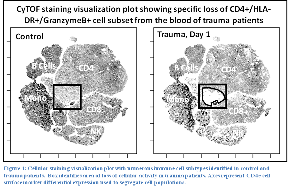
Results: Staining profiles of mononuclear cells using our lymphoid CyTOF panel revealed a specific loss of a unique subset of CD4 T cells that express MHC class II, GranzymeB, PD-1, and Perforin (Figure 1). Staining total leukocyte cells with a myeloid panel showed a complete loss of cells showing eosinophil/basophil staining phenotype and marked reduction in NK cells. As expected, neutrophils were dramatically increased in trauma patients. Other lymphoid populations were unchanged.

Conclusion: These preliminary results demonstrate the feasibility of CyTOF for large-scale profiling of circulating immune cells in trauma patients. Our pilot study of myeloid and lymphoid markers identified specific trauma-induced cellular phenotypic changes that are consistent with movement of certain immune cell types out the circulatory system and possibly towards affected tissues or organs.

04.03 Effect of Factor XIII in an ex vivo assay of hemostatic bandage adhesion

D. J. Zhou1,2, R. Spretz1,3, G. Larsen3,4, W. H. Velander4, M. A. Carlson1,2 1University Of Nebraska Medical Center,Department Of Surgery,Omaha, NE, USA 2VA Nebraska-Western Iowa Healthcare System,Omaha, NE, USA 3LNK Chemsolutions, LLC,Lincoln, NE, USA 4University Of Nebraska-Lincoln,Department Of Chemical & Biomolecular Engineering,Lincoln, NE, USA

Introduction:
Bandage adhesion to bleeding tissue in the setting of traumatic coagulopathy can be improved with fibrin glue (FG). The objective of this study was first to establish an ex vivo assay of bandage adhesion to liver and then to test our hypothesis that use of FG containing recombinant Factor XIII (rFXIII) would improve the adhesion strength (AS) of bandage glued to liver compared to FG without rFXIII.

Methods:
Customized FG (0.2 mL; 9 mg/mL plasma-derived fibrinogen, pdFI, + 106 U/mL r-thrombin + 0.36 mg/mL rFXIII) or commercial FG (0.2 mL Tisseel; Baxter), an FG that contains ~75 mg/mL pdFI and only trace pdFXIII, was applied to a 1×2 cm interface between custom electrospun polycaprolactone (PCL) mesh and a fresh porcine liver strip, and the interface was compressed with a 170 g weight for 5 min at 37°C (default setup). A T-peel adhesion test was performed with an Instron 5943 tensiometer with a 10 N load cell. Force vs. displacement data were used to calculate AS (N/cm), defined as average force during the peel divided by the interface width. AS data were compared with ANOVA (α<0.05) and unpaired t-tests (p<0.05).

Results:
Using the default setup, AS of custom FG was ~2-fold greater by gluing the PCL mesh to the capsular surface of the liver vs. raw parenchyma (Fig. 1A), so use of the liver capsule was incorporated into the default setup. Neither capsular surface wetness (patted dry vs. pre-wet with PBS) nor prolonged compression time (5 vs. 10 min) affected AS (Fig. 1B). There appeared to be decreased AS with lower temperature during compression (25 vs. 37?C), but this was not significant (Fig. 1B). Decreasing FG volume by 50% (0.05 vs. 0.1 mL/cm2) resulted in a lower AS (Fig. 1B). Increasing FG volume beyond 0.1 mL/cm2 was ineffective secondary to glue spillage during compression. Removing rFXIII from the default setup decreased AS by ~50%, but doubling the [rFXIII] did not increase AS (Fig. 1C). AS of customized FG vs. commercial FG was not different (Fig. 1C).

Conclusion:
An ex vivo adhesion test of synthetic resorbable mesh applied to porcine liver with customized FG was optimized with respect to liver surface qualities, adhesion compression time and temperature, and FG quantity. AS was augmented by rFXIII in the FG. The customized FG produced AS similar to that of commercial FG, despite the former having only ~1/8 the pdFI. The AS equivalence between these two FGs likely was a result of the added rFXIII to the customized FG, suggesting that efficacy testing of rFXIII addition to biologic hemostatic devices may be warranted.

09.17 Impact of Socioeconomic Status on Frailty in Elderly Trauma Patients

K. M. Ibraheem1, P. Rhee1, T. Orouji Jokar1, N. Kulvatunyou1, T. O’Keeffe1, A. Tang1, R. Latifi1, G. Vercruysse1, J. Mohler1, M. Fain1, B. Joseph1 1University Of Arizona,Trauma Surgery,Tucson, AZ, USA

Introduction: Frailty is an important determinant of outcomes in elderly patients after trauma. However the impact of social factors such as racial background and socioeconomic status on frailty remains unknown. The aim of this study was to assess the impact of socioeconomic and racial disparity on frailty in elderly trauma patients.

Methods: A prospective study of all geriatric (age ≥ 65 yrs.) trauma patients was performed. Frailty was assessed using 50 variable modified Rockwood frailty index. Frailty index ≥ 0.25 was used as cutoff point for frail status. Primary outcome measure was predictors of frailty in elderly trauma patients. Factors significant on univariate analysis were used for regression model. Multivariate logistic regression was performed.

Results: A total of 271 patients with calculated frailty index were enrolled. Mean age was 77.7±8.2 years, 60% were male, median ISS was 11[9-17], and 43.6% of the population were frail. Uninsured elderly trauma patients were five times more likely to be frail (p=0.001). (Table 1) On sub-analysis of insured patients, patients with Medicare were 3 times more likely to be frail compared to patients with private insurance (OR [95% CI]: 2.9 [1.3-6.1]; p=0.005).

Conclusion: Current models assessing frailty do not incorporate socioeconomic status of an elderly as a contributing factor in frailty. However, results of our study suggest that there is a significant effect of socioeconomic disparity on frailty status of elderly trauma patients. Further attention is required on outreach strategies to provide effective health care to uninsured elderly patients.

03.19 The Microbiome of a Hartmann’s Pouch vs Mucous Fistula is Not Influenced by Exposure to Luminal Flow

A. Trecartin1, J. Debelius2, M. Wieck1, R. Spurrier3, R. Knight2, T. Grikscheit1 1Children’s Hospital Los Angeles,Pediatric Surgery,Los Angeles, CA, USA 2University Of California – San Diego,Pediatrics And Computer Science & Engineering,San Diego, CA, USA 3Cedars-Sinai Medical Center,Surgery,Los Angeles, CA, USA

Introduction:
Pediatric surgical emergencies often result in high levels of antibiotic use and intestine may be left in discontinuity postoperatively with either uni-directional [Hartmann’s pouch (HP)] or bi-directional [mucous fistula (MF)] luminal flow. We hypothesized that phylogenetic diversity would not vary between the two despite severe perturbations including antibiotics, surgery and disease.

Methods:
Pediatric surgical specimens including HP (5) and MF (8) were swabbed under sterile conditions for 16s rRNA characterization. A Kruskal-Wallis test was used to compare HP/MF groups with each other and with a larger data pool encompassing a wide range of surgical diagnoses to obtain alpha and beta-diversity. Weighted and unweighted UniFRAc distances were calculated using permanova and anosim tests. UniFRAc distances were measured between the HP or MF (5 pairs each) and the specimen in continuity with the same patient’s fecal stream. A g-test was also used to identify elevated operational taxonomic units (OTUs).

Results:
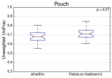
There was no significant difference in alpha-diversity (p=0.38) between HP/MF or beta-diversity (p=0.07) between HP/MF and all samples (figure). There was no difference between HP and MF paired samples for both weighted and unweighted UniFRAc distances (p=0.76 and 0.74). Also, no statistically significant difference was found in levels of OTUs between groups.

Conclusion:
There is no statistically significant difference between HP and MF despite changes in luminal flow in the context of drastic environmental shifts such as antibiotic use, surgical diseases and host factors. Consequently, the effect of luminal flow on the microbiome should not alter the surgeon’s choice of operation.

03.20 ASCL1 Knockdown Suppresses Neuroblastoma Growth through Induction of Apoptosis

S. S. Erickson1, M. Balamurugan1, S. Kunnimalaiyaan1, T. C. Gamblin1, M. Kunnimalaiyaan1 1Medical College Of Wisconsin,Surgical Oncology,Milwaukee, WI, USA

Introduction: Neuroblastoma is a tumor of the developing sympathetic nervous system and one of the leading causes of childhood cancer deaths. Despite recent therapeutic advances, about 60 % of patients with high-risk neuroblastoma experience relapse. The carcinogenic mechanisms driving these cancers are poorly understood, so advances in this area are much needed. The transcription factor ASCL1 is expressed in the early stages of neurogenesis and plays an important role in neuronal fate determination. It has been shown to be highly expressed in neuroendocrine tumors, including neuroblastoma. Results from the overexpression (in non-endocrine airway epithelial cells of transgenic mice) and depletion (in lung cancer cells) of ASCL1 suggest an oncogenic role for this protein. Its downregulation may therefore be a potential treatment strategy for neuroblastoma. In this study, we examined the biological consequences of ASCL1 depletion in neuroblastoma cells.

Methods: NGP cells were transfected with a doxycycline-inducible shRNA sequence against ASCL1 to create an NGP-ASCL1 knockdown (KD) cell line. As a control, non-specific, no-target scrambled sequence containing plasmid was transfected. These cells were selected on puromycin media. Western analysis was used to study the effects of doxycycline treatment on levels of ASCL1, apoptotic proteins, and neuroendocrine tumor markers for up to eight days. The proliferation of both control and doxycycline-treated cells was measured by MTT assay.

Results: Doxycycline treatment induced RFP expression and decreased ASCL1 protein levels in NGP-ASCL1 KD cells. The amount of ASCL1 reduction was dependent on the amount of doxycycline added to the media. ASCL1 knockdown was associated with decreased cellular proliferation at the 4 day (21 % reduction, p=.005), 6 day (48 %, p=.001), and 8 day (61 %, p<.001) time points. Knockdown cells also exhibited lower levels of the neuroendocrine tumor marker CgA, the cell cycle driver cyclin D1, and the pro-survival proteins Mcl1 and survivin.

Conclusion: The controlled system we have developed for regulated expression of ASCL1 allows direct analysis of loss?of?function, with ASCL1 reduction being dependent on the amount of doxycycline added to the media. ASCL1 knockdown in NGP neuroblastoma cells inhibits their proliferative abilities, apparently through an apoptotic pathway. Our data lend further support to the idea that ASCL1 is an attractive molecular target in neuroblastoma.

03.21 PIM Kinase Inhibition Decreases Tumorigenicity of Neuroblastoma Cells

L. L. Stafman1, A. M. Waters1, E. F. Garner1, J. E. Stewart1, E. A. Beierle1 1University Of Alabama,Birmingham, AL, USA

Introduction:
Neuroblastoma, a tumor of neural crest origin, is the most common extracranial solid tumor of childhood. For those with advanced or metastatic disease, this tumor carries a dismal prognosis. Given the current limited treatment options, novel therapies are needed. Proviral insertion site in Moloney murine leukemia virus (PIM) proteins are a family of serine/threonine kinases which has been implicated in tumorigenesis in many solid tumors including glioblastoma, but not studied extensively in neuroblastoma. We hypothesized that neuroblastoma cells would express PIM and that treatment of these cells with AZD1208 (AZD), a PIM-specific inhibitor, would lead to decreased cellular proliferation, migration, and invasion.

Methods:
Four human neuroblastoma cell lines – SK-N-AS, SK-N-BE(2), SH-EP and WAC2 – were utilized. PIM protein expression was determined by immunoblotting of whole cell lysates. PIM inhibition was accomplished with AZD, a competitive inhibitor that binds to the unique ATP pocket in PIM proteins. Proliferation was measured using CellTiter96 assays. Migration and invasion assays were completed with Transwell plates. Data reported as mean ± SEM. Student’s t-test was used to compare data between groups, with p ≤ 0.05 considered significant.

Results:
PIM protein expression was detected in all 4 neuroblastoma cell lines with immunoblotting. Inhibition of PIM with AZD (5µM, 12 hours) significantly decreased cellular proliferation in all 4 cell lines compared to control (vehicle, 12 hours) (Figure 1). In addition, PIM inhibition (AZD, 1µM) significantly inhibited cellular migration in the SK-N-AS (5872 ± 240 vs. 5000 ± 200 cells, control vs. AZD, p=0.03), SK-N-BE(2) (6580 ± 256 vs. 4182 ± 200 cells, control vs. AZD, p=0.0003), SH-EP (17388 ± 451 vs. 15960 ± 487 cells, control vs. AZD, p=0.04) and WAC2 (18200 ± 576 vs. 1660 ± 372 cells, control vs. AZD, p=0.03). Finally, AZD-induced PIM abrogation also significantly inhibited cellular invasion in the SK-N-AS (16,763 ± 559 vs. 14,868 ± 385 cells, control vs. AZD, p=0.03), SK-N-BE(2) (8872 ± 161 vs. 4909 ± 57 cells, control vs. AZD, p<0.0001) SH-EP (13,947 ± 396 vs. 6668 ± 203 cells, control vs. AZD, p<0.0001), and WAC2 (6944 ± 377 vs. 5707 ± 268 cells, control vs. AZD, p=0.04) cell lines.

Conclusion:
All four neuroblastoma cell lines utilized expressed PIM. The PIM inhibitor AZD1208 had a significant impact upon these neuroblastoma cells, resulting in decreased proliferation, migration, and invasion. These results indicate that further exploration of the PIM kinase pathway in neuroblastoma should be undertaken.

03.16 Developmental Expression of Intestinal Alkaline Phosphatase Activity in Premature Infants

S. M. Koehler1, K. Fredrich1, M. Christensen1, T. Nghiem-Rao2, D. Gourlay1 2Children’s Hospital Of Wisconsin,Neonatology,Milwaukee, WI, USA 1Children’s Hospital Of Wisconsin,Pediatric Surgery,Milwaukee, WI, USA

Introduction: Intestinal alkaline phosphatase (IAP) is an enzyme secreted from the mucosa of the gut which exerts a number of beneficial effects on the gut, including the enhancement of barrier function, reducing excessive inflammation, maintaining commensal bacteria and detoxifies lipopolysaccharide. The premature gut has a permeable intestinal barrier and an immature microbiome which predispose the premature neonate to necrotizing enterocolitis (NEC). Minimal research has focused on the activity of IAP in the premature gut, thus we sought to determine normal IAP activity levels for premature infants.

Methods: This is an observational prospective trial involving 10 neonates, ranging in age from 26 to 37 weeks estimated gestational age, in the neonatal intensive care unit at Children’s Hospital of Wisconsin (CHW). Inclusion criteria included: capable of per rectum stooling, parents/guardians consent to the study, and born at or transferred to CHW within 72 hours of life. Patients were excluded if they had life threatening congenital anomalies, congenital intestinal obstruction, or colostomy/ileostomy due to causes other than NEC. If possible, the first stool was collected and then weekly thereafter. The stool was homogenized and the amount of protein per volume determined. An AP activity assay was performed using a p-Nitrophenyl phosphate (pNPP) colorimetric assay. Absorbance of all samples was measured in triplicate at 405nM. The AP activity was measured in the presence of IAP and tissue nonspecific alkaline phosphatase inhibitors. A standard curve of pNPP dilutions was used to quantify the amount of AP activity.

Results: AP activity in all stool samples of premature infants is almost completely inhibited by phenylalanine, an IAP specific inhibitor, but minimally inhibited by a tissue non-specific alkaline phosphatase inhibitor. This cohort of premature infants has a heterogeneous amount of AP activity. For the majority of patients, there is a substantial drop in the AP activity beginning at approximately day of life 4. This decline was followed by an increase in AP activity over the following 2-3 weeks which generally reached and just exceeded the AP activity in the first stool. Only one patient, who had stool samples at the appropriate time points, did not follow this trend of increasing AP activity after an initial drop and this patient was being treated with meropenem for bacteremia.

Conclusions: AP activity in the stool of premature infants is almost exclusively due to IAP as it is inhibited by phenylalanine. The activity of IAP, while initially higher, declines rapidly within days of birth and then rises slowly over the following 3 weeks. The relatively high initial level of AP activity could explain the decreased incidence of NEC in the early postnatal period. Larger studies will be necessary to determine if the heterogeneity of the baseline IAP levels has a physiologic significance.

03.17 Identification of Lactobacillus Strains in the Early Postnatal Rat Microbiota

M. Isani1, A. Grishin1, H. Ford1 1Children’s Hospital Los Angeles,Los Angeles, CA, USA

Introduction:
Necrotizing enterocolitis (NEC) affects premature infants and its etiology remains largely unknown. Colonization of the neonatal gastrointestinal (GI) tract with opportunistic pathogens, such as Coronobacter muytjensii, is thought to be a prime initiating event. Opportunistic pathogens compromise the gut barrier, leading to bacterial translocation, inflammation, and, ultimately, intestinal necrosis. Lactobacillus biotics have been shown in a number of studies to protect against NEC. However, results of Lactobacillus trials remain inconclusive due to the use of different species/strains and doses in different trials. Moreover, an important question of whether the lactobacilli used as probiotics are colonizing the intestine remains unanswered. We propose that an efficient probiotic strain should not only protect the intestinal epithelium, but should also be capable of colonizing the GI tract. To identify naturally occurring colonizing lactobacilli, we sought to isolate and characterize the strains of this genus in the intestines of 4 day old rats.

Methods:
Animal experiments were approved by CHLA IACUC. Neonates were obtained from timed pregnant Sprague Dawley rats purchased from Charles River or Harlan Labs. The neonates were kept in a temperature and humidity controlled incubator, and fed with formula for 4 days. Care was taken not to introduce extraneous bacteria during handling and feeding. To enumerate and isolate the lactobacilli, the content of the large intestine was serially diluted and plated on MRS agar. After 2 day incubation at 37oC, the emerging colonies were classified according to their appearance, and numbers in each class counted. Pure cultures were established for each class and maintained as frozen stocks. The lactobacilli were identified as Gram+ or Gram+/- non-spore-forming rods. The species identity was established by sequencing a variable region of the 16S rRNA gene and by biochemical tests.

Results:
Seven morphologically unique strains of lactobacilli were isolated from 138 rat pups belonging to 18 litters (Figure 1). Among the identified species, there were L. reuteri and L. murinus.

Conclusion:
Various lactobacilli are common first GI tract colonizers in neonatal rats. Availability of the pure cultures of the natural colonizing Lactobacillus strains will allow us to experimentally evaluate their ability to colonize the intestine and protect against NEC.