02.17 Lung-Selective Delivery of PAN-PI3K Inhibitor-Loaded Nanoparticles as Treatment for CRC Metastasis

P. Rychahou1,2, Y. Bae1,4, Y. Zaytseva1, E. Y. Lee1,3, H. L. Weiss1, B. M. Evers1,2 1University Of Kentucky,Markey Cancer Center,Lexington, KY, USA 2University Of Kentucky,Department Of Surgery,Lexington, KY, USA 3University Of Kentucky,Department Of Pathology And Laboratory Medicine,Lexington, KY, USA 4University Of Kentucky,Department Of Pharmaceutical Sciences,Lexington, KY, USA

Introduction: Colorectal cancer (CRC) is the second leading cause of cancer deaths in the US. The phosphatidylinositol 3-kinase (PI3K)/Akt signaling pathway is important for CRC progression and metastasis; inhibitors have been developed that are being evaluated in clinical trials with limited success due to systemic toxicity. The purpose of our study was to: (i) determine expression of pAkt (Ser473), Akt1 and Akt2 in primary and metastatic CRCs, and (ii) develop an effective nanocarrier for lung-selective delivery of pan-PI3K inhibitors as targeted therapy of CRC lung metastasis.

Methods: (1) To determine the expression of PI3K/Akt pathway components, we obtained primary CRCs (n=12) and CRC lung metastases (n=10). All samples were tested for pAkt (Ser473), Akt1 and Akt2 expression by immunohistochemistry (IHC) and blindly scored by a pathologist. (2) Polymeric nanoparticles were constructed and loaded with either fluorescent dye (Alexa 547) or pan-PI3K inhibitors (either PX-866 or wortmannin). Lung selective accumulation of fluorescently-labeled nanoparticles was confirmed by confocal imaging of frozen tissue sections from lung, liver and spleen. Selective PI3K inhibition in lung tissue was confirmed by western blot of protein extracts from lung, liver, spleen and kidney after intravenous administration of PX-866-loaded nanoparticles.

Results: (1) Increased pAkt (Ser473) expression was detected in 80% of primary CRCs and CRC lung metastases; 60% of the samples demonstrated markedly elevated expression of Akt2. (2) Treatment with PX-866, an irreversible pan-PI3K inhibitor currently in the clinic, inhibited cell cycle progression and induced apoptosis in patient-derived CRC organotypic cultures, CRC stem cell lines and pik3ca mutant CRC cells. (3) Importantly, in vivo treatment with pan-PI3K-loaded nanoparticles demonstrated a marked suppression of lung metastasis growth using a clinically-relevant CRC lung metastasis model.

Conclusion: We demonstrate, for the first time, safe and efficient delivery of drug-loaded nanocarriers to lung metastases, suggesting that lung selective PI3K inhibition is a viable treatment strategy for CRC lung metastasis.

02.18 Novel Small Molecule ML327 Sensitizes Colon Cancer Cells to the TRAIL Ligand

C. Padmanabhan1, C. W. Lindsley5,6, A. G. Waterson5,6, R. D. Beauchamp1,2,3,4 1Vanderbilt University Medical Center,Surgery,Nashville, TN, USA 2Vanderbilt University Medical Center,Cell And Developmental Bioogy,Nashville, TN, USA 3Vanderbilt University Medical Center,Cancer Biology,Nashville, TN, USA 4Vanderbilt University Medical Center,Ingram Cancer Center,Nashville, TN, USA 5Vanderbilt University Medical Center,Chemistry,Nashville, TN, USA 6Vanderbilt University Medical Center,Pharmacology,Nashville, TN, USA

Introduction:

Colorectal cancer (CRC) is the third most common cancer diagnosis and the third most common cause of cancer related death in the United States. The inability of chemotherapy to eradicate quiescent cancer stem cells is a leading hypothesis for cancer recurrence. New therapies that do not rely on cellular proliferation for cytotoxic effect must be developed. The tumor necrosis factor related apoptosis inducing ligand (TRAIL), which selectively induces apoptosis in cancer cells irrespective of cell proliferation, was thought to be such a therapy but the anti-cancer effect identified in preclinical models was not realized in clinical trials due to resistance. Our lab has identified a novel small molecule (ML327) that sensitizes colorectal cancer cell lines to TRAIL. We hypothesize this effect is due to loss of the anti-apoptotic protein c-FLIP, a potent inhibitor of the extrinsic apoptosis pathway initiated by TRAIL.

Methods:

CRC cell lines were pre-treated with ML327 or vehicle control at a 10-μM concentration for 24 hours. TRAIL was then added at 100 ng/mL for 4 hours. Cells were lysed and Western blot was performed with antibodies against cFLIP and cleaved caspase 3. Cells were also fixed with 70% ethanol and stained with propidum iodide for FACS analysis. Cells were then treated with ML327 at a 10-μM concentration for up to 4 hours and lysed at hourly increments. Western blot was performed with antibodies against cFLIP.

Results:

24 hours of ML327, but not vehicle, pre-treatment increased TRAIL-induced cleaved caspase 3 protein levels (Fig. A). cFLIP protein level was abundant in vehicle treated cells but was nearly undetectable after 24 hour ML327 treatment (Fig. B). These findings were associated with increased cell death after TRAIL exposure as confirmed by a statistically significant increase (p = 0.002) in the Sub G0 population on FACS analysis (Fig. C). ML327-induced reductions in cFLIP protein occurred quickly, with reduced levels evident by 1 hour and complete loss by 4 hours (Fig. D).

Conclusion:

ML327 is a novel small molecule that sensitizes CRC cells to the TRAIL ligand as evident by increased apoptosis and subsequent cell death. This sensitization is associated with loss of cFLIP. Ongoing analysis will determine whether this is a cause and effect association and further elucidate the exact mechanism of cFLIP loss. It will be of interest to determine the in vivo efficacy of ML327 induced TRAIL sensitization. With these experiments, we hope to develop a novel therapeutic that will induce cell death in cancer cells irrespective of cell proliferation.

02.12 Notch-2 and -4 May Mediate Vemurafenib Drug Resistance in Melanoma

J. Sheldon1, G. Khaushik1, P. Dandawante1, S. Anant1, J. M. Mammen1 1University Of Kansas,Surgery,Kansas City, KS, USA

Introduction:
The incidence of melanoma has increased dramatically over the last few decades making it one of the fastest growing malignancies in the United States. Melanoma expresses a plastic and aggressive phenotype, lacking the majority of regulatory mechanisms due to the aberrant activation of various signaling pathways. In this context, aberrant activation of the Notch signaling pathway in melanoma has also been reported by our laboratory as well as by others. Also of importance, the oncogenic mutation predominantly at codon 600 in the BRAF gene (present in nearly 40–50% of melanoma patients) has changed treatment options for melanoma over the past decade. Unfortunately, recent studies report that the majority of patients treated with the BRAF inhibitors vemurafenib and dabrafenib eventually develop drug resistance. The mechanisms of resistance are still poorly understood. In the present study, we explore the role of Notch signaling in vemurafenib induced drug resistance in melanoma cells.

Methods:

For our experiments, we used various melanoma cell lines (SKMEL-28, UACC275 and A2058 cells). The hexoseaminidase assay was used to determine cell proliferation and to calculate the IC50 (drug concentration causing 50% growth inhibition) in drug sensitive and drug resistant cell lines. Drug-resistant cells were developed by repeatedly growing cells in culture media with increasing doses of drug with time. The surviving daughter resistant cells were compared to the parental sensitive cells using proliferation assay. The IC50 for these paired cell lines was used to determine the increase in resistance. Protein expression studies in cells were done by using standard immunoblotting techniques.

Results:
We observed an increased IC50 value of vemurafenib in drug resistant UACC275 cells. IC50 values at the 72hr time point for UACC-RV-10, UACC-RV-10(1), UACC-RV-15(1) and UACC-RV-15(2) (UACC275 isogenic cells) were ~15, 14, 25 and 22 µM respectively as compared to the IC50 value for parental wild type UACC275 cells of ~0.50 µM. We observed a similar pattern of increased IC50 values in other melanoma cell lines (B16/F10, SKMEL-28, A2058, M14, and melanoma patient derived cells). After several passage of culture in vemurafenib, we also noted changes in cell morphology with cells becoming small and round compared to their usual elongated and stretched appearance. We further evaluated the expression pattern of various Notch receptors in drug sensitive cells as compared to drug resistant isogenic cell lines. Levels of cleaved Notch-2 (10 fold) and -4 (3 fold) were significantly higher in drug resistant cells.(n=5)

Conclusion:

A reductionist model of vemurafenib resistance can be developed using the UACC275 cell line. Both Notch-2 and -4 levels are higher in vemurafenib resistant melanoma cells in this model. Notch signaling may directly or indirectly play an important role in the development of vemurafenib drug resistance in melanoma cells.

02.13 Platinum Agents, Triplatin and TriplatinNC, Suppress 4T1 Murine Breast Cancer Lung Metastasis

S. C. DeMasi1, S. J. Katner2, E. Katsuta1, H. Aoki1, E. J. Peterson2, N. P. Farrell2, K. Takabe1 1Virginia Commonwealth University School Of Medicine And Massey Cancer Center,Division Of Surgical Oncology, Department Of Surgery,Richmond, VA, USA 2Virginia Commonwealth University And Massey Cancer Center,Chemistry Department,Richmond, VA, USA

Introduction: Recent Phase II and III clinical trials reported that Cisplatin is effective in certain metastatic breast cancer, however with severe side effects. New polynuclear platinum analogs, Triplatin and TriplatinNC, were developed to overcome the pitfalls of the former platinum agents; drug resistance and severe toxicity (Farrell et al, Chem Soc Rev 2015). Pharmacokinetics limited the clinical efficacy of Triplatin in Phase II clinical trials. Development of TriplatinNC resolved the PK issue. With less cytotoxic properties it is expected to be more clinically useful if it is in fact as effective as Triplatin.

Methods: MTT assay determined the IC50 of murine breast cancer 4T1-luc2 cells, and tube formation assay compared tube length of HUVEC cells when exposed to treatment. 4T1-luc2 cells were orthotopically implanted in female Balb/C mice 76 weeks of age. Tumor size was measured by caliper and bioluminescent imaging. Mice were randomized based on tumor size on day 1. Triplatin (0.3mg/kg), TriplatinNC (25mg/kg) or Saline was given ip on Day 1, 5, 9 and mice were sacrificed on Day 20. Lung metastasis was evaluated by ex vivo imaging. Pancreatic cancer peritoneal carcinomatosis, generated by ip injection of 1 million panc02-luc cells into C57/Blk6 mice, was used as a distant metastasis model.

Results:IC50 of 4T1-luc cells at 24, 48, and 72h were 89, 25, and 9µM for Triplatin, and 61, 24, and 8µM for TriplatinNC. Tube formation at 10µM concentration of Triplatin and TriplatinNC, using the student’s t-test a significant difference was found between groups, p=0.005 and p=0.002. Additionally 0.1µM TriplatinNC was statistically significant, p=0.003. In 4T1-luc2 implanted model, Triplatin and TriplatinNC suppressed tumor size to 20% and 43% of the control respectively, by Day 20. Bioluminescent imaging on Day 7 demonstrated reduced tumor growth in Triplatin and TriplatinNC to 54% and 57% of the control, respectively. However, imaging data turned out to be inaccurate after day 10 due to ulceration and necrosis of the tumor. Triplatin and TriplatinNC significantly reduced the amount of lung metastasis to 14% and 21% of the control, respectively. In pancreatic cancer peritoneal carcinomatosis model, Triplatin reduced carcinomatosis to 14% and 35% of the control, TriplatinNC by 39% and 39% of the control, in female and male mice on Day 9, respectively.

Conclusion: Both Triplatin and TriplatinNC effectively suppressed 4T1-luc2 breast cancer cell growth, and angiogenesis in vitro. Both agents demonstrated growth suppression of the primary tumor. However, striking effects were observed in advanced models, both lung metastasis and peritoneal carcinomatosis. New platinum compounds with anti-angiogenic properties warrant further investigation for advanced metastatic cancer.

02.14 Serum Sphingosine-1-phosphate is Elevated in Breast Cancer Patients with Lymph Node Metastasis

J. Tsuchida1, M. Nagahashi1, K. Moro1, T. Niwano1, K. Tatsuda1, C. Toshikawa1, M. Hasegawa1, Y. Koyama1, T. Kobayashi1, S. Kosugi3, H. Kameyama1, H. Aoki2, K. Takabe2, T. Wakai1 1Niigata University Graduate School Of Medical And Dental Sciences,Division Of Digestive And General Surgery,Niigata, NIIGATA, Japan 2Virginia Commonwealth University School Of Medicine And The Massey Cancer Center,Division Of Surgical Oncology,Niigata, NIIGATA, Japan 3Uonuma Kikan Hospital,General Surgery,Minami Uonuma, NIIGATA, Japan

Introduction: The bioactive lipid mediator sphingosine-1-phosphate (S1P) has emerged as a key regulatory molecule in cancer progression. S1P exerts its regulatory functions after it is secreted out of cells and by binding to specific G protein-coupled receptors. We previously demonstrated that S1P is a crucial mediator of breast cancer-induced angiogenesis and lymphangiogenesis, generation of new blood and lymphatic vessels, and promote metastasis to the lymph nodes and to the lung. Although important roles of S1P in breast cancer progression has been repeatedly reported in experimental models, the research field suffers from very few data on human patients due to the difficulty in accurate measurements of S1P levels in the clinical samples. In this study, we measure the levels of sphingolipids including S1P in serum from the patients with breast cancer utilizing state-of-the-art mass spectrometry.

Methods: A retrospective analysis was conducted on 49 patients who were pathologically diagnosed with stage I or II breast cancer. The serum from the patients were obtained at the time of diagnosis, prior to any treatment. Sphingolipids were measured by liquid chromatography-electrospray ionization-tandem mass spectrometry (LC-ESI-MS/MS). The levels of sphingolipids including S1P were analyzed with patients’ clinical demographics.

Results: The levels of sphingolipids including sphingosine, dihydro-sphingosine, S1P, dihydro-S1P were detected successfully in the serum from 49 breast cancer patients. Despite our expectation, no significant differences in serum S1P levels were identified by age, hormone receptors (ER, PgR) and HER2 status, Ki-67 index, nuclear grade, lymphatic and vascular invasion of the tumor, pT, and pStage. Interestingly, however, S1P levels in patients with pathologically proven lymph node metastasis (median 2198, range 1575–3462 pmol/ml) was significantly higher than that in patients without lymph node metastasis (median 1977, range 1309–2746 pmol/ml) (P = 0.029). In contrast, levels of sphingosine in patients with lymph node metastasis showed trend to be lower compared to patients without lymph node metastasis (P = 0.073).

Conclusion: Our results indicate an important role of S1P during the process of lymph node metastasis of breast cancer patients. It may also implicate that serum S1P level may have a role as a biomarker for lymph node metastasis. Further study is required to investigate the mechanisms how S1P promotes the lymph node metastasis in human patients and whether S1P levels correlate with lymph node metastasis. This work was supported by the Japan Society for the Promotion of Science (JSPS) Grant-in-Aid for Scientific Research Grant Number 15H05676 and 15K15471 for M.N and 15H04927 for W.T. M.N. is supported by the Uehara Memorial Foundation, Nakayama Cancer Research Institute, Takeda Science Foundation, and Tsukada Memorial Foundation.

02.15 The Autophagic Response in a Mouse Model of HPV-associated Anal Carcinogenesis.

E. Carchman1, K. Matkowskyj2, P. Lambert3 1University Of Wisconsin,General Surgery,Madison, WI, USA 2University Of Wisconsin,Pathology,Madison, WI, USA 3University Of Wisconsin,Oncology,Madison, WI, USA

Introduction: The incidence and death rates from anal cancer are increasing 2% a year. The human papillomavirus (HPV) is the major etiologic risk factor for the development of squamous cell carcinoma (SCC) of the anus, with greater than 90% of anal cancers being HPV positive (high-risk subtypes HPV16 and 18). With HPV infection, two viral oncoproteins (E6 and E7) are expressed and their intracellular molecular changes enhance cellular growth and inhibit cell death, promoting an environment for carcinogenesis. We have developed a double transgenic mouse model (K14E6/K14E7 mice) of anal carcinogenesis in which the two oncoproteins are expressed in the stratified squamous epithelium. Topical treatment with the carcinogen dimethylbenz[a]anthracene (DMBA) induces histologic changes progressing from dysplasia to SCC of the anus. Using this model, we have previously described the up-regulation of the mammalian target of rapamycin (mTOR) pathway. mTOR is an upstream inhibitor of the protective cellular pathway called autophagy. Autophagy is important in the removal of damaged organelles/proteins through lysosomal degradation to maintain cellular health. The goal of this project is to examine the autophagic response throughout anal carcinogenesis. There are two autophagic proteins that are examined in this proposal, first is LC3 that is attached to the autophagosome membrane and second is the autophagic-specific substrate p62.

Methods: Cohorts (3-14 mice/cohort) of K14E6/K14E7 mice were treated topically with DMBA for 5, 10, 15 or 20 weeks. Control mice were not treated with DMBA for the same time points. Anal tissue was collected at the end of treatment, fixed, and serially sectioned. Every 7th section was stained with hemotoxylin and eosin and analyzed by a trained pathologist for the presence of dysplasia or SCC. Immunofluorescence (IF) was performed to detect autophagic proteins, LC3 and p62. Electron microscopy (EM) was performed at each time point.

Results:IF demonstrated an increase in LC3 protein levels throughout neoplastic progression, whereas p62 levels were noted to be high in dysplastic lesions and low in invasive SCC. EM demonstrated an accumulation of lysosomes and mitochondria as lesions progressed from low-grade to high-grade dysplasia, and normalization of lysosome numbers and late autophagosome appearance in SCC.

Conclusion:This study provides evidence of late autophagic flux inhibition during anal carcinogenesis in an HPV mouse model as indicated by increased p62 levels in the setting of increased LC3. This indicates that autophagy is induced, but then the autophagosome is inhibited from fusing to the lysosome resulting in an accumulation of p62. In anal SCC, the levels of p62 expression decreased with the continued increase in LC3 expression, demonstrating that autophagy is able to go to completion in established tumors. These findings support our hypothesis that inhibition of autophagy plays an important role in anal cancer development.

02.09 Xanthohumol Reduces Notch 1 and Inhibits Proliferation in Cholangiocarcinoma

D. Walden1, K. Sokolowski1, S. Kunnimalaiyaan1, C. Gamblin1, M. Kunnimalaiyaan1 1Medical College Of Wisconsin,Department Of Surgical Oncology,Milwaukee, WI, USA

Introduction: Cholangiocarcinoma (CCA) remains the second most prevalent hepatic neoplasm in the United States. Novel therapies are imperative as aggressive metastasis, chemo-resistance, and limited treatment options precipitate a 5-year survival to less than 10%. We have shown that Notch1 alteration reduces CCA cell growth in vitro; therefore, suggesting the importance of Notch1 in carcinogenesis. More recently, we have identified xanthohumol (XN), a prenylated chalcone from hop plants, inhibits both hepatocellular and pancreatic carcinoma through Notch1 reduction. Despite this investigation, the role of XN in CCA remains unclear. We hypothesize that XN inhibits CCA proliferation through reducing Notch1 signaling.

Methods: Notch isoform expression in CCA cell lines (CCLP-1 and SG-231) was determined via Western analysis. The anti-proliferative effect of XN on CCA cell lines was assessed through 3-(4, 5-dimethylthiazol-2-yl)-2, 5-diphenyltetrazolium bromide (MTT), real-time imaging and colony forming assays. To determine mechanism of action, cell lysates were analyzed via Western blot for Notch1 signaling pathway members and apoptotic markers.

Results: Western analysis revealed that basal levels of the Notch1 isoform were highly expressed indicating an integral relationship in CCA carcinogenesis. Statistically significant (p-value<0.01) reductions in cell viability, when compared with control, were observed with XN treatment at concentrations of 5, 10, and 15µM by approximately 23%, 46%, and 56% for CCLP-1 and 9%, 25%, and 38% for SG-231, respectively. The colony formation assay showed apparent growth reductions formation starting at 5 µM XN. Both cell lines demonstrated a dose dependent decrease in Notch1 expression. Reduced viability was mediated through apoptosis evidenced by decreases in cyclin D1 and survivin proteins.

Conclusion: Xanthohumol effectively inhibits CCA growth through the inhibition of Notch. Based on these findings along with reports in other organ-specific cancers, this provides further evidence of the importance of Notch signaling in carcinogenesis as well as XN as a novel anti-carcinogenic treatment.

02.10 Minnelide Inhibits Cancer Cell-Stellate Cell Cross Talk and Reduces Pancreatic Cancer Growth

S. Modi1, X. Zhao1, A. Nomura1, V. Dudeja1, S. Banerjee1, A. K. Saluja1 1University Of Minnesota,Surgery,Minneapolis, MN, USA

Introduction: Pancreatic cancer (PDA) is an aggressive disease which is resistant to conventional therapeutic approaches. Although there are multitudes of reasons for its aggressiveness, recent studies suggest that cancer stroma and stellate cells play a major role in tumor aggressiveness. PDA is extremely desmoplastic; almost 90% of the tumor is stroma. Pancreatic stellate cells (PSCs) contribute extensively to this desmoplastic reaction. PSCs are activated by cancer cells, and vice versa leading this feedback loop to create a niche for pancreatic cancer in the primary and metastatic lesions. Triptolide (TPL) is a terpenoid compound with therapeutic potential against various cancers including PDA. The aim of this study is to evaluate whether triptolide and its water soluble prodrug, Minnelide (currently in Phase I trials), could inhibit the cancer cell-stellate cell cross talk to effectively suppress pancreatic cancer growth.

Methods: In vitro, we used human PSCs and two human PCC lines: S2-VP10 and MIA PaCa-2. The effect of various chemotherapeutic drugs (Gemcitabine, Paclitaxel, TPL, and TRAIL) on the viability of these cells was evaluated using the WST-8 assay. The PCC-PSC interaction was studied by co-culturing the cells in transwell chambers. Effects of pretreatment of one cell type with TPL were studied on the other cell type in a co-culture setup. . Viability, migration and invasion were measured using the WST-8 and the Boyden chamber assay respectively. Expression of various ECM components (collagen, fibronectin), activation (αSMA, vimentin), and invasion markers (N-cadherin, MMPs) was estimated using qPCR and Western Blot.

In vivo, quantification of desmoplastic reaction in the spontaneous pancreatic cancer murine model (KPC) was done by measuring collagen and αSMA expression in the Minnelide and saline treated tumors. Kaplan-Meier survival curves were obtained for same model comparing the two groups.

Results: Viability of PCCs was significantly decreased by various drugs while PSCs were only affected by TPL treatment. The proliferation of MIA PaCa-2 and S2-VP10 was markedly augmented in co-culture with untreated PSCs while TPL pre-treated PSCs did not show this effect. The invasive phenotype of PCCs, as suggested by their migration and invasion, was significantly augmented by co-culture with untreated PSCs while co-culture with TPL pre-treated PSCs failed to do so. TPL treatment resulted in decreased expression of αSMA, fibronectin HSF-1, HSP 70 and 47 in PSCs and reduced collagen production as compared to other drugs.

Minnelide treated tumors in KPC mice had significantly decreased collagen and αSMA content. Minnelide treatment also increased the survival of these mice significantly as compared to saline arm

Conclusion: Our findings indicate that triptolide/Minnelide effectively targets ductal cancerous compartment and its associated stroma leading to disruption of the tumor-stroma interaction in pancreatic cancer.

02.11 Triptolide Inhibits Gallbladder Cancer Growth by Inducing Cell Cycle Arrest via SP1 Inhibition

K. Majumder1, B. Giri1, N. Arora1, S. Modi1, V. Dudeja1, A. Saluja1, S. Banerjee1 1University Of Minnesota,Surgery,Minneapolis, MN, USA

Introduction: Gallbladder cancer (GBC) is the 5th most common gastrointestinal cancer in the US. Despite a dismal 5 year survival of advanced GBC (Stage III= 8%), therapeutic regimes for the same are ill-defined. Current treatment strategies for unresectable disease offer a survival benefit of less than a year. Triptolide, a derivative of a Chinese herb and its water soluble pro-drug Minnelide, which was developed by our laboratory, has been previously shown to be effective against various solid organ cancers. We have previously show that one of the mechanisms of triptolide/Minnelide’s anti-cancer action is SP1 (Specificity protein 1, a transcriptional factor) inhibition. Minnelide is currently in Phase I clinical trial against advanced gastrointestinal malignancies.

Methods: The effect of triptolide on cell viability of three patient derived GBC cell lines SNU 308, NOZ, and GBD1 was assessed. Cells were treated with triptolide (0-200nM) and protein and mRNA were extracted and cell cycle analysis was performed using flow cytometry. Xenograft mouse models using GBC cell lines (GbD1and NOZ) in athymic nude mice were used to analyze the efficacy of Minnelide against GBC.

Results: In vitro, triptolide treatment (0-200 nM) resulted in a significant dose and time dependent decrease in cell viability in all three cell lines (p<0.05). As opposed to our previous studies in pancreatic cancer cells in which triptolide induced apoptotic cell death, western blotting did not demonstrate apoptosis. Flow cytometry analysis revealed a G1/S phase arrest following treatment with triptolide as compared to control treated cells in all three cell lines.

Triptolide treatment results in a significant dose and time dependent decrease in the protein and mRNA levels of transcription factor SP1 in all three cell lines. As a proof of principle, GBC cell lines were treated with SP1 inhibitor mithramycin (0-200nM) and treatment resulted in a significant dose and time dependent decrease in viability of all three cell lines. Moreover, on flow cytometry analysis, mithramycin treatment led to a G1/S phase arrest.

In vivo, tumor weights ( GbD1: 1.5± 0.3 vs 0.7 ± 0.1 vs 0.6 ± 0.1 p< 0.05) as well as tumor volumes ( GbD1: 1515 ± 329 vs 510 ± 133 vs 419 ± 49, p < 0.05) were significantly smaller with Minnelide treatment as compared to saline treatment. [Saline vs Minnelide 0.21 mg/kg/day vs Minnelide 0.42 mg/kg/day]

Conclusion: Minnelide markedly reduces tumor growth in animal model of gallbladder cancer. Our mechanistic studies demonstrate that triptolide/ Minnelide induces cell cycle arrest via SP1 inhibition. Given that Minnelide is already in Phase I clinical trials against advanced GI cancers, it can emerge as a potential therapeutic option for advanced gallbladder cancer.

02.05 Mapping of Notch1 Promoter by HDAC Inhibitors Treatment and the Gene Activation in NE Cancers

H. Jin1,2, M. Roy1, A. Dammalapati1, A. Harrispn1, A. Ma1, R. Jaskula-Sztul1,2, H. Chen1,2 1University Of Alabama,Surgery,Birmingham, AL, USA 2University Of Alabama,Surgery,Birmingham, Alabama, USA 3University Of Wisconsin,Surgery,Madison, WI, USA

Introduction: ~~It is known that Notch signaling is minimally active in neuroendocrine (NE) cancer cells and the induction of Notch isoforms alter the malignant neuroendocrine phenotype. Although the induc¬tion of Notch1 by Histone Deacetylase Inhibitors (HDACi) appeared to be the result of increased Notch1 expression at the transcriptional level, the effects of HDACi on the Notch1 promoter regulation have not been determined thus far. The aim of our study is to investigate the molecular mechanism of HDACi activation on the Notch1 pathway.

Methods: ~~We functionally characterized the Notch1 promoter using deletion mapping. The mapping started with the truncated genomic DNA fragment fused with a luciferase reporter, transfected into BON cell, a carcinoid cell line, screened for luciferase activity. Protein-DNA binding was then performed by electrophoretic mobility shift assay (EMSA).

Results:~~Two HDACi, Valproid Acid and Tailandepsin-A, were shown to induce luciferase activity controlled by a small distal region of Notch1 promoter, from -80 to +1 of the start codon ATG. Further, we identified a functional DNA motif that is responsible for HDACi induction located at -75 to -55 of the Notch1 promoter region. Thus, an in vitro assay, EMSA revealed the transcription factor-DNA complex firmed in the flanking sequence.

Conclusion:~~We have identified the DNA motif located in the Notch1 promoter region that is responsive to HDACi. This understanding of how HDACi act on the Notch1 promoter may lead to the development of future novel therapies for neuroendocrine cancers.

02.07 Role of Sphingosine-1-phoshate of the Host in Progression of Pancreatic Cancer Carcinomatosis

H. Aoki1,2, M. Aoki1,2, P. Mukhopadhyay1,2, C. C. Barnett3, S. Spiegel1,2, K. Takabe1,2 1Virginia Commonwealth University,Division Of Surgical Oncology, Department Of Surgery,Richmond, VA, USA 2Virginia Commonwealth University,Department Of Biochemistry & Molecular Biology,Richmond, VA, USA 3University Of Colorado Denver,Department Of Surgery,Aurora, CO, USA

Introduction: In the U.S., pancreatic cancer is the 4th leading cause of cancer death in both gender. The 5-year relative survival rate is approximaly 6% for all stages. Up to 60% of the patients are found to have distant metastatic disease at the time of diagnosis, at which median survival is around 6 months. The most common sites for distant metastases are the liver (80%), peritoneum (60%), lung and pleura (50-70%), and adrenal glands (25%). Prognosis of patients with peritoneal carcinomatosis (PC), dissemination of cancer cells throughout the abdominal cavity, are particularly poor with median survival of only 6 weeks. This poor overall 5-year survival rate has not significantly changed over the past 5 decades, which reflects the fact that there is no effective treatment available for this condition. Sphingosine-1-phosphate (S1P), a bioactive lipid mediator produced by sphingosine kinase 1 (SphK1) and sphingosine kinase 2 (SphK2), plays critical roles in many aspects of cancer progression, such as cell proliferation, migration, angiogenesis and lymphangiogenesis. We have recently published that S1P link inflammation and cancer in colitis-associated cancer progression (Cancer Cell 2013). Given the fact that inflammation is known to be essential for establishment and progression of PC, where cancer cells need to adhere to the peritoneum and form a nodule, we hypothesized that S1P levels regulated by SphK1 and SphK2 in the host animal may have different mechanism in promoting progression of pancreatic cancer PC depending upon its levels.

Methods: Murine pancreatic adenocarcinoma panc02-luc cells were intraperitoneally injected into SphK1 wild type (WT) or knockout (KO), or SphK2 WT or KO mice to generate PC model. Tumor burden was quantified using bioluminescence imaging. Survival was assessed by Kaplan-Meier analysis. PC nodules were harvested 14 days after injection and analyzed. The proliferation was assessed by Ki-67 staining and apoptosis was evaluated by TUNEL assay. We also compared mRNA expression by RT-PCR.

Results: Circulating S1P levels were lower in SphK1 KO mice and higher in SphK2 KO mice compared with respective littermate WTs probably due to compensatory elevation of SphK1. Panc02-luc cells developed significantly less tumor burden, less inflammatory cell infiltration, and less cancer cell proliferation, but with no difference in apoptosis in PC of SphK1 KO mice. These results suggest that host S1P promotes PC progression by stimulation of proliferation of cancer cells. Interestingly, SphK2 KO mice developed less tumor burden, longer survival with elevated CD4 and CD8 lymphocyte infiltrates in PC.

Conclusion: Our results implicate an intriguing possibility that S1P levels in the host may have different mechanisms in promoting progression of pancreatic cancer PC depending upon its levels.

02.08 Obstructive Jaundice Aggravate Pancreatic Cancer via Sphingosine 1 Phosphate Receptors.

H. Aoki1,2, M. Aoki1,2, E. Katsuta1,2, L. J. Fernandez1, P. Mukhopadhyay1,2, J. Yang3, H. Zhou3, S. Spiegel2, K. Takabe1,2 1Virginia Commonwealth University,Division Of Surgical Oncology, Department Of Surgery,Richmond, VA, USA 2Virginia Commonwealth University,Department Of Biochemistry & Molecular Biology,Richmond, VA, USA 3Virginia Commonwealth University,Department Of Microbiology And Immunology,Richmond, VA, USA

Introduction: Pancreatic cancer (PC) remains one of the deadliest types of cancers. Despite the recent improvement in survival of patients with small resectable tumors, prognosis of unresectable PC that collectively represents over 80% of individuals remains dismal with the 5-year survival of 5% and median survival is within ten months. The classical clinical sign of pancreatic head cancer is painless jaundice. There have been numerous publications regarding the clinical benefit of bile drainage in obstructive jaundice, however, the effect of it on pancreatic cancer biology, such as tumor growth, has never been thoroughly investigated. We have recently published that conjugated bile acids (CBAs) binds to sphingosine 1-phosphate receptor 2 (S1PR2), and it activate nuclear SphK2 that epigentically regulate gene expression (Nagahashi, Takabe, Zhou et al, Hepatology 2015). Indeed, it was reported that CBAs promote growth of cholangiocarcinoma through S1PR2. Thus, we hypothesized that CBAs from obstructive jaundice aggravate the pancreatic cancer progression via S1PRs.

Method: Expression of S1P receptors (S1PR1-5) in murine (panc02-luc) and human (MiaPaca-2) pancreatic cancer cell lines were determined by real-time RT-PCR. Cells were treated with CBAs with or without JTE-013 (S1PR2 antagonist) and viable cells were quantified using WST-8. Panc02-luc cells were implanted in the left lobe of the liver of C57/Bl6 mice with or without obstructive jaundice, created by left and middle bile duct ligation with cholecystectomy. Tumor burden was quantified using bioluminescence imaging on the indicated days and tumors were harvested on day 18.

Result: Hielarchial cluster analysis of pancreatic adenocarcinoma in The Cancer Genome Atlas (N=183) demonstrated that S1PR2, S1PR5 and SphK1 gene expressions have strong positive correlations. Among 5 S1P receptors, S1PR2 is the only one expressed in panc02-luc cells and S1PR2 and S1PR5 are expressed in MiaPaca-2 cells. CBA-mediated cell growth was inhibited by JTE-013, but JTE-013 alone was also found to be growth inhibitory even in the absence of CBA. In obstructive jaundice mice group, significant increase in tumor burden was observed by bioluminescence. Left lobe+tumor / body weight ratio was significantly larger in obstructive jaundice group.

Conclusion: Conjugated bile acid signaling via S1PR2 promotes pancreatic cancer cell growth. Obstructive jaundice aggravated pancreatic cancer, and further study is warranted to investigate the possibility of targeting S1PR2 as a potential new therapeutic for pancreatic cancer treatment.

02.01 PV-10 Induces Potent Immunogenic Apoptosis in Colon Cancer Cells

N. M. Kunda1, J. Qin1, G. Qiao1, B. Prabhakar2, A. V. Maker1,2 1University Of Illinois At Chicago,Division Of Surgical Oncology, Department Of Surgery, College Of Medicine,Chicago, IL, USA 2University Of Illinois At Chicago,Department Of Microbiology & Immunology, College Of Medicine,Chicago, IL, USA

Introduction:
We have previously demonstrated that human and murine colon cancer cells undergo near complete cell death in vitro and in vivo upon direct exposure to PV-10 (10% rose bengal disodium), a synthetic dye currently in clinical trails for intralesional therapy of in-transit melanoma. Occasional bystander responses have raised the possibility that PV-10 induced cell death can generate an anti-tumor immune response. The mechanism of PV-10 cell death in not known, and it is critical to determine, if rose bengal disodium is to be used as an immunotherapeutic strategy for solid tumors.

Methods:
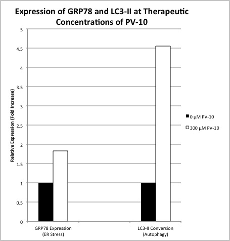
Murine colon cancer CT-26 cells were treated with PV-10 concentrations of 0 – 300 μM for 24 hours. For cell cycle studies, cells were fixed with 80% ethanol and DNA staining was performed using DAPI. DNA content was measured with FACS and cell cycle distribution was analyzed. GRP78 and LC3 were analyzed using Western blot. Protein levels of GRP78 were measured to evaluate ER (Endoplasmic Reticulum) integrity, and the conversion of LC3-I to LC3-II was determined as an indicator of stress-induced autophagy.

Results:
At clinical concentrations of PV-10, the fraction of cells in the G2-M phase increased from 21%-43% (p=0.05), and the fraction of cells in the G1 phase decreased correspondingly from 58% to 30% (p=0.06), consistent with cell cycle growth arrest. The fraction of cells in the sub-G1 phase increased over 4-fold from 3% to 14% (p=0.04), consistent with DNA degradation and apoptosis. GRP78 expression increased almost 2-fold from 1 to 1.83 upon PV-10 exposure, indicative of ER stress. Similarly, LC3-II expression increased almost 5-fold from 1 to 4.53 with PV-10 exposure, consistent with autophagy-induced cell death (Figure 1).

Conclusion:
Treatment of colon cancer cells with PV-10 induced cell cycle arrest, apoptosis, autophagy, and significant ER stress; consistent with immunogenic apoptosis. In order for cytotoxic agents to act as potential immunotherapeutic strategies in the treatment of solid tumors, immunogenic cell death targeting the endoplasmic reticulum (ER), leading to ER stress may be critical. Therefore, based on these results, further evaluation of PV-10 as a potential agent to stimulate immunologic cell death in solid tumors is warranted.

02.02 The Role of Intestinal Alkaline Phosphatase in Inflammatory Bowel Disease

S. A. Morrison1, S. Hamarneh1, H. Sturgeon1, D. Hu1, H. Huo1, W. Zhang1, K. Economopoulos1, S. Gul1, F. Adiliaghdam1, J. Ramirez Decrescenzo1, R. Hodin1 1Massachusetts General Hospital,Surgery,Boston, MA, USA

Introduction: Inflammatory Bowel Disease (IBD) remains a chronic disease of major clinical significance secondary to its increasing prevalence and morbidity worldwide. While the exact mechanistic pathogenesis of IBD remains elusive, its origins are known to be both complex and multi-factorial; involving variants in gene expression, exposure to environmental agents, and a dysregulated immune response. At the heart of suspected disease expression lies the tenet that aberrant gut-derived immune factors play a central role in pathogenesis. This work is based on the hypothesis that potential decreased expression of the brush border enzyme, Intestinal Alkaline Phosphastase, known for its anti-inflammatory properties, may contribute to disease development in these populations.

Methods: Colonic biopsy samples were obtained from 17 patients with Inflammatory Bowel Disease, as well as from 10 healthy controls. In IBD patients, specimens were obtained both from inflamed and non-inflamed areas. Inflammatory cytokine levels and IAP mRNA was determined by quantitative reverse transcription-polymerase chain reaction.

Results:When compared with tissue from healthy controls, IAP mRNA expression was decreased in both inflamed and non-inflamed samples taken from patients with Ulcerative Colitis (UC), with the most significant decrease seen in inflamed tissue( Healthy Controls: 1.15, UC non-inflamed 0.85 (p=0.07), UC inflamed 0.19 (p=0.01)) . In a smaller sample size, tissue samples from Crohn’s patients did not reveal a significant difference in IAP expression when compared to healthy controls (Healthy Controls: 1.15, Crohn’s disease inflamed and non-inflamed: 1.16, 1.02). Inflammatory cytokines TNF-alpha and IL-8 were higher in IBD patients, specifically in inflamed tissue, with expression in Ulcerative Colitis patients being particularly high, correlating to lower levels of IAP, a gut derived anti-inflammatory enzyme (IL-8: HC 0.82, CD non-inflamed and inflamed 8.4 and 276 (p=0.2 and 0.002), UC non-inflamed and inflamed 22.5 and 3441 (p=0.06 and 0.007) TNF-a: HC 0.91, CD non-inflamed and inflamed 1.76 and 6.24 (p=0.05 and 0.006), UC non-inflamed and inflamed 9.17 and 47.3 (p=0.002 and p=0.01)).

Conclusion: Decreased levels of IAP expression, correlating with increased cytokine levels in the inflamed mucosa of patients with Ulcerative Colitis may indicate a role for the enzyme in disease pathogenesis and could lay groundwork for use of this agent as a therapeutic modality in future clinical studies.

02.03 Interleukin-17 Stimulation Induces Stem Cell Marker Expression in Colon Organoids

G. Karagkounis1,2, J. Zhao3, X. Li3, M. F. Kalady1,2 1Cleveland Clinic,Stem Cell Biology And Regenerative Medicine,Cleveland, OH, USA 2Cleveland Clinic,Colorectal Surgery,Cleveland, OH, USA 3Cleveland Clinic,Immunology,Cleveland, OH, USA

Introduction:
Our group has previously shown that interleukin-17 (IL-17) supports colorectal cancer stem cells, enhancing their viability and growth. Studying these effects in normal colonic mucosa is challenging due to the limited availability of in vitro models. Recently developed complex culture systems allowing the maintenance and expansion of multicellular structures derived from colonic mucosa, also known as colon organoids or epithelioids, have allowed new insight in colon tumorigenesis and response to inflammation. The goal of this study was to use colon organoids to investigate whether IL-17 promotes stemness in normal colonic mucosa.

Methods:
Freshly isolated mucosa without evidence of malignancy was collected from human colon according to an established IRB-approved protocol. Samples from different areas of the colon were cultured independently and expanded in media containing R-Spondin, Noggin, EGF and Wnt3a. Each cultured sample was split in two grossly equal fractions that were subsequently treated with either IL-17 (100ng/ml) or vehicle control. After 24 hours, the organoids were harvested and mRNA was isolated. Real-time quantitative PCR (RT-qPCR) was used to measure mRNA expression levels for interleukin-6 (IL-6), a known target of IL-17 used as positive control, as well as stem-cell markers ALDH1, and CD44. Paired t-test was used to compare expression levels between stimulated and control organoids.

Results:
Organoid cultures were established successfully and expanded for 10 days before experiments were performed. IL-17 stimulation resulted in significant upregulation of IL-6 expression compared to control (fold change 18.1, p=0.003). In addition, IL-17 induced CD44 (fold change 1.5, p=0.04), and ALDH1 (fold change 1.7, p=0.03).

Conclusion:
In this proof-of-concept study, inflammatory stimulation in the form of IL-17 induced stem-cell associated genes in normal colonic mucosa. Colon organoids enable in vitro studies of tumorigenesis and allow the assessment of mucosal response to various stimuli. These findings highlight the potential of this model and provide an opportunity for further studies to elucidate the role of IL-17 in the development of cancer.

02.04 Comparison of Baicalein with Metformin on mTOR Pathway Inhibition in Triple-Negative Breast Cancer

A. H. Choi1, Q. Xing1, J. Yan1, W. Wen1, E. S. Han2, J. H. Yim1 1City Of Hope National Medical Center,Division Of Surgical Oncology,Duarte, CA, USA 2City Of Hope National Medical Center,Division Of Gynecologic Oncology,Duarte, CA, USA

Introduction: The mammalian target of rapamycin (mTOR) signaling pathway controls fundamental cellular processes, including cell proliferation and metabolism. mTOR phosphorylates ribosomal protein S6 kinase 1 (S6K1), activating ribosomal protein S6 (S6), which is involved in cellular proliferation and glucose metabolism. Metformin has been shown to disrupt the mTOR pathway and is currently the subject of an ongoing phase III randomized trial as adjuvant treatment in breast cancer (NCT01101438). The flavone baicalein, which is present in the herb thyme and in asian herbal remedies, appears to be non-toxic in animals and humans. We have previously demonstrated that baicalein effectively inhibits cancer cell proliferation by inhibiting mTOR signaling. The objective of this study was to compare the effect of baicalein with metformin in triple-negative breast cancer.

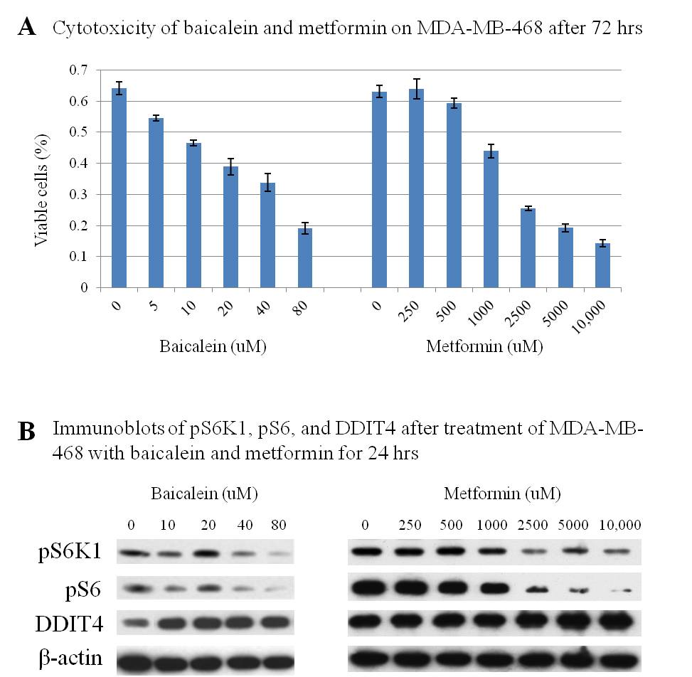
Methods: MDA-MB-468, a triple-negative breast cancer cell line, was treated with baicalein (0-80 uM) and metformin (0-10,000 uM) for 24-72 hours. Total protein lysates were obtained for immunoblot analysis of the mTOR pathway mediators S6K1 and S6, as well as the mTOR inhibitor DNA Damage-Inducible Transcript 4 (DDIT4). Cytotoxicity after 72 hours of treatment was evaluated by MTT assay.

Results: Baicalein demonstrated dose-dependent growth inhibition of MDA-MB-468 in micromolar concentrations. By comparison, metformin required over a 60-fold higher concentration to achieve similar cytotoxicity (Figure 1A). Dose-dependent inhibition of phospho-S6K1 (pS6K1) and phospho-S6 (pS6) were observed following treatment with both baicalein and metformin. However, baicalein-induced phosphorylation blockade was more effective at a far lower concentration. The activity of DDIT4, an mTOR inhibitor, increased with higher concentrations of baicalein (Figure 1B).

Conclusion: Baicalein effectively inhibits growth of triple-negative breast cancer in vitro. Dose-dependent pS6K1 and S6 inhibition, as well as increased DDIT4 levels, were observed at micromolar concentrations. Baicalein, a flavone that appears to be non-toxic in humans, may represent a novel treatment for triple-negative breast cancer.

09.19 Predictors of Inpatient Mortality After Colon Resection for Cancer in the Geriatric Population

E. He1,3, A. N. Kothari1,3, M. De Jong2, R. Yau1,3, J. Eberhardt1, T. Saclarides1, P. C. Kuo1, D. Hayden1 1Loyola University Medical Center,Surgery,Maywood, ILLINOIS, USA 2Loyola University Chicago,Health Sciences Division,Maywood, ILLINOIS, USA 31:Map Surgical Analytics Group,Maywood, IL, USA

Introduction:
As the population ages, more surgeons will find themselves operating on older patients who require special preoperative evaluation and discussion of postoperative risks and expectations. We hypothesize that different age groups within the geriatric population have differing patient predictors for mortality that may help to guide their care.

Methods:
The Health Care Utilization Product State Inpatient Database (HCUP SID) for Florida from 2008-2013 was queried using ICD-9-CM codes. All patients older than 65 years who underwent elective colon resection for cancer were included. Mixed effects logistic regression was performed to determine the impact of patient demographics, comorbidities as well as post-operative complications on inpatient mortality.

Results:
12,945 patients ≥65 underwent colon resection during our study period. Patients were divided into three age groups: 65-79 (n=8411), 80-89 (n=4038), ≥90 (n=496). The average age for each group was 72.1, 83.7, and 91.2 years. Patients ≥90 had more comorbidities than the youngest cohort: anemia (47.2% vs. 28.9%, p<0.001), congestive heart failure (15.5% vs. 5.4%, p<0.001), chronic renal failure (12.5% vs. 6.7%, p<0.001), and malnutrition (9.7% vs. 3.8%, p<0.001). The youngest group had a higher incidence of chronic lung disease (18.6% vs. 15.7%, p<0.001). The average length of stay for each group was 6.9, 7.9, and 8.1 days (p<0.001). Post-operatively, the oldest patients were more likely to experience pulmonary failure (7.1%, p<0.001), MI (1.6%, p<0.001), acute renal failure (7.1%, p<0.001), and pneumonia (4.0%, p<0.001) compared to those aged 65-79 and 80-89. Mortality was 1.0%, 2.5%, and 3.6% in each group respectively (p<0.001). We examined the impact of the above characteristics on the odds of mortality following surgery for each age group using multivariable modeling. A representative sample of the most significant patient-level determinants is reported in Table 1.

Conclusion:
We have demonstrated significant variability of patient-related factors affecting mortality between age groups within the geriatric population. Respiratory failure in the immediate post-operative period is a significant predictor for mortality in all age groups. Post-operative ARF was associated with increased mortality in the two younger age groups while post-operative MI was only significant in those patients ages 65-79. These findings can be used to develop a more sophisticated prediction tool that will impact clinical decision-making and may improve outcomes for older patients undergoing elective colon cancer surgery.

18.18 Design, feasibility and outcomes of locally-led rural pediatric surgery outreach in Africa

A. Muzira1, N. Kakembo1, P. Kisa1, J. Sekabira1, E. Christison-Lagay2, M. Langer4, T. Fitzgerald3, M. Ajiko5, A. Kintu1, R. Kabuye1, D. Namuguzi1, R. Nassanga1, D. Ozgediz2 1Mulago Hospital,Surgery Department,Kampala, , Uganda 2Yale University School Of Medicine,New Haven, CT, USA 3Texas Tech University At El Paso,El Paso, TEXAS, USA 4Maine Medical Center,Portland, MAINE, USA 5Soroti Hospital,Soroti, , Uganda

Introduction:
Pediatric surgery access is limited in rural low-income sub-Saharan Africa. Uganda has two full-time clinical pediatric surgeons, and no physician anesthetists work outside the capital. A high burden of untreated pediatric surgical conditions exists in rural areas. Rural outreach with a primarily Ugandan team working with international collaborators was planned to improve surgical access.

Methods:
Needs assessments were conducted in public regional hospitals in rural Soroti (east) and in Masaka (west). Collaborative objectives included 1) skills transfer to local clinicians; 2) reducing operative backlog; and 3) community sensitization to pediatric surgical conditions. Patient recruitment was done through local providers who screened cases before outreaches, in addition to public radio announcements. Visiting teams were composed of primarily Ugandan providers. All operations were done with mixed teams of surgical and anesthesia providers to facilitate skills transfer. Patient follow-up occurred both by host clinicians and through regular communication (phone and email), referral to the capital as needed, and return visits as needed.

Results:
One-week outreach trips were made to Soroti (5/2012, 1/2013) and Masaka (8/2013, 2/2015). 103 patients (median) were screened/outreach and 83 cases completed/outreach (median). All operations were done under general anesthesia with frequent use of regional blocks. All anesthesia and surgery was done by a combination of visiting team and host providers. The most common operations were hernia/hydrocele repair, orchiopexy, colostomy, PSARP/pull through, and hypospadias repair. There was one post-operative death from dehydration due to enteritis. There were three major complications: wound dehiscence requiring re-operation and two patients with prolonged extubation. All made a full recovery and were discharged uneventfully. Follow up for major cases such as PSARP and pull through were more commonly performed by the host team in Soroti, and through follow up in the capital, for the patients in Masaka (located closer to the capital).

Conclusion:
Rural pediatric general surgery outreach can be performed safely in a very austere environment with primarily local providers. Preparatory planning with host clinicians, interdisciplinary teams, and careful case selection are essential for success. If skills transfer is a primary objective, joint participation in operations is important for hands-on experience of local clinicians. Longer-term follow-up, particularly for major cases, should be determined based on local personnel and practical barriers for follow up in the capital. Ongoing work will examine longer-term outcomes, impact, and cost-effectiveness of these programs.

18.19 Barriers to Trauma Registry Implementation in Rural Gujarat, India: Results from a Qualitative Study

A. E. Jaffe1, K. Vishwanathan2, S. Raithatha2, S. Nimbalkar2, H. P. Santry1 1University Of Massachusetts Medical School,Surgery,Worcester, MA, USA 2Charutar Arogya Mandal (CAM),Pramukhswami Medical College,Karamsad, GUJARAT, India

Introduction: Trauma accounts for >5 million deaths annually, with a large burden occurring in low- and middle-income countries (LMICs). In India, trauma is the most frequent cause of death for Indians <40 years, and 13-18% of all deaths are due to trauma. Trauma registries are an integral part of advanced trauma systems and have been shown to help reduce mortality in developed countries. Such registries are increasingly being implemented in LMICs. We conducted interviews with local stakeholders to identify barriers to registry implementation at Charutar Arogya Mandal (CAM) medical complex in Karamsad, Gujarat, India.

Methods: We conducted a 3-month pilot trauma registry to assess feasibility of a permanent registry for continuous trauma quality improvement at CAM where approximately 40% of emergency room patients are victims of trauma. The pilot consisted of a hardcopy registry form created in conjunction with key stakeholders at CAM. The form gathered basic demographic information, mechanism of injury, physiologic parameters, a brief description of injuries, outcome from emergency department and 2 week outcome for those admitted. Trauma Counselors (social workers) posted in the emergency department were responsible for filling out the forms with the assistance of Casualty Medical Officers (CMOs), surgical residents, and emergency room nurses. Following the pilot phase we conducted 16 interviews with staff (5 CMOs, 3 surgery and orthopedic surgery residents, 4 trauma counselors, and 4 nurses) who were directly or indirectly involved with the pilot registry to gather qualitative data on challenges, barriers, and success to implementation. Interviews were conducted in English, audio recorded and transcribed for review. Inductive analysis was used to identify themes representing barriers to registry implementation.

Results: Only 19% of people interviewed had previous knowledge of a trauma registry prior to the pilot. All respondents saw benefits to having a trauma registry at the hospital and were willing to help contribute to a registry. 50% of respondents encountered some barrier to filling out the registry form, with the most commonly mentioned barrier being ‘high patient volume.’ ‘Role in patient care’ was also identified by 50% as a barrier with trauma counselors being less suited for data collection compared to CMOs and residents. 100% thought that incorporating the form into the official medical record and replacing existing paperwork would provide the greatest benefit and reduce ‘duplication of effort.’

Conclusion: Trauma registries are a centerpiece of sophisticated trauma systems. However, implementation of trauma registries in LMICs must take into account barriers that may be structure, process, financial, or personnel related. Interviews with staff involved in the pilot phase of trauma registry can provide important qualitative data for successful planning of the next steps for implementation.

18.20 Availability of Running Water at 430 Hospitals Providing Surgical Care in 19 Countries

S. Chawla1, S. Gupta2, B. T. Stewart3,4, E. B. Habermann5, A. L. Kushner6,7,8 1Mayo Clinic,Mayo Medical School,Rochester, MN, USA 2University Of California, San Francisco-East Bay,Department Of Surgery,Oakland, CA, USA 3University Of Washington,Department Of Surgery,Seattle, WA, USA 4Komfo Anokye Teaching Hospital,Department Of Surgery,Kumasi, , Ghana 5Mayo Clinic,Robert D. And Patricia E. Kern Center For The Science Of Health Care Delivery,Rochester, MN, USA 6Johns Hopkins Bloomberg School Of Public Health,Department Of International Health,Baltimore, MD, USA 7Columbia University,Department Of Surgery,New York, NY, USA 8Surgeons OverSeas,New York, NY, USA

Introduction: Although 2 billion people now have access to safe drinking water since the Millennium Development Goals were introduced in 1990, this success has not extended to health facilities. A reliable source of clean, running water should be considered essential when providing surgery. However, in many low and middle-income countries (LMICs) this is not the case. A lack of data for advocacy hinders efforts to improve water infrastructure. The goal of this study was to review the surgical capacity literature and document the availability of water at healthcare facilities.

Methods: Using PRISMA guidelines, a systematic search of PubMed for all reports of surgical capacity in LMICs was performed in July 2015. Additionally, references from these reports were examined for potentially useful records. Data were abstracted for studies that reported access to water at health facilities using one of three assessment tools: (a) Surgeons OverSeas Personnel, Infrastructure, Procedure, Equipment and Supplies (PIPES), (b) WHO Emergency and Essential Surgical Care, and (c) Harvard Humanitarian Initiative. When multiple studies assessing surgical capacity in a given country were available, the one with the largest number of facilities assessed was used.

Results: Of the 72 paper identified, 19 were published between 2008 and 2015 included for analysis. These papers assessed 430 hospitals in 19 LMICs spanning 5 continents: Africa (11 reports; 58% of reports); Asia (4; 21%); Central America (1; 5%); South America (2; 11%); and Oceania (1; 5%). Assessed facilities per country varied, ranging from 9 in Guyana and the Solomon Islands to 48 in Tanzania. Three countries, Afghanistan, Somalia, and Ghana, did not include facility-specific assessment of water availability. The median percent of reliable water availability was 61% (IQR 22-100%).

Conclusion: Less than two thirds of hospitals providing surgical care in 19 LMICs had running water. Governments and non-governmental organizations need to increase efforts to improve water infrastructure at healthcare facilities to promote safe essential surgical care and health more broadly. Future research could document the effect of the lack of running water on surgical outcomes.