63.15 Chest radiograph after fluoroscopic guided line placement: no longer necessary

B. G. Dalton1, K. W. Gonzalez1, M. C. Keirsey1, D. C. Rivard1, S. D. St. Peter1 1Children’s Mercy Hospital- University Of Missouri Kansas City,Kansas City, MO, USA

Introduction: Obtaining a chest radiograph after central line placement in the operating room is a historical standard. Retrospective studies at our institution and others have found these to be low yield. After our retrospective investigation, we changed our practice to avoid obtaining a routine post-operative film. In this study, we examine the impact of our clinical change on chest radiograph utilization, adverse events, and cost benefit.

Methods: After obtaining institutional review board approval, we reviewed the charts of patients undergoing central venous catheter placement by the pediatric surgery or interventional radiology service between January 2010 and July 2014 was performed. Outcome measures included CXR within 24 hours of catheter placement, reason for chest radiograph, complication, and complication requiring intervention.

Results: In the study population 622 catheters were placed under fluoroscopy. A chest radiograph was performed in 118 (19%) patients within 24 hours of the line placement with 25 (4%) of these patients being symptomatic in the recovery room. One patient required a chest tube for shortness of breath and pleural effusion. Four symptomatic patients (0.6%) were found to have a pneumothorax, none of which required intervention. There were no re-operations due mal-position of the catheter. In the 504 patients with no postoperative chest x-ray, there were no adverse outcomes. At our institution, the current average charge of a chest radiograph is $283, thus we produced savings of $142,632 over the study period without adverse events.

Conclusion: After placement of central venous catheter under fluoroscopic guidance, a chest radiograph is unlikely to be helpful in an asymptomatic patient.

63.11 The Management and Outcomes of Cervical Neuroblastic Tumors

J. R. Jackson1, H. Tran2, J. Stein1, H. Shimada4, A. M. Patel3, A. Marachelian2, E. S. Kim1 3Children’s Hospital Los Angeles,Otolaryngology,Los Angeles, CA, USA 4Children’s Hospital Los Angeles,Pathology,Los Angeles, CA, USA 1Children’s Hospital Los Angeles,Pediatric Surgery,Los Angeles, CA, USA 2Children’s Hospital Los Angeles,Hematology/Oncology,Los Angeles, CA, USA

Introduction:

Neuroblastoma is a neural crest malignancy of childhood that arises from the sympathetic nerve chain from the neck to the pelvis. While studies have shown that extra-abdominal neuroblastoma (pelvic and thoracic) is associated with favorable biological and clinical characteristics, little has been published with regard to the management and outcomes of cervical neuroblastic tumors. Cervical neuroblastoma represents 2-4% of all neuroblastomas, and current practice is to resect as much tumor as possible without incurring injury to nearby vital structures. In this study we sought to determine the characteristics of these tumors and the effect that the extent of resection has on the overall survival and rate of complications in these patients.

Methods:

We performed a retrospective review of 325 children who were identified to have a neuroblastic tumor at Children’s Hospital Los Angeles over a 15-year period (1/1990–2/2015). Data collected from the medical record included location of tumor, age at diagnosis, age at resection, extent of resection, chemotherapy course, INSS stage, INPC histological classification, and MYCN amplification. Outcome variables included postoperative complications and overall survival.

Results:

Thirteen patients (13/325 – 4%) were identified to have cervical neuroblastic tumors (Table 1); 10 patients (77%) with neuroblastoma (NB), 1 patient with ganglioneuroblastoma (GNB)(7.7%), 2 patients with ganglioneuroma (GN)(15.4%). Median age at diagnosis was 5 months (range 1 mo-15 yrs). One 15 year old had high-risk stage 4 NB with unfavorable histology while the other 9 NB patients were infants <12 months of age. The remaining 3 patients (18-66 mos of age) had differentiating pathology (GN, GNB). All but one had favorable histology and none had MYCN amplification. Six of 10 NB patients underwent resection (5 gross total resection (GTR) and 1 partial resection) while the other 4 underwent biopsy followed by chemotherapy or observation. After GTR, 1 NB patient required prolonged intubation necessitating tracheostomy and another developed eyelid ptosis. The GNB patient, also post-GTR, developed Horner syndrome. The 2 GN patients underwent GTR as well, which resulted in permanent injury to 5 cranial nerves and eyelid ptosis. At latest follow-up, there has been 1 death secondary to relapsed disease.

Conclusion:

Cervical neuroblastic tumors represent favorable lesions with good outcomes similar to other extra-abdominal neuroblastic tumors. In our study, survival was excellent regardless of extent of tumor resection. Based on our data, with the high incidence of complications following GTR, we recommend a minimally agressive surgical approach in managing children with cervical neuroblastic tumors.

63.12 Pediatric Surgical Specialists Are Unaware if Out-of-pocket Cost Influenced Preoperative Decisions

H. Jen1, C. Calkins2, R. Dasgupta3, S. Shah4, S. Safford5, I. Bernstein6, M. Langham7, L. Chen8 1Tufts Medical Center,Boston, MA, USA 2Medical College Of Wisconsin,Milwaukee, WI, USA 3Cincinnati Children’s Hospital Medical Center,Cincinnati, OH, USA 4Children’s Mercy Hospital – University Of Missouri Kansas City,Kansas City, MO, USA 5Virginia Tech Carilion Clinic Children’s Hospital,Roanoke, VA, USA 6University Of Texas Southwestern Medical Center,Dallas, TX, USA 7University Of Tennessee Health Science Center,Memphis, TN, USA 8Baylor University Medical Center,Dallas, TX, USA 9American Academy Of Pediatrics Section On Surgery Committee On Delivery Of Surgical Care,N/A, N/A, USA

Introduction: The Affordable Care Act has increased insurance coverage for children in the United States, but variation in out-of-pocket expenses for families with health insurance seeking pediatric surgical care for their children has also increased. Little is known about pediatric surgical specialists’ experience and attitude toward patients' insurance deductibles or whether pediatric surgical specialists take their patients’ out-of-pocket expenses into account when planning care. A national survey of pediatric surgical specialists was conducted to define the their experience with patient insurance concerns.

Methods: Members from the American Academy of Pediatrics Sections on Plastic Surgery, Surgery and Urology were asked to participate in an anonymous online survey to assess the relationship between surgeon experience with patients’ insurance plans and resource utilization. A 6-item Likert-type scale was used to assess surgeon experience with patients’ insurance concerns. A higher score on the scale associates with increased empathy and concern towards patients’ insurance status. Analysis of variance (ANOVA) was used to investigate practice pattern differences.

Results: Two hundred and eighteen out of 973 (21%) surgeons representing 38 states completed the survey. Almost half of the surveyed surgeons did not know if cost was a determinant for their patients’ choice in surgical facility (43%), or if parents compared provider costs prior to the visit (50%). If the family brought up cost or insurance coverage as an issue, the majority of surgeons would consider cheaper diagnostic modalities (84%) and adjust surgery schedules to decrease patient deductibles (94%). The minority (34%) of surgeons never considered cost as a determinant when scheduling multistage operations. The 6-item Likert-type patient insurance experience scale score did not differ significantly among surgical subspecialties, practice sizes, practice types or states of practice. Surgeons who scored lower on this scale tended to recommend laboratory and radiologic testing at their own facility, citing test reliability and accessibility to results as the top reasons for this practice (p<0.05). The majority of the surgeons surveyed (84%) would consider patient cost in their recommendation of a test or therapy if medically appropriate.

Conclusion: Pediatric surgical specialists are currently unaware if out-of-pocket cost influences patients’ preoperative decisions, but are sympathetic to the issue of out-of-pocket costs if families raised cost as an issue during their visit. As the financial burden of health care shifts to patients and families, the influence of this burden on health care choices by consumers and providers may affect both access to care and surgical outcomes.

63.13 An Evaluation of eHealth Utilization in Pediatric Surgery – What is the Parent’s Perspective?

A. Eguia2, B. Freemyer2, D. Pham2, E. Hamilton2, K. Tsao2, M. Austin1,2 1University Of Texas MD Anderson Cancer Center,Houston, TX, USA 2University Of Texas Health Science Center At Houston,Houston, TX, USA

Introduction: eHealth is the use of digital information and online communication to improve a person’s health or health care. Previous studies have shown that patients face many barriers when attempting to utilize e-Health including factors related to socioeconomic status, language, age, and education. We hypothesized that barriers exist that significantly impact a parent’s ability to access and feel comfortable with using eHealth.

Methods: We performed a cross-sectional study which included 24 non-randomly selected parents of 21 pediatric surgical patients. After obtaining informed consent, semi-structured interviews were conducted in an outpatient clinic by one of two co-authors (AE and BF). The interviews were conducted in each participant’s primary language (11 English and 10 Spanish) and participants were asked about access to eHealth, mechanism(s) of use and challenges faced in accessing and using eHealth. The interviews were recorded and transcribed, and the qualitative data were analyzed using thematic analysis. English-speaking participants (ESP) and Spanish-speaking participants (SSP) were compared.

Results: All participants, except one SSP, had access to the Internet at home. Compared to SSP, ESP were more likely to use the Internet to learn about their own (73% vs 38%) or their child’s health (80% vs 50%). In both groups, parents who used eHealth were more likely to look up information regarding their child’s health versus their own health. ESP tended to use computers and cell phones, whereas SSP were more likely to use only tablets or cell phones. Challenges to using eHealth for ESP included the vast amount of knowledge available, uncertainty of resource credibility and poor Internet connections. Among SSP, non-Spanish websites, inadequate access and lack of knowledge on how to use the Internet presented challenges to utilizing eHealth. In both groups, most participants viewed the potential for email communication with their child’s physician as positive (85%).

Conclusion: While most parents report access to the Internet, both English-speaking and Spanish-speaking parents face challenges in utilizing eHealth. In this pilot project, we identified several key differences between ESP and SSP. We will use our results to inform content in developing a survey to identify and characterize comfort level and barriers that might hinder a parent’s ability to navigate eHealth. This will lay the foundation for the development of a program that can facilitate patient and parent access to and comfort engaging in eHealth.

63.08 Pancreaticoduodenectomy Outcomes in the Pediatric, Adolescent, and Young Adult Population

S. A. Mansfield1, J. P. Walker2, J. H. Aldrink3 1Ohio State University,Department Of General Surgery,Columbus, OH, USA 2Ohio State University,Division Of Gastroenterology, Hepatology And Nutrition,Columbus, OH, USA 3Nationwide Children’s Hospital,Division Of Pediatric Surgery,Columbus, OH, USA

Introduction:
Pancreatic malignancy and chronic pancreatitis are rare in the pediatric, adolescent, and young adult (AYA) population, making pancreas resections an infrequent procedure in this demographic. Only case reports and small case series exist in the literature describing surgical outcomes and complications in this population. The aim of this study is to review the surgical complications and outcomes of pediatric and AYA patients undergoing pancreaticoduodenectomy at our institution.

Methods:
All pediatric, adolescent, and young adult patients (≤30 years) undergoing pacreaticoduodenectomy over a 15-year period (1998-2013) were identified for inclusion in this single-center, observational cohort study. Retrospective chart review was performed to identify pertinent preoperative, perioperative, and postoperative data, including indications for procedure, duration of hospital stay, pathologic data, 30-day mortality, complications, and re-operation data. Overall survival and disease-free survival was calculated using Kaplan-Meier curves.

Results:
Twenty-one patients with a median age of 25 years (range 11-30 years) underwent pancreaticoduodenectomy during the study period and comprised the cohort. Indications for surgery included chronic pancreatitis in 3 and mass/malignancy in 18. The most common post-operative histologic diagnoses were chronic pancreatitis (5, 23.8%), solid pseudopapillary neoplasm (5, 23.8%), and adenocarcinoma (4, 19.0%). For tumor resections, all surgical margins were negative. Six patients required reoperation, with a median time to reoperation of 26 months (range 4.4-136.1). Three reoperations were required in patients with chronic pancreatitis, all due to recurrent or continued pain. Other indications for re-operation included stricture of hepaticojejunostomy (n=1), primary choledocholithiasis (n=1) and upper gastrointestinal bleeding (n=1). The most common postoperative complication was intraabdominal abscess (3, 14.3%). Pancreatic leak occurred in only one patient. Thirty-day mortality was 0% for all patients. There were no recurrences or disease-related deaths in patients with solid pseudopapillary neoplasm. Patients with adenocarcinoma had a median survival of 15.6 (range 9-142) months.

Conclusion:
This is the largest series of pancreaticoduodenectomy procedures reported in the pediatric and AYA population. As in adults, surgical resection remains the mainstay of treatment for neoplasms of the pancreas and complicated chronic pancreatitis for the pediatric and AYA groups. Given how rare these pancreatic conditions are in this age group, cohort studies such as this may help improve therapies for this unique patient population.

63.09 Successful Non-operative Management of Esophageal Perforations in the Newborn

E. A. Onwuka1, P. Saadai1, L. A. Boomer2, B. C. Nwomeh1 1Nationwide Children’s Hospital,Pediatric Surgery,Columbus, OH, USA 2LeBonheur Children’s Hospital,Pediatric Surgery,Memphis, TENNESSEE, USA

Introduction:

Esophageal perforation in neonates occurs most often in cases of extreme prematurity. Common etiologies include orogastric (OG) tube placement, endotracheal intubation, and endoscopy. Mortality as high as 29% has been reported. Treatment over the last decade has leaned towards non-operative management with nil per os (NPO), total parenteral nutrition (TPN), antibiotics, and radiographic examination prior to the re-institution of oral feeds. To date, treatment duration for non-operative management has not been well studied, therefore neonates may experience unnecessarily prolonged periods of enteral feed disruption and antibiotic exposure. The purpose of this study was to review cases of esophageal perforation in neonates to assess the outcomes of non-operative management.

Methods:

A retrospective chart review was performed of patients under one year of age with ICD-9 code 530.4 for esophageal perforation treated at our institution between the years of 2009 and 2015. Data collected included demographic information, etiology of perforation, treatment course, time to resumption of enteral feeds, length of antibiotic use, time to subsequent radiographic resolution, and mortality.

Results:

Twenty-nine patients met study criteria. The etiologies of perforation were orogastric tube placement (n=26) and esophageal dilation for stricture (n=1). Three patients with a primary surgical diagnosis (diaphragmatic hernia, esophageal atresia, non-accidental trauma) were analyzed separately. Of the 26 patients with a non-surgical etiology for esophageal perforation, the average post-conceptual age at time of diagnosis was 27 ± 3.3 weeks. All 26 patients were managed non-operatively for the esophageal perforation. All were kept NPO with TPN and were placed on broad-spectrum antibiotics. Enteral feeds were resumed after a median of 8 days [Interquartile Range (IQR): 7-11]. Median antibiotic duration was 7 days (IQR: 7-9.8), and the median time to follow-up esophagram was 7 days (IQR: 7-9.8). Twenty-five of 26 patients (96%) demonstrated radiological resolution of perforation on initial follow-up esophagram, with only one requiring a second study. Five patients expired during the study period, but no deaths were related to the diagnosis of esophageal perforation.

Conclusion:

In this largest reported sample of neonates treated for esophageal perforation, non-operative treatment with NPO, TPN, antibiotics, and follow-up esophagram was successful. In addition, all but one neonate demonstrated radiographic resolution of perforation by the time of initial esophagram. This data suggests that further investigation of a shorter duration for non-operative management and time to contrast study may be warranted, thus reducing the morbidities associated with enteral feed interruption and antibiotic administration.

63.10 Low Rates of VACTERL Screening in Children with Anorectal Malformations

V. A. Lane1,2, E. J. Ambeba1, J. N. Cooper1, D. L. Lodwick1, M. A. Levitt2,4, D. J. Chisolm3,5,6, R. J. Wood2, P. C. Minneci1, K. J. Deans1 1Nationwide Children’s Hospital,Center For Surgical Outcomes Research,Columbus, OH, USA 2Nationwide Children’s Hospital,Center For Colorectal And Pelvic Reconstruction,Columbus, OH, USA 3Nationwide Children’s Hospital,Pediatrics And Public Health,Columbus, OH, USA 4Ohio State University,Surgery,Columbus, OH, USA 5Ohio State University,Pediatrics,Columbus, OH, USA 6Ohio State University,Public Health,Columbus, OH, USA

Introduction:

The VACTERL (Vertebral, Anal, Cardiac, TracheoEsophageal, Renal, Limb) association is a group of congenital anomalies that often occurs among patients diagnosed with anorectal malformation (ARM). Despite the existence of recommended VACTERL screening practices for ARM patients, it is unclear whether such screening is routinely implemented by clinicians. Missed and delayed diagnoses may delay medical intervention and contribute to increased morbidity. Our objective was to examine VACTERL screening practices throughout the United States in children born with ARM.

Methods:

We conducted a retrospective cohort study using the 2005-2009 Medicaid Analytic eXtract (MAX), a health care utilization database that contains Medicaid enrollment and utilization claims. We included 17 states with claims data usable for research. Patients born between January 1, 2005 and December 31, 2008 who had an ICD-9 diagnosis code for ARM and at least 12 months of continuous enrollment in Medicaid were included. In order to minimize disease misclassification, patients were excluded if there was no record of a definitive ARM procedure within the first year of life. VACTERL screening tests were identified based on ICD-9 and CPT procedure codes and included echocardiogram, spinal radiographs, spinal cord assessment (spinal ultrasound [US] and/or spinal MRI), renal US, and limb radiographs.

Results:

A total of 2,278 children with an ARM diagnosis code were identified, of which 406 children underwent a definitive ARM procedure within the first year of life. Males comprised over half of the cohort (57%). Overall, 6% of children did not have any VACTERL screening test. Eighty percent had 2 or more screening tests; only 3% of the sample had all 5 screening tests. Seventy-seven percent of children had an echocardiogram; 20% had a spinal radiograph; 57% had some type of spinal cord assessment (spinal US only, 30%; spinal MRI only, 14%; both spinal US and spinal MRI, 13%); and 77% had a renal US. In addition, limb (upper and/or lower) radiographs were performed in 16% of children (Table).

Conclusion:

In this population-based study, we identified a low rate of VACTERL screening in patients with ARM, especially for vertebral anomalies. Under-screening for associated anomalies may lead to delayed diagnosis of significant anomalies that may require either medical or surgical intervention; thus improving screening rates could enhance care of children with these conditions.

63.05 The Return of the Bilateral Neck Exploration for the Treatment of Primary Hyperparathyroidism

B. De Rienzo-Madero1, C. Toledo-Toral1, G. Kraus-Fischer1, D. Kajomovitz-Bialostozky1, E. Luque1, E. Moreno1, M. Muñoz1, F. Cordera2, R. Arrangoiz2 1Centro Médico ABC,General Surgery,México, D.F., Mexico 2Centro Médico ABC,Surgical Oncology,México, D.F., Mexico

Introduction:
Primary hiperparathyroidism (PHPT) is a benign disease with malignant potential. It is more common in women in the fifth decade (incidence: 1 in 75). 95% of patients are symptomatic, with the most common symptoms being neuro-psychiatric in nature. Surgery is the gold standard for the management of PHPT. We propose the use of a radio-guided parathyroidectomy with a bilateral neck exploration, biopsy of the 4 parathyroid glands in-vivo, and the evaluation of their functionality ex-vivo in order to determine which should be excised. We describe the feasibility and efficacy of this technique at our hospital in Mexico City.

Methods:
We present a retrospective observational study from a prospectively maintained cohort of 36 consecutive patients with the diagnosis of PHPT who underwent surgical intervention at our institution by two surgeons. The diagnosis was confirmed with serum calcium, 25 OH vitamin D, and parathyroid hormone (PTH) levels. The NIH criteria were used to determine surgical candidates. For the procedure, patients underwent a 99m-Tc sestamibi scan 1 to 2 hours prior to surgery, a bilateral neck exploration through a 2 to 2.5 cm incision in which the 4 parathyroid glands were identified was performed. A biopsy in-vivo was taken from each of the glands and their ex-vivo functionality was evaluated using a gamma probe. The radioactivity of each gland was compared to the basal count taken before the start of the procedure, the hyperfunctioning glands were excised. Additionally, intraoperative PTH was measured in accordance to the Miami criteria. Patients were followed-up clinically and biochemically at 1 week, 3 weeks, 8 weeks and 1 year postoperatively.

Results:
Symptomatic PHPT was diagnosed in 100% of the patients. The average serum calcium was 10.13+0.6 mg/dl, and PTH 93.82+43.45 ng/ml. 52.7% of our patients had a negative 99m-Tc sestamibi scan and 44.4% had a positive scan for a single adenoma. Pathology confirmed that 19.4% of patients had a single adenoma, 36.1% a double adenoma, 33.3% a triple adenoma, and 11.1% hyperplasia. Compared to the gamma probe basal counts in the neck, adenomas presented on average 71%, and hyperplasic glands 20% of the basal value. All patients had a decrease in serum calcium and PTH postoperatively. There was a failure rate (persistent or recurrent PHPT) of 5.5%, and no patient presented with significant associated complications.

Conclusion:
The radio-guided bilateral neck exploration with evaluation of the functionality ex-vivo of the 4 parathyroid glands offers a similar cure rate to the traditional bilateral neck exploration and has a greater cure rate than unilateral parathyroidectomy, with a similar morbidity rate to the latter. We show that this as a safe and effective technique for the management of PHPT and is associated with reduced hospital costs. This technique offers excellent clinical and esthetic results and represents a feasible alternative to the traditional parathryoidectomy.

63.06 Complications Following Pediatric Thyroidectomy are Rare at a High Volume Center

T. M. MADKHALI1, A. I. Salem1, D. F. Schneider1, R. S. Sippel1, H. Chen1,2 1University Of Wisconsin-Hospital & Clinics,Endocrine Surgery,Madison, WI, USA 2University Of Alabama,Surgery,Birmingham, Alabama, USA

Introduction: Thyroid disease requiring surgery is relatively rare in children but can have significant life-long health implications. Published data indicates higher postoperative complications following thyroidectomy when compared to the adult population reaching more than 50%. However, this percentage is much less when thyroidectomy is performed in high volume centers. We sought to assess the incidence of postoperative complications in pediatric patients in a high volume thyroid surgery center

Methods: A retrospective review of patients younger than 19 years who underwent thyroidectomy at our institution between July 1994 and July 2014. The primary outcomes were the incidence of postoperative hypocalcemia, hoarseness, hematoma, and surgical site infection. Hypocalcemia was defines as plasma calcium level < 8 mg/dl, parathyroid hormone level < 10 pg/ml, or the need of oral calcium and activated vitamin D (calcitriol) supplementations to prevent hypocalcemia symptoms

Results: While more than 3,290 thyroidectomies were performed on adult patients in our institution, around 126 pediatric patients underwent 131 thyroid operations in the same time frame. For the latter group, the average age was 13 ± 5 years with female gender predominance (77%). Nodular diseases (37%) and hyperthyroidism (34%) were the most common indications for thyroidectomy in pediatric age group. Subtotal/total thyroidectomy was performed in 97 patients (74%). 33 patients developed 35 complications (27%), mainly due to transient hypocalcemia in 28 patients (21%) and transient hoarseness in 5 patients (4%). Only 1 patient (<1%) experienced permanent hypocalcemia. None of the patients developed permanent hoarseness, hematoma, or surgical site infection

Conclusion: In our institution, thyroidectomy in pediatric patients can be considered as a safe procedure with a low postoperative complication rate

63.07 Surgical Approaches to Achalasia in Children Undergoing Esophagomyotomy: An IPEG Survey

J. Gould1, R. Rentea1, S. St. Peter1 1Children’s Mercy Hospital- University Of Missouri Kansas City,Pediatric Surgery,Kansas City, MO, USA

Introduction: Achalasia is a rare neurodegenerative disorder of the esophagus characterized by dysmotility. Surgical repair consists of esophagomyotomy, often in conjunction with a reflux-reducing procedure. Given the rarity of this surgery in the pediatric population, we sought to determine which surgical techniques and studies are being performed for the surgical treatment of pediatric achalasia.

Methods: Data of surgeon workup and technique preferences treating esophageal achalasia were collected as part of a comprehensive online-based survey sent to members of the International Pediatric Endosurgery Group (IPEG).

Results: The survey was completed by 191 surgeons. Of the 191 surgeons polled, 141 perform esophagomyotomies for achalasia.The number of procedures performed per surgeon were; 1-2 (15%); 3-5 (34%); 6-10 (28%); 11-20 (15%); >20 (8%). The majority of responders approach the operation laparoscopically (90%) while robotic and open approaches were used with equal frequencies at 5% each. None of the surgeons employed peroral endoscopic myotomy. Work up prior to esophageal myotomy most frequently consisted of a diagnostic esophagram (94%) or manometry (73%). Only 60% required an EGD and few requested T cruzi studies.

No preference between circumferential, isolated anterior, or anterior and lateral division of the phrenoesophageal ligament for mobilization of the esophagus was demonstrated. Placement of anchoring sutures between the crus and esophagus after myotomy were performed equally. There was a predominant preference for hook cautery (68%) over harmonic shears (21%), ligasure (13%) and other devices (8%) for muscle division. Intraoperatively 57% had endoscopy and 50% had post operative esophagram prior to initiation of enteral feeding. Fundoplication accomplished by the Thal/Dor approach was performed most frequently (81%) followed by the Toupe (13%) and Nissen (3%). Five percent of patients did not obtain any type of fundoplication to prevent post operative GERD. Diet restrictons were provided in 76% of post-operative patients.

Conclusion: Given the relative infrequency of achalasia in the pediatric population, no established treatment protocol exists. Most existing recommendations are based on those established in the adult population. We have identified current practices as a first step in developing more standard, and ultimately more effective, treatment pathways.

63.03 Intaoperative Parathyroid Hormone Level: Factors Affecting the Drop

B. D. Graffree1, R. Martin1, A. Quillo1 1University Of Louisville School Of Medicine,Surgical Oncology/Surgery/Medicine,Louisville, KY, USA

Introduction: Parathyroid hormone (PTH) monitoring during minimally invasive parathyroidectomy (MIP) has become a staple in optimizing cure by confirmation of the removal of the hyperfunctioning parathyroid tissue. However, insufficient PTH drop within 10 minutes is noted in a number of patients. The aim of this study was to evaluate potential factors affecting the time period in which a fifty percent PTH drop is observed.

Methods: Patients included in this study were those who underwent MIP by a single surgeon between between December 2011 and April 2015 with a single parathyroid gland removed and by definition whose PTH values eventually dropped by 50% and to within normal range. This data was collected under IRB protocol after patient consent in a prospective clinical outcomes database. Patients were then grouped as follows: 1) 50% PTH drop at 10 min after gland excision 2) 50% drop at 15 min 3) 50% drop greater than 15 min. These groups were compared according to pre-operative PTH and calcium, age, glomerular filtration rate (GFR), and weight of adenoma. Statistical analysis was performed by Oneway Anova.

Results: A total of 88 patients fit the selection criteria. There were 62 (70%) patients that experienced a PTH drop at ten minutes; 18 (20%) patients with PTH drop at 15 minutes; and 9 (10%) patients with PTH drop greater than 15 minutes. Of the factors analyzed, statistical significance was only shown in the postoperative PTH values (p=0.001). However, patients with a 50% PTH drop at ten minutes displayed a higher mean GFR than the other groups. This difference approached statistical significance (p=0.0783).

Conclusion: Factors such as age, pre-operative PTH and calcium have no significant effect on the time required for a patients PTH to drop by 50%, indicating a successful operation. Because GFR was near statistical significance, it is plausible to consider GFR levels in deciding how long a surgeon should wait intra-operatively to draw PTH levels. Although postoperative PTH was shown to be statistically significant between the groups, there is very little use for this in recommendations for clinical protocol. Future study should involve a larger patient size in order to provide a more accurate assessment of factors that could cause this difference, if any.

63.04 Encapsulated FVPTC: Are these Tumors Really Benign?

Z. Aburjania3, D. Elfenbein4, E. Weinlander4, C. Montemayor5, R. Lloyd5, D. Schneider4, R. Sippel4, H. Chen3 3University Of Alabama,Department Of Surgery,Birmingham, Alabama, USA 4University Of Wisconsin,Department Of Surgery,Madison, WI, USA 5University Of Wisconsin,Pathology,Madison, WI, USA

Introduction: Follicular variant papillary thyroid cancer (FVPTC) is a well differentiated thyroid cancer thought to be slightly more aggressive than papillary thyroid cancer. Total thyroidectomy is the common treatment for FVPTC. However, recent studies suggest that the encapsulated form of FVPTC (eFVPTC), a subtype reported to behave more like a benign lesion, can be treated with thyroid lobectomy alone. The objective of this study was to determine if the eFVPTC behaves less aggressively than the non-encapsulated variant.

Methods: A prospectively collected endocrine surgery database was reviewed for all patients with either type of FVPTC on surgical pathology between 1999-2012. Histology was re-reviewed by a pathologist to determine if the FVPTC was encapsulated (eFVPTC) versus non-encapsulated (FVPTC).

Results:Of the 68 patients with FVPTC, 27(40%) had eFVPTC while the remaining 41(60%) had FVPTC. The mean age was 48 ± 1.8 years and 63% were female. Sixty-four (94%) underwent total thyroidectomy while the remaining patients had thyroid lobectomy alone. Fifty-four (84%) patients who had a total thyroidectomy received radioactive iodine. In comparing the groups, eFVPTC was more common in females than in males (49% vs. 24%, p=0.043). Five (7%) patients had cervical lymph node (LN) involvement, and the mean age of those patients was 34 ± 2.3 years compared with the patients without (vs 49.9 ± 6.8, p < 0.0001). The eFVPTC group had lower rates of cervical LN involvement (4% vs 10%, p=0.6411). Patients were followed for median of 3 years (range: 0-13). Recurrence occurred in only 2 patients: one with eFVPTC and one with FVPTC. None of the patients had distant metastasis and no patients died of their disease.

Conclusion: Encapsulated FVPTCs appear to have a lower rate of cervical lymph node metastases compared to non-encapsulated tumors, but recurrent disease may be seen in both subtypes of FVPTCs.

62.20 Challenges of Investigating a Learning Curve: Institutional Experience with High Resolution Anoscopy

A. Najafian1, E. B. Schneider1, E. C. Wick2, J. K. Canner1, J. Wolf2, S. H. Fang2 1Johns Hopkins University School Of Medicine,Johns Hopkins Surgery Center For Outcomes Research/ Department Of Surgery,Baltimore, MD, USA 2Johns Hopkins University School Of Medicine,Division Of Colorectal Surgery/Department Of Surgery,,Baltimore, MD, USA

Introduction:
Learning curves have been widely used to evaluate the impact of training and experience on performance of a new procedure. However, there are many unmeasurable factors that may influence a learning curve. This study aimed to investigate an approach to the learning curve as an academic institution starts a high-resolution anoscopy (HRA) practice.

Methods:
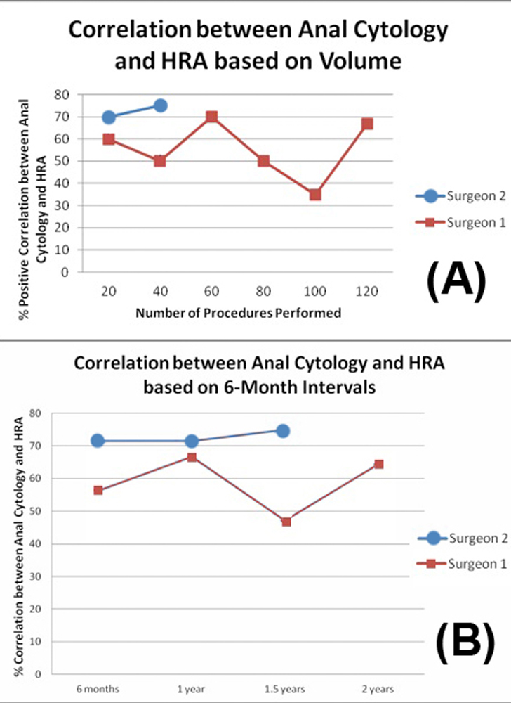
Following IRB approval, a total of 161 HRAs performed on 103 patients by two surgeons over two years at an academic institution were retrospectively reviewed. The two colorectal surgeons had completed the American Society for Colposcopy and Cervical Pathology (ASCCP) approved colposcopy and HRA course. Anal pap smears were obtained concurrently with each HRA performed and the concordance of HRA and Pap smear was examined, based on both volume and duration of practice.

Results:
The mean age was 45.5 years (range 23-79) and 70 (68.0%) patients were male. Seventy-eight percent (80/103) of the patients were HIV (Human Immunodeficiency Virus)-positive and 11% (11/103) of the patients had CD4 counts less than 200. Fifty-three percent of all HRAs performed were positive for anal dysplasia in the presence of abnormal anal cytology. The concordance of anal cytology and HRA improved as case volume increased (from 60% to 67% for surgeon 1 [p=0.24], and from 70% to 75% for surgeon 2 [p=0.72]) (Fig-1-A). Similar results were seen after evaluating the learning curve based on the duration of practice (from 56% to 64% for surgeon 1 [p=0.37], and from 71% to 75% for surgeon 2 [p=0.97]) (Fig-1-B). Interestingly, the changes in concordance were not consistent over time for surgeon 1, who had a peak of 70% vs. a nadir of 35%.

Conclusion:
Although both surgeons demonstrated a non-statistically significant improvement in HRA-cytology concordance over time, the unusual learning curve pattern for surgeon 1 may hint to some challenges that might have impacted the learning curve, including lack of a gold standard for which to compare HRA results and multiple patient-related factors. In order to better evaluate the learning curve, we need to consider all these limitations and be able to control all the potential patient related factors that may impact the surgeon’s performance.

63.01 The consensus and controversial points of new IPMN guideline

H. Ito1, T. Ochiai1, S. Matsumura1, Y. Mitsunori1, A. Aihara1, D. Ban1, A. Kudo1, M. Tanabe1 1Tokyo Medical And Dental University,Heptobiliary And Pancreatic Surgery,Bunkyo-ku, Tokyo, Japan

Introduction:

International consensus guideline for management of Intraductal Papillary Mucinous Neoplasms (IPMN) was revised and published in 2012. Despite widespread acceptance of this guideline, the validity and problem have not been well-studied. We aim to evaluate the clinical utility of this new?guideline with our cases and make clear consensus and controversial points.

Methods:

This is a retrospective study of 105 patients who were consulted for IPMN from Jan 2006 to Oct 2014 at our department. Diagnosis was performed with dynamic enhanced CT and MRI. Pathological analyses were according to Who Classification of Tumours of the Digestive System 4th edition.

Results:

A total of 15 patients undergoing resection for IPMN with new guideline were identified from Apr2013 to Oct 2014. Of these, 11 patients had a component of MD/Mix-IPMN. Carcinoma was found in 5/11(45%) patients and the following were the details. Invasive carcinoma was found in 4/11(36%) patients and high grade dysplasia in 3/11(27%) patients. We present a case report with noninvasive carcinoma which grew 3mm to 8mm of main pancreatic duct with 16mm cyst at the pancreatic head for one year. Of these, 4 patients had a component of BD-IPMN. Invasive carcinoma was found in only one case with high risk stigmata of jaundice. Our previous study from Jan 2006 to Mar 2013 revealed that invasive carcinoma was found in 6/24(25%) patients and high grade dysplasia in 7/24(29%) patients with MD/Mix-IPMN. While, invasive carcinoma was found in 3/29(10%) patients and high grade dysplasia was found in 5/29(17%) patients with BD-IPMN. Next we investigated 37 patients who were continuously observed. 12 patients of these had a component of MD/Mix-IPMN including 2 cases with high-risk stigmata and 10 cases with worrisome features. The other 25 patients had a component of BD-IPMN including 7 cases with worrisome features. All these observing patients have no aggressive growth of tumor for about two years.

Conclusion:

It was consensus point new guideline made the surgical indication stricter compared to the old one. From now we tend to avoid surgical treatment for IPMN, so another predicting factor of malignancy should be considered through continuous observation.

63.02 Should Vitamin D Deficiency be Corrected Prior to Parathyroidectomy?

R. W. Randle1, C. J. Balentine1, E. Wendt1, D. F. Schneider1, H. Chen2, R. S. Sippel1 1University Of Wisconsin,Department Of Surgery,Madison, WI, USA 2University Of Alabama,Department Of Surgery,Birmingham, Alabama, USA

Introduction:

Vitamin D deficiency is common in patients presenting with hyperparathyroidism (HPT), but the importance of replacement prior to surgery is controversial. We aimed to evaluate the impact of low vitamin D on the extent of resection and post-operative hypocalcemia for patients undergoing parathyroidectomy for primary HPT.

Methods:

We identified patients with primary HPT undergoing parathyroid surgery between 2000 and 2015 using a prospectively maintained database. Patients with normal (30ng/mL or greater) vitamin D (25-OH) levels were compared to those with levels less than 30ng/mL.

Results:

The study included 1015 (54%) patients with normal vitamin D and 872 (46%) patients with low vitamin D undergoing parathyroidectomy for primary HPT. Lower vitamin D was associated with higher preoperative parathyroid hormone (PTH) compared to normal vitamin D (median 90 vs 77pg/mL, p<.001). Calcium (median 10.5 vs 10.4mg/dL, p<.001) was also higher while phosphate (median 2.8 vs 2.9mg/dL, p<.001) was lower in patients with low vitamin D, indicating more severe disease. Despite higher preoperative and baseline PTH levels in the low vitamin D group, 10 and 15 minute post-excision PTH was similar (Figure) resulting in a greater overall drop in PTH (median drop 78 vs 72%, p<.001) and similar post-operative calcium (median 9.3 vs 9.3mg/dL, p=.13) compared with the normal vitamin D group. A subgroup analysis in patients with severely low (≤10ng/mL) vitamin D (n=67) also revealed higher preoperative PTH, similar post-excision PTH, greater drop in PTH (median drop 81 vs 72%, p<.001), and similar post-operative calcium (median 9.3 vs 9.3, p=.39) compared to those with normal vitamin D. To achieve similar cure rates, patients with low vitamin D were less likely to require 4-gland exploration (19 vs 23%, p=.009), conversion from a focused approach to 4-gland exploration (11 vs 15%, p=.01), removal of more than 1 gland (20 vs 30%, p<.001), and subtotal parathyroidectomy (8 vs 12%, p=.003) than patients with normal vitamin D. Despite undergoing a more focused operation, patients with low vitamin D had similar rates of persistent (1.5 vs 2.0%, p=.43) and recurrent (1.7 vs 2.6%, p=.21) HPT. Also, at the time of parathyroidectomy both groups had equally low rates of both transient (2.3 vs 2.3%, p=.97) and permanent (0.2 vs 0.4%, p=.52) hypocalcemia.

Conclusion:

Restoring vitamin D in deficient patients should not delay the appropriate surgical treatment of primary HPT. Even though low vitamin D may be a marker for more severe primary HPT, deficient patients are more likely to be cured with the excision of a single adenoma and no more likely to suffer persistence, recurrence, or hypocalcemia than patients with normal vitamin D.

62.17 Virtual Reality Simulation for Residents: A Trainee Experience in Damage Control Endovascular Skills

W. Teeter1, M. L. Brenner1,2, M. R. Hoehn2, D. S. Stein1, T. Scalea1 1University Of Maryland,Division Of Trauma And Critical Care,Baltimore, MD, USA 2University Of Maryland,Division Of Vascular Surgery,Baltimore, MD, USA

BACKGROUND: The use of catheter-based techniques is increasing in the field of trauma. Virtual reality simulation (VRS) is a well-established means of endovascular skills training, and other simulation skills are now mandatory for board-eligibility in general surgery. Training for emerging endovascular damage control skills in trauma, including resuscitative endovascular balloon occlusion of the aorta (REBOA), may be obtained by residents through VRS.

Methods: Fifteen trainees in either an ACGME-approved General Surgery or Surgical Critical Care Fellowship at one institution received didactic and instructional sessions on REBOA. The subjects performed the procedure 6 times. Subjects were excluded if they had taken a similar endovascular training course, had post-graduate training in endovascular surgery, or had performed the procedure in the clinical setting. Performance metrics were measured on a Likert scale, and included procedural time; accurate placement of guide wire, sheath, and balloon; correct sequence of steps; economy of motion; and safe use of endovascular tools. A pre- and post-course test and questionnaire were completed by each subject.

Results: Fifteen subjects, with a mean PGY level of 4.9 years (SD±0.95) participated in the study. Significant improvements in knowledge (p < 0.0001, CI 95%), as assessed by a standardized exam, were observed at the completion of the course. Procedural task times significantly improved from a mean of 207 seconds (SD ± 19.9) to 107 seconds (SD ± 20.6)[(p < 0.0001, CI 95%] (Fig 1). No correlation was observed with endovascular experience in residency, number of endoluminal catheters placed per week, or other parameters. All trainees strongly agreed that the course was beneficial, and the majority would recommend this training to other trainees.

Conclusion: Damage control endovascular skills can be effectively acquired using VRS. Significant improvements in procedural time and knowledge can be achieved regardless of previous endovascular experience or area of training. Novice interventionalists such as surgical trainees can add a specific skill set (REBOA) to their existing core competencies. Use of this procedure in the clinical setting will determine if VRS for REBOA training confers validation metrics such as transfer of skills.

62.18 Surgical Resident Experience with Ethical Controversy

M. P. Kuncewitch1, J. M. Nicastro1, G. F. Coppa1, W. Doscher1 1North Shore University And Long Island Jewish Medical Center,Surgery,Manhasset, NY, USA

Introduction: Throughout surgical training residents may find themselves in situations in which they disagree with the clinical decision-making of the attending surgeon. We sought to survey the experiences and opinions of surgical residents of all PGY levels at our large surgical residency program with regard to moral and ethical controversy. Our purpose was to understand the frequency of such situations, what role residents believe they play in confronting them, and what recourse residents believe they have to address ethically controversial situations in

Methods: A survey was distributed to the 60 residents in our general surgery residency program. The survey asked our residents to anonymously respond to a questionnaire with 12 multiple-choice questions covering the incidence of ethically controversial situations as well as resident feelings towards these situations and any subsequent action taken by the resident. An additional question asked residents for their PGY year. At the close of the survey residents were given the opportunity to anonymously comment and extrapolate their experience.

Results:Forty-seven out of our 60 residents (78.3% responder rate) completed the survey. Sixty-six percent of residents reported at least one instance in which they faced a moral objection to an attending’s decision to operate (or refuse to operate) on a patient, while 48% reported taking part in intra-operative decision making that they felt clearly deviated from appropriate care. Sixty-one percent of residents said they felt comfortable raising concerns with an attending decision on moral or ethical grounds, however only 27% of residents felt assured that this would not result in any form of retaliation. Sixty-one percent of residents felt that at least half of their attending would be receptive to a resident voicing their patient-care decisions or moral or ethical grounds. A majority of residents (71%) felt that a senior resident was the most appropriate person to consult when they didn’t feel comfortable voicing their concerns with a particular attending.

Conclusion:Surgical residency training can challenge residents with difficult decision-making regarding patient care on both moral and ethical grounds. A majority of residents in our general surgery residency reported facing situations in which they disagreed with an attending on such grounds. Variability exists in how residents chose to respond and the comfort level with which they do so.

62.19 Does Operative Case Volume Affect Surgery In-Training Exam Scores?

C. S. Schoolfield1, G. Nightengale1, R. H. Kim1, N. Samra1, Q. Chu1, W. W. Zhang1, T. Tan1 1Louisiana State University Health Sciences Center,Shreveport, LA, USA

Introduction:
In the era of duty hour restrictions, there is a concern that increased time spent by residents in operative cases could lead to decreased time available for studying and therefore adversely impact their ABSITE scores. The objective of this study is to determine if a residents’ number of operative cases correlates with their American Board of Surgery In-Training Exam (ABSITE) scores and could be used as a predictor of future success.

Methods:
A retrospective review was performed on ABSITE scores and operative case logs from a university hospital-based general surgery residency program from 2008-2015. All preliminary and categorical residents were included. Excluded was any incomplete data or records. The data was grouped as a collaborative ABSITE percent correct score and then in groups who scored above and below the 30th percentile. The data was analyzed using linear regression analysis.

Results:
A total of 125 ABSITE scores from 29 residents were reviewed. The overall mean percent correct was 72.4%. Thirty nine scores were <30th percentile ranking with a mean percent correct score of 66.4%. Overall, there was no correlation between number of operative cases and ABSITE scores (R2=.005, p=.4). When those ≥30th percentile on the ABSITE were grouped together, there was a significant positive correlation in number of case logs and ABSITE scores (p=.02). When data was broken down to groups <30th percentile there was however not a significant correlation between case logs and ABSITE scores (p=.61).

Conclusion:
At an academic medical center, there was not significant correlation between general surgery residents’ operative case numbers and their ABSITE scores. An increase in operative cases, and therefore assumed increased time in the operating room, does not appear to have a negative effect on resident academic performance as measured by ABSITE scores.

62.14 Impact of Hepatopancreatobiliary and MIS Fellowships on General Surgery Resident Experience

R. M. Minter1, B. D. Schirmer2, R. Rosenthal3, M. Arregui4, L. Swanstrom5 1University Of Michigan,Surgery,Ann Arbor, MI, USA 2University Of Virginia,Surgery,Charlottesville, VA, Virgin Islands, U.S. 3Cleveland Clinic Florida,Surgery,Weston, FL, USA 4Nagan, Arregui, And Davis, MD Inc,Surgery,Indianapolis, IN, USA 5The Oregon Clinic,Surgery,Portland, OR, USA

Objective: Evaluate the impact of Fellowship Council(FC) accredited Hepatopancratobiliary(HPB) and Advanced GI Minimally Invasive Surgery(GIMIS) fellowships on General Surgery resident case experience.

Methods: HPB(n=12) and GIMIS(n=53) fellow case volume data were quantified in programs with affiliated residencies 2010-2012. Using ACGME chief resident defined category data for the residencies affiliated with these fellowships, liver and pancreas experience was quantified in programs with HPB fellowships, and basic laparoscopy, complex laparoscopy, upper endoscopy(UEND), and colonoscopy experience in programs with GIMIS fellows. Resident data were benchmarked against national standards.

Results: Data are derived from 12/16(75%) HPB and 53/60(88%) GIMIS FC accredited fellowships. Comparative data for resident performance in institutions with HPB and GIMIS fellowships are shown in the Table. Median UEND and colonoscopy volumes for 2010-12 were 43 and 54 for residents (national average 48.4 UEND; 65 colonoscopy), and 55 and 1 for GIMIS fellows.

Conclusions: HPB fellowships do not have a deleterious impact on affiliated General Surgery residents’ liver and pancreas experience, and resident endoscopy experience is not threatened by the presence of a GIMIS fellow. Resident case volumes in institutions with GIMIS fellows are comparable to the national average within the ACGME complex laparoscopy domain, but lower within the basic laparoscopy domain. As GIMIS fellows are performing few basic laparoscopic cases, this is most likely due to fewer cases being performed in these institutions versus fellow impact.

62.15 Back to Basics—The Importance of Enterostomal Therapy Education for General Surgery Residents

J. J. Tackett1, A. L. Fonseca1, W. E. Longo1 1Yale University School Of Medicine,Department Of Surgery,New Haven, CT, USA

Introduction:
General surgery residents’ perceived knowledge base and comfort with intestinal stomas and enterostomal therapy are profiled in the results of a national survey.

Methods:
An anonymous survey was distributed to 734 U.S. general surgery residents through permission of their residency program directors. This anonymous survey explored the existence of formal didactics and training in ostomy creation and stomal care, examined residents’ perceived knowledge base of clinical indications for enterostomal care approaches, and elicited residents’ comfort with performing enterostomal surgeries and mitigating complications. Responses were recorded on a Likert scale. Chi squared test was applied when appropriate.

Results:
Surveys were distributed to 734 residents across the U.S. through their program directors. 218/734 respondents completed the survey (30%): 40% from the Northeast, 22% from the Midwest, 22% from the South, 16% from the West, and 82% with direct university affiliation. Only 12% of respondents stated that they had experienced formal enterostomal therapy training and only 15% had attended a lecture on the subject. Most respondents (86%) stated they routinely worked with enterostomal therapists during the care of patients with ostomies. Only 11% of graduating chiefs felt ‘very confident’ in their knowledge base of clinical indications for enterostomal care and just over half (61%) felt ‘very comfortable’ with the surgical procedures of ostomy creation and closure. Overall, a mere 6% of all residents felt ‘very comfortable’ dealing with common ostomy complications.

Conclusion:
In an era of surgical subspecialization and advanced nursing practice in enterostomal therapy, general surgery residents lack confidence in either their knowledge base or comfort when approaching enterostomal therapy and stomal complications. Resident education across the country should be reformed and formalized to enhance training in these principles that are essential to general surgery practice.