70.17 Limited Utility of Gene Expression Classifier in Surgical Patients with Bethesda III Thyroid Nodules

T. M. Vaghaiwalla1, G. A. Rubio1, M. LoPinto1, Z. F. Khan1, A. R. Marcadis1, J. I. Lew1 1University Of Miami,Division Of Endocrine Surgery, The DeWitt Daughtry Family Department Of Surgery,Miami, FL, USA

Introduction: With implementation of the Bethesda System for Reporting Thyroid Cytopathology (BSRTC), Bethesda III FNA results remain a clinical dilemma for clinicians and surgeons alike. Gene expression classifier (GEC) testing was developed to further stratify patients with Bethesda III nodules as benign or suspicious for cancer. Given the known variability of GEC testing between institutions, this study evaluates the utility of this genetic testing in patients with Bethesda III nodules at a single institution.

Methods: A retrospective review of prospectively collected data of 663 consecutive patients with index thyroid nodules who underwent FNA and thyroidectomy was performed. FNA results were based on the BSRTC system, and GEC testing was later utilized in Bethesda III patients as benign or suspicious for malignancy. Patients with Bethesda III nodules underwent initial thyroid lobectomy for definitive diagnosis unless there was a history of radiation exposure, familial thyroid cancer, obstructive symptoms, bilateral nodules and/or patient preference for which total thyroidectomy was performed. Bethesda III nodules were subdivided into malignant or benign groups based on final pathology. Among patients who underwent GEC testing, final pathology was compared to initial GEC results.

Results: Of 663 patients who underwent FNA, 129 patients had Bethesda III nodules of which 66 (51.2%) had malignancy (Papillary thyroid cancer, n=54; Follicular thyroid cancer (FTC), n=10; Medullary carcinoma, n=1; Lymphoma, n=1) on final pathology. Of the remaining Bethesda III patients, 63 (48.8%) had benign pathology (Adenomatoid nodule, n=18; Hürthle Cell adenoma, n=2; chronic lymphocytic thyroiditis, n= 6; Follicular adenoma n= 10; Multinodular hyperplasia (MNH), n=26; Cyst, n=1). A total of 108 patients with Bethesda III nodules without GEC testing had a malignancy rate of 52.8% (57/108) and benignity rate of 47.2% (51/108). Of 21 patients with Bethesda III nodules who underwent GEC testing, 38.9% (7/18) had suspicious results and malignancy on final pathology with a sensitivity of 77.8% (7/9). Of 3 patients with benign GEC results, 2 (66.7%) had malignancy (FTC, n=2; MNH, n=1) on final pathology. Overall, patients with Bethesda III nodules and suspicious GEC results had a malignancy rate of 38.9% compared to a rate of 52.8% in patients with Bethesda III nodules without GEC testing.

Conclusion: In surgical patients with Bethesda III nodules, there was a greater than expected malignancy rate. However, in patients with Bethesda III nodules and GEC testing, malignancy rates were as predicted. GEC testing may have limited utility in surgical decision making, as this patient population is already highly selected for malignancy due to other factors. Surgeons should assess their local institutional experience to determine whether there is added utility of GEC testing for Bethesda III nodules in their everyday clinical practice.
70.19 A Novel Magnetic Resonance Imaging Protocol for Pancreatic Cystic Lesions

R. K. Schmocker1, S. B. Reeder2, E. R. Winslow1, S. M. Weber1 1University Of Wisconsin,Surgery,Madison, WI, USA 2University Of Wisconsin,Radiology,Madison, WI, USA

Introduction: Cystic pancreatic tumors present a diagnostic dilemma, as initial imaging often fails to provide a definitive diagnosis; therefore serial imaging is needed to follow the lesion over time. CT scan has traditionally been used to follow these lesions, however, given its excellent depiction of fluid containing lesions, and its lack of ionizing radiation, MRI has been increasingly utilized to assess these lesions. We sought to evaluate an initial experience and costs utilizing a novel pancreatic cyst-specific protocol for MRI.

Methods: A single center retrospective study was performed by searching an electronic imaging database for MRI pancreas studies from 2006-2014. After identification of the studies, the reports were examined to confirm that the indication for imaging was pancreatic cystic lesions. Retrospective chart review was used to assess demographics, imaging profiles (presence of other serial imaging – ex: CT), and the presence of interventions. Reimbursed costs of the studies were determined using the CMS physician fee data for the imaging procedures including both the professional and technical components, however contrast cost was not included.

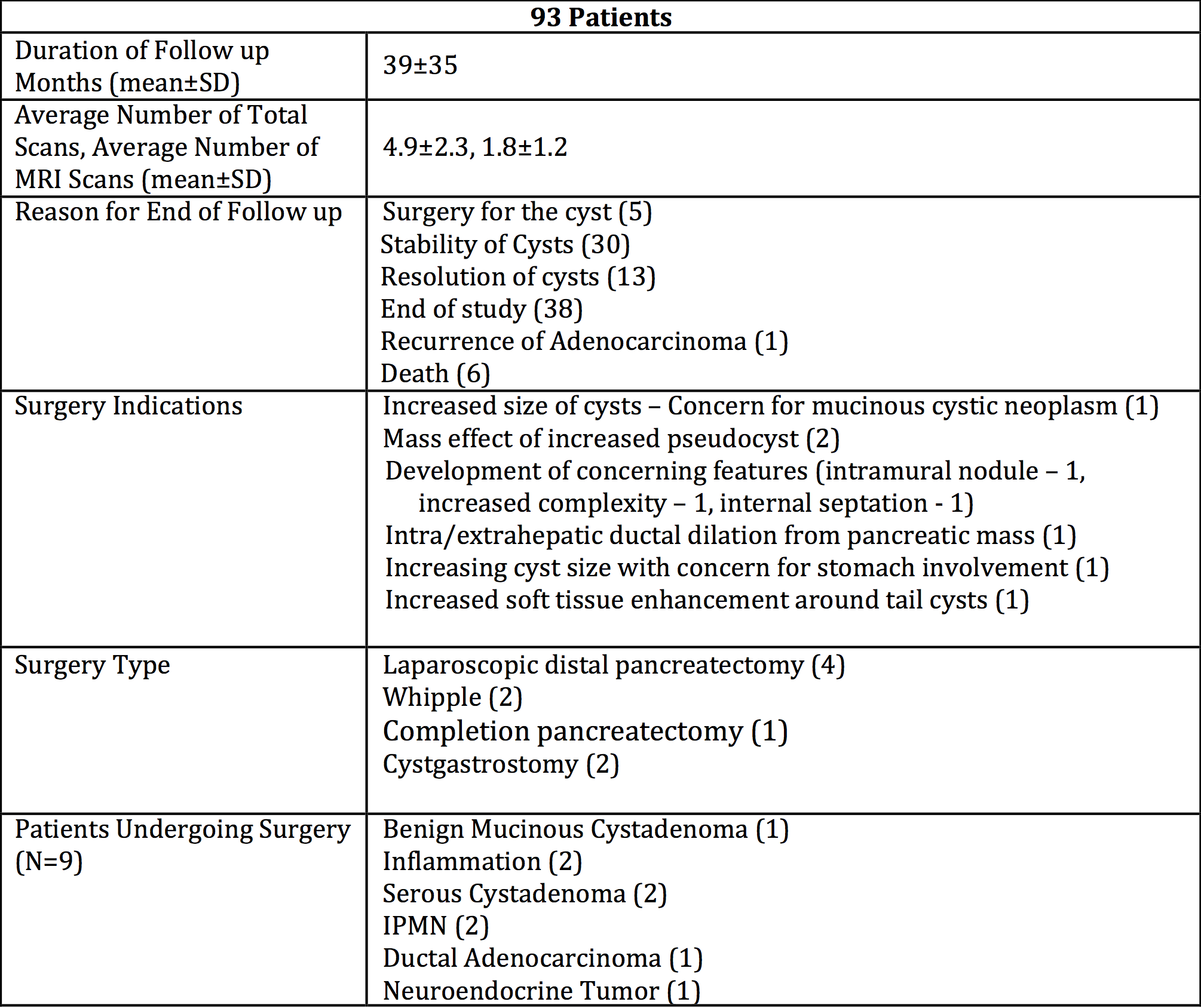
Results: 93 patients underwent an MRI pancreas protocol (average age: 65±13, % female: 61.5%). All patients had one contrasted imaging scan (MRI or CT) during the study period. 9 of these patients underwent a surgical intervention for a pancreatic cyst – 5 with a definitive operation, while 4 underwent surgery, but required surveillance of cysts in the remaining pancreas. Surgery was most often indicated due to concerning features or size (n=6). An MRI pancreas was used to diagnose cyst change for 3/5 patients that had a definitive operation, while CT scan identified a change in imaging characteristics for the other 2 patients. 43 patients underwent EUS/FNA during the study period. The average follow-up was 39 months, with the time between studies being 8.2 months. Patients had, on average, a total of 4.9 scans over the 39 months (1.7 CT scans, 1.3 MRI w/contrast, and 1.8 MRI pancreas protocol scans) or 1.5 scans per year. MRI pancreas was more expensive than CT w/Contrast ($333 vs. $230), but with the absence of radiation and contrast related complications. Additionally, if the average patient were exclusively followed with CT scans; the overall cost savings would be minimal ($189).

Conclusion: Our initial experience with the use of a novel pancreas protocol MRI is favorable, with minimal increase in cost. The lack of ionizing radiation and intravenous contrast with the MRI pancreas potocol, is an important consideration for patients undergoing serial imaging. The use of MRI for patients with pancreatic cystic disease shold be investigated further.

70.16 Causes and Predictors of Early Mortality Following Pancreatic Resection, Recognize Then Prevent

C. Mosquera1, T. L. Fitzgerald1, E. E. Zervos1 1East Carolina University Brody School Of Medicine,Division Of Surgical Oncology,Greenville, NC, USA

Introduction: Pancreas resection is among the highest risk procedures in terms of post-operative mortality. The purpose of the study was to determine predictors and causes of early and late post-operative mortality in patients undergoing pancreatectomy.

Methods: A prospective pancreas surgery registry at a very-large volume hospital was queried to identify early (30-day) and late (90-day) postoperative mortality between 2008 and 2015. Logistic regression was undertaken to determine predictors of each. Causes of death were categorized as: hemorrhagic, cardiorespiratory, sepsis and disease progression. Sepsis was sub-categorized as related (presence of infected intra-abdominal collections, abscess, fistula, and surgical site infection) or unrelated to surgery (pneumonia, CLABSI, CAUTI). Disease progression signified withdrawal of care in an otherwise viable patient due to perceived poor prognosis as a result of documented recurrent or persistent disease.

Results: A total of 312 patients underwent resection (68% Whipple, 32% Distal Pancreatectomy). Early postoperative mortality occurred in 12 (3.8%) patients and late in 24 (7.7%) patients with a total postoperative mortality of 11.5%. Early deaths occurred during index admission in 83% while 95.5% of late deaths occurred outside of the hospital or in patients that were discharged and returned to hospital and died. (Table)

On univariate analysis, age, smoking history, intensive care unit requirement (ICU), presence of complications, operative estimated blood loss, body mass index, discharge destination, presence of tachycardia, elevated WBC at discharge and type of insurance predicted any postoperative mortality. On multivariate analysis, only ICU (OR 20.3 p 0.0003) and discharge destination of home and SNF compared to rehabilitation facility remained significant (p<0.05). Private insurance was a protective factor compared to Medicare, Medicaid and uninsured (p<0.05). Sepsis arising from surgical complications was the primary source of early postoperative mortality while disease progression was the most common cause of late mortality.

Conclusion: Surgical complications remain the primary source of early postoperative mortality, which are largely unavoidable. Late postoperative mortality may be mitigated by minimization and early detection of hospital-acquired infection through adherence to best care guidelines and neo-adjuvant strategies to identify patients with low risk of disease progression in which further treatment is non futile.

70.11 Hepato-Pancreatectomy: Outcomes of Synchronous Hepatic and Pancreatic Resection

T. B. Tran1, J. N. Leal1, M. M. Dua1, B. C. Visser1, J. A. Norton1, G. A. Poultsides1 1Stanford University,Surgery,Stanford, CA, USA

Introduction: Simultaneous resection of both the liver and the pancreas remains controversial due to the high morbidity and questionable oncologic benefit. The purpose of this study is to evaluate the short-term morbidity and mortality after combined resection of the liver and pancreas, as well as to determine whether or not these aggressive operations are associated with long-term survival.

Methods: Consecutive patients who underwent synchronous hepatectomy and pancreatectomy (SHP) at a single instution were retrospectively evaluated. Extent of liver resection was categorized into major hepatectomy (defined as lobectomy or trisectionectomy) and minor hepatectomy (less than 3 liver segments). Preoperative patient features, perioperative outcomes, and long-term survival following synchronous hepatectomy and pancreatectomy (SHP) were evaluated.

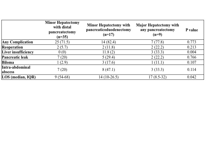
Results: From 2005 to 2014, 61 patients underwent SHP. Diagnoses include 34 pancreatic neuroendocrine tumors (PNET), 6 sarcomas, 3 cholangiocarcinomas, 3 adrenocortical carcinomas, 3 cystic pancreatic neoplasms, 2 renal cell carcinomas, 2 colon cancers, 2 recurrent gastric cancers, 2 gastrointestinal stromal tumors, 2 peritoneal mesotheliomas, 1 appendiceal cancer, and 1 ovarian cancer. Patients were further categorized into 3 groups based on the extent of SHP (Table). There were no differences in patient demographics, comorbidities, ASA status, estimated blood loss, need for blood transfusion, 30 or 90-day mortality. Furthermore, the incidence of major complications was similar between the three groups, except for liver insufficiency (defined as peak postoperative bilirubin of 7 or more), which affected one-third of patients who underwent major hepatectomy with any pancreatectomy (p=0.004). Length of stay correlated with extent of SHP (p=0.042). Patients with PNET had more favorable prognosis compared to all other malignant histologies combined (5-year overall survival 47% vs. 26%; p=0.002).

Conclusion: Combined hepatectomy with pancreatectomy is a technically challenging operation. In carefully selected patients, combined hepatectomy and pancreatectomy can be associated with long-term survival.

70.12 Incidence and Pattern of Port-Site Metastasis Following Cholecystectomy in GBCA: A Systematic Review

D. Berger-Richardson1, T. Chesney1, M. Englesakis3, S. Cleary1,3, A. Govindarajan1,2, C. Swallow1,2 1University Of Toronto,Division Of General Surgery,Toronto, ONTARIO, Canada 2Mount Sinai Hospital,Toronto, ONTARIO, Canada 3University Health Network,Toronto, Ontario, Canada

Introduction: Laparoscopic cholecystectomy for the treatment of symptomatic cholelithiasis was rapidly adopted in the early 1990’s. However, there were early reports of port-site metastasis (PSM) when incidental gallbladder adenocarcinoma (GBCA) was discovered in the specimen after surgery. In a review of cases up until 1999, the estimated incidence of PSM in incidental GBCA was reported to be 14%. Recognition of this phenomenon may have prompted changes in surgical technique, and we questioned whether the incidence of port-site metastasis has changed since then. Furthermore, direct contact between the specimen and wound edges during extraction is one of the possible mechanisms of PSM. Determining the incidence of both extraction and non-extraction port PSMs may provide insight into this hypothesis. Hypothesis: 1) The risk of PSM following resection of GBCA has decreased over the past fifteen years. 2) Extraction ports are more likely to harbour recurrence than non-extraction ports.

Methods: A systematic review of articles related to wound recurrence of GBCA was conducted by two independent reviewers. Inclusion criteria for our first objective of determining the modern incidence of PSM were English language papers reporting the presence or absence of PSM following a minimum of 5 cholecystectomies harbouring GBCA published from 2000 – 2014, the 15 year period following the previous review. Inclusion criteria for our second objective of determining the location of the port site involved include English language papers reporting the presence of PSM following laparoscopic cholecystectomy in the setting of GBCA, without limiting this search temporally or by study size.

Results:5448 abstracts were screened. 23 published case series met criteria for estimating the modern incidence of PSM in GBCA. PSM were found in 73 of 728 patients (incidence=10%). Results from the two largest studies using prospectively collected databases (n= 114 and n= 96) had PSM incidences of 14% and 15% respectively. With respect to the pattern of PSM, data was extracted from 98 papers meeting inclusion criteria. More than 305 individual port sites have been reported to harbour recurrence in 236 patients with GBCA (number of PSM/ person = 1-4). Of those with decipherable location (extraction vs non-extraction) (n=190) based on description within the paper, 53% occurred at extraction ports compared to 47% at non-extraction ports (p=0.526).

Conclusion:Despite the implementation of techniques for risk reduction, the incidence of PSM following gallbladder cancer resection is stable at 10-15%. Recurrence at non-extraction ports may indicate mechanism(s) other than direct contact with the wound during extraction. Studies into mechanisms of PSM are warranted. Preoperative imaging studies should be carefully reviewed to minimize inappropriate laparoscopic resection of gallbladder cancer.

70.14 Gender Differences in Length of Hospital Stay After Elective Major Operations for Colon Cancer

C. E. Cauley1,2, D. Chang1,3, L. Bordeianou1, A. B. Haynes1,2,3 1Massachusetts General Hospital,Department Of Surgery,Boston, MA, USA 2Ariadne Labs At Brigham And Women’s Hospital And The Harvard T.H. Chan School Of Public Health,Boston, MA, USA 3Codman Center Center For Clinical Effectiveness In Surgery,Boston, MA, USA

Introduction:
Length of stay (LOS) after elective colectomy has markedly decreased with the advent of enhanced recovery pathways, yet there remains substantial variability in postoperative stays. This study aims to assess the influence of gender on LOS after elective colectomy for colon cancer.

Methods:
Patients undergoing elective colectomy for colon cancer between 1998 and 2011 were identified from the Nationwide Inpatient Sample. Multivariate logistic regression modeling was used to determine if gender was a significant predictor of extended postoperative LOS (> 8 days) controlling for potential confounders, including year of operation, race, age, comorbidity, complications, and hospital factors.

Results:
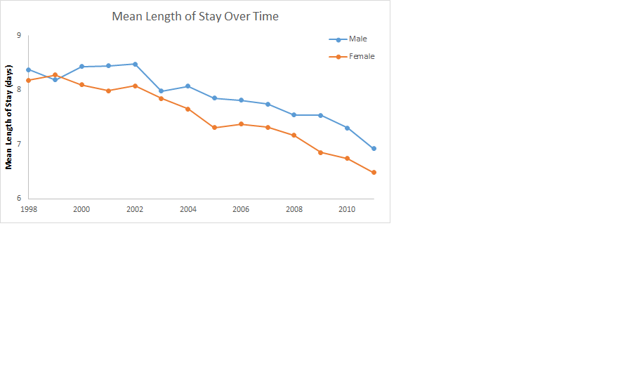
The cohort consisted of 88,778 patients, 40,526 (45.65%) men and 48,252 (54.35%) women. The mean LOS was 7.7 days (SE = 0.020), which decreased from 8.3 to 6.6 days over the study period, with a mean stay of 7.9 days (SE = 0.031) versus 7.5 days (SE = 0.025) for men and women, respectively (p<0.001). Multivariate logistic regression analysis revealed lower odds of extended LOS in women (OR 0.90; 95% CI: 0.87-0.94). This gender difference was greater in patients over 60 years old, and has continued despite decreasing LOS in more recent years.

Conclusion:
Men have a longer length of stay after elective colectomy for colon cancer, controlling for patient and operative characteristics. This difference in healthcare utilization between genders despite increasing standardization of clinical care should be explored further to determine if factors such as social support influence this phenomenon.

70.15 Very Long Acyl Chain (C24:0 and C22:0) Ceramides are Associated with Obesity and Breast Cancer Progression

K. Moro1, M. Nagahashi1, J. Tsuchida1, T. Niwano1, K. Tatsuda1, C. Toshikawa1, M. Hasegawa1, Y. Koyama1, T. Kobayashi1, S. Kosugi2, H. Kameyama1, H. Aoki3, K. Takabe3, T. Wakai1 1Niigata University Graduate School Of Medical And Dental Sciences,Digestive And General Surgery Niigata University,NIigata, NIIGATA, Japan 2Uonuma Kikan Hospital,Division Of Digestive And General Surgery,Minami-Uonuma, NIIGATA, Japan 3Virginia Commonwealth University School Of Medicine And The Massey Cancer Center,Surgical Oncology,Richmond, VA, USA

Introduction:
It is well established that obesity is associated with poor prognosis of breast cancer patients. Obesity evokes chronic inflammation, which stimulates cancer progression. Ceramide is a key metabolite in both anabolic and catabolic pathways of sphingolipids, and the very long fatty acyl chain (C24:0 and C22:0) ceramides are elevated both in the tissues and in the circulation of obesity, insulin resistance and diabetes. One of the mechanisms of obesity-mediated inflammation is due to release of ceramide from adipocytes after lipolysis. It has been reported that the ceramides and other obesity-related factors trigger activation of inflammasome that result in the secretion of inflammatory cytokines such as IL-1 beta, IL-6, and TNF-alpha under the obese condition. However, serum levels of ceramides in breast cancer patients associated with obesity have not been elucidated to date. In this study, we examine the levels of ceramides in breast cancer patients to reveal the association of the sphingolipids with obesity and breast cancer progression.

Methods:
A retrospective analysis was conducted of 59 patients with breast cancer, in whom the disease was diagnosed as stage I, II or III pathologically. The serum from the patients were obtained when the diagnosis was made and any treatment had been done before the blood collection. Ceramides (C14:0, C16:0, C18:1, C18:0, C20:0, C22:0, C24:1, C24:0, C26:0) were measured by liquid chromatography-electrospray ionization-tandem mass spectrometry . The levels of ceramides were analyzed with clinicopathological data of the patients.

Results:

Body mass index (BMI) was significantly associated with the pathological stage (P < 0.05). The levels of ceramides were detected successfully in the serum from 59 breast cancer patients. The levels of ceramide were not associated with clinical demographics of the patients including age, hormone receptors (ER, PgR) and HER2 status, Ki-67 index, nuclear grade, lymphatic and vascular invasion of the tumor. Interestingly, however, levels of C24:0 ceramide in patients with high BMI (> 25) was significantly higher than that in patients with normal BMI (< 22) (P < 0.05). Further, levels of C24:0 ceramide were significantly elevated with pathological stage (P < 0.05). C22:0 ceramide also showed trends of similar associations with BMI and pathological stage, albeit they were not statistically significant.

Conclusion:

Our results show an elevation of very long fatty acyl chain (C24:0 and C22:0) ceramides in serum of obesity-related breast cancer. Further studies are needed to elucidate the mechanism, and possible association with the prognosis. This work was supported by the Japan Society for the Promotion of Science Grant-in-Aid for Scientific Research Grant Number 15H05676 and 15K15471 for M.N and 15H04927 for W.T. M.N. is supported by the Uehara Memorial Foundation, Nakayama Cancer Research Institute, Takeda Science Foundation, and Tsukada Memorial Foundation.

70.08 Outcomes of Non-elective Gastric Cancer Surgery Following Admission Through the Emergency Department

I. Solsky1,2, P. Friedmann1,2, P. Muscarella1,2, H. In1,2 1Montefiore Medical Center,Department Of Surgery,Bronx, NY, USA 2Albert Einstein College Of Medicine,Department Of Surgery,Bronx, NY, USA

Introduction: Outcomes following non-elective surgery for gastric cancer are poorly defined. Gastric cancers are mostly asymptomatic and the presence of symptoms generally signals more advanced disease. Studies suggest that emergent cancer surgery for gastrointestinal (GI) cancers are associated with later cancer stages and worse outcomes. Our objective was to compare outcomes of patients who underwent non-elective gastric cancer surgery following an admission through the emergency department (ED) with patients receiving elective surgery or surgery after planned admission using a US representative database.

Methods: Nationwide Inpatient Sample (NIS) was used to examine adults admitted with gastric cancer who underwent gastric cancer surgery over five years (2007-2011). NIS is an all-payer database designed to yield national estimates of hospital inpatient stays. Demographics and outcomes were compared between those who had their surgery performed non-electively after an ED admission with those who did not. Multivariable logistic regression was used to examine predictors of being discharged to home.

Results: 9,279 patients who underwent gastric cancer surgery were included for analysis. 1,143 (12%) underwent non-elective surgery following an ED admission. These patients were more likely to be female (42% vs. 35%), non-white (61% vs. 43%), elderly ≥75 years (40% vs. 26%), admitted to an urban non-teaching hospital (46% vs. 25%), in the lowest quartile for median household income (31% vs. 25%), and have one or more comorbidities (87% vs. 70%). They were less likely to have private insurance (19% vs. 37%). They had a longer median length of stay (16 vs. 9 days), number of days to surgery (5 vs. 0), were more likely to die during their hospitalization (8% vs. 3%) and less likely to be discharged home (63% vs. 82%). On multivariable logistic regression analysis, we found that having non-elective surgery following an ED admission was independently associated with a lower likelihood of being discharged to home [OR: 0.49 (95% CI: 0.42 – 0.57)]. On sensitivity analysis, our findings remained unchanged regardless of whether specific complications were included in the model.

Conclusion: Nationally, 12% of all gastric cancer surgeries are done following an admission through the ED. This tends to occur more frequently in vulnerable populations. Our finding that patients undergoing non-elective surgery following an ED admission had worse outcomes than those of patients getting surgery electively has implications towards the design of future studies to help improve outcomes for these patients. Outcomes after gastric cancer surgery may be improved by a) developing programs to detect gastric cancer in patients prior to the development of symptoms prompting presentation to the ED, b) improving access to health care in vulnerable populations, and c) encouraging elective scheduling of surgery for stable gastric cancer patients.

70.09 Prognostic Factors In Anaplastic Thyroid Neoplasms In The Adolescent And Young Adult Population

I. I. Maizlin1, G. McGwin2, M. Goldfarb3, K. W. Gow5, S. A. Vasudevan6, J. J. Doski4, A. B. Goldin5, M. Langer7, J. G. Nuchtern6, E. A. Beierle1 1Children’s Hospital Of Alabama, University Of Alabama,Division Of Pediatric Surgery,Birmingham, Alabama, USA 2University Of Alabama,School Of Public Health,Birmingham, Alabama, USA 3John Wayne Cancer Institute/Providence St. John’s Medical Center,Department Of Surgery,Santa Monica, CA, USA 4University Of Texas Health Science Center At San Antonio, San Rosa Children’s Hospital,Department Of Surgery/Pediatric Surgery Division,San Antonio, TX, USA 5Seattle Children’s Hospital,Division Of General And Thoracic Surgery,Seattle, WA, USA 6Baylor College Of Medicine,Division Of Pediatric Surgery,Houston, TX, USA 7Maine Medical Center,Division Of Pediatric Surgery,Portland, ME, USA

Introduction: The aggressive nature and rarity of anaplastic thyroid cancer (ATC) makes it difficult to determine patient outcomes, especially in single-institution studies with small cohorts and short follow-up. Furthermore, considering that ATC increases in frequency with age, no investigations of large data sets exist that have examined the adolescent and young adult (AYA) population. Therefore, we utilized the National Cancer Data Base to determine the importance of age as a prognostic factor in anaplastic thyroid cancer.

Methods: All AYA patients (≤39 yo) with diagnosis of ATC were reviewed from the National Cancer Data Base from 1998 to 2012. They were then compared to older patient groups [40-65 yo (middle age) and ≥ 66 yo (elderly)] with same diagnosis. Log-rank test was used to compare survival functions between age groups and chi-squared tests were used to compare tumor size, method of diagnosis, time from diagnosis to definitive treatment, presence of comorbidities (based on Charlson/Deyo Score), and type of surgery performed. Cox proportional hazards models were used to estimate hazard ratios (HR) and their associated p-values. A secondary analysis was performed to evaluate whether surgical intervention was associated with survival in AYA patients.

Results:Out of total of 3,154 patients with ATC, 39 patients fit the criteria of AYA. Median survival in the AYA group was 118.5 months compared to 61.1 months (p=0.06) in middle and 66.2 months (p=0.06) in elderly groups. Compared to the AYA group, the HR for middle age and elderly groups were 1.96 (p=0.02) and 1.73 (p=0.06), respectively. The age groups demonstrated similar tumor size (>4cm in 84.6% vs. 85.7% vs. 83.8%) at time of diagnosis. AYA group had a significantly lower prevalence of comorbid conditions (12.5%) when compared to other age groups (21.2% and 27.6% respectively). AYA group had greater mean period between diagnosis and definitive treatment (19.4 ±3.6 days vs. 16.3 ±0.8 vs. 15.1 ±0.5 days) and a higher rate of FNA prior to intervention (97.3% vs. 92.0% vs. 87.8%). Consequently, following adjustment for confounders (tumor size, comorbidities, method of diagnosis and surgery) mortality in the middle- and older-aged groups was approximately 50% higher compared to the AYA group [HR=1.52 (p=0.16) and HR=1.56 (p=0.14), respectively], though these differences were not statistically significant. Within the AYA group there was no significant difference in survival between surgery employed as initial treatment (followed by radiation and chemotherapy) and non-surgical (radiation and chemotherapy) intervention only.

Conclusion:While anaplastic thyroid cancer is an aggressive tumor with overall poor prognosis, adolescents and young adults appeared to have increased survival compared to older populations. Survival rates in the AYA population were not affected by surgical intervention as initial therapeutic modality, compared to radiation and chemotherapy alone.

70.10 Pancreatic Cancer Disparities in an Underserved Population: A Need for Accessible Healthcare

C. Mosquera1, J. Lee1, S. D. Kachare1, T. L. Fitzgerald1, E. E. Zervos1 1East Carolina University Brody School Of Medicine,Division Of Surgical Oncology,Greenville, NC, USA

Introduction: The 29 county subregion comprising eastern North Carolina (ENC) is characterized by rurality, poverty and cancer outcome disparities when compared to the rest of the state. This study was undertaken to identify factors in this unique region contributing to disproportionately poor outcomes for pancreatic adenocarcinoma (PCA).

Methods: All patients diagnosed with PCA from 1996-2015 in ENC were identified through a central tumor registry. Logistic regression was undertaken to determine demographic, tumor, treatment and socioeconomic factors that contribute to observed outcome disparities.

Results:916 patients with PCA were identified, 93% of which arose from counties whose median household income is below the federal poverty limit. Compared to the rest of the state, PCA incidence in ENC (per 100k) is significantly higher (12.5 ±1.4 vs. 11.6 ± 1.7, p<0.03) as is the death rate (11.8±1.7 vs. 10.5±1.5, p<0.001). Multivariate analysis identified 7 factors independently predictive of poor survival (Table1). African Americans comprised 40% of these patients and were over-represented in each category that predicted poor outcomes.

Conclusion: Improved access to healthcare in ENC would positively impact 4 of 7 factors that are associated with poor survival in PCA; 3 are immutable. In this underserved population, Medicaid expansion or full implementation of the Affordable Care Act carries the greatest potential to erase these disparities.

70.06 A Clinically Applicable Muscular Index Predicts Morbidity and Survival in Resectable Pancreatic Cancer.

D. Delitto1, S. M. Judge1, R. L. Nosacka1, T. J. George1, S. M. Wallet1, G. A. Sarosi1, R. M. Thomas1, K. E. Behrns1, S. J. Hughes1, A. R. Judge1, J. G. Trevino1 1University Of Florida,Gainesville, FL, USA

Background: The relationship between myopenia, nutritional status, and long-term oncologic outcomes remains incompletely defined in patients with resectable pancreatic cancer (PC). Additionally, more advanced technologies to quantify myopenia, including complex volumetric analysis and densitometric algorithms, are not always available in the clinical setting. We sought to reliably quantify prognostic indicators of preoperative cachexia as a predictor of clinical outcomes with routine imaging applicable to any office or hospital site.

Methods: Preoperative CT scans were electronically available and suitable for analysis in 73 of 82 consecutive patients with PC undergoing pancreaticoduodenectomy (PD) between November, 2010 and February, 2014. The psoas index was computed from cross-sectional areas of psoas muscles normalized to vertebral body area at L3. Correlation and proportional hazards analyses were performed to identify relationships between muscularity, preoperative nutritional markers, clincopathologic parameters and long-term survival.

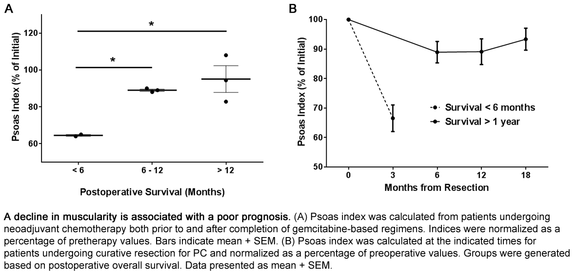
Results: Psoas index correlated strongly with preoperative hemoglobin and albumin levels (P = .001 and .014, respectively), identifying a pattern of preoperative frailty. High psoas index, albumin and hemoglobin levels significantly correlated with improved long-term survival (HR 0.014, P < .001; HR 0.43, P < .001 and HR = 0.80, P = .014). However, on multivariate analysis, psoas index proved to be the only independent predictor of survival (HR 0.021; P = .003). Notably, rapid declines in psoas index during neoadjuvant chemotherapy were associated with poor postoperative outcomes, as were declines in psoas index during the postoperative period.

Conclusions: The data indicate that the psoas index, a measurement available in any clinical setting, is a statistically powerful predictor of survival in PC, when compared to tumor grade and stage as well as previously validated nutritional parameters.

70.07 Positive Margins Contribute to the Survival Paradox between Stage 2B/C and Stage 3A Colon Cancer

Q. D. Chu1, M. Zhou2, K. Medeiros2, R. H. Kim1, X. Wu2 1Louisiana State University Health Sciences Center-Shreveport,Surgical Oncology,Shreveport, LA, USA 2Louisiana State University Health Sciences Center,Louisiana Tumor Registry & Epidemiology And School Of Public Health,New Orleans, LA, USA

Introduction: We found a persistence of a survival paradox between Stage 3A and Stage 2B/C colon cancer who had optimal treatment. The underlying reasons are elusive. We hypothesized that positive surgical margins contribute significantly to this paradox.

Methods: We evaluated a cohort of 16,471 patients with stage 3A or stage 2B/C with ≥ 12 lymph nodes (LNs) retrieved (N=5,670) from 709,583 patients diagnosed with colon cancer in 2003-2012 from the National Cancer Data Base. All received chemotherapy. Patients with Stage 3A were further subdivided into those with < 12 LNs retrieved (N=3,195) and those with ≥ 12 LNs retrieved (N=7,606). Univariate and multivariate survival analysis were employed.

Results: The 5-year overall survival (OS) rate was 70.8% for stage 2B/C ≥ 12 LNs, 81.6% for stage 3A with < 12 LNs, and 85.6% for Stage 3A with ≥ 12 LNs (P<0.0001). Patients with stage 2B/C had significantly higher rate of positive surgical margins compared to stage 3A (19% vs 1%; P<0.0001). Significant predictors (P<0.01) of poor OS include stage 2B/C, community cancer program, advanced age, African-American ethnicity, Medicaid, low education level, high comorbidity index, and positive surgical margins.

Conclusion: Positive surgical margins contribute to the survival paradox between optimally treated Stage 2B/C and Stage 3A colon cancer patients.

70.04 Impact of Time from Initial Biopsy to Definitive Excision When Residual Melanoma is Present

A. Nadler1, K. J. Ruth2, J. M. Farma1, S. S. Reddy1 1Fox Chase Cancer Center,Surgical Oncology,Philadelphia, PA, USA 2Fox Chase Cancer Center,Biostatistics,Philadelphia, PA, USA

Introduction: The time from initial biopsy (bx) to definitive excision, or surgical interval (SI), does not appear to affect outcomes for melanoma. However, the impact of a prolonged SI where residual melanoma (RM) may be present in the final specimen is less clear. This study was undertaken to assess whether SI as it relates to the presence of RM in the final specimen affects prognosis.

Methods: A retrospective review of our institutional melanoma database from 2009 and 2011 was performed. Fisher exact tests and Kruskal Wallis tests were used to compare characteristics by RM status. Survival was estimated with Kaplan Meier methods and compared with the log-rank test. Cox proportional hazards regression was used to adjust for covariates.

Results: Of 240 patients in the database, 179 treated for non-metastatic cutaneous melanoma were included. The median age was 61 years and 51% were male (n=92). At initial bx, 60% (n=108) had a shave bx, 14% (n=25) had a punch bx, and 26% (n=46) had an excisional bx. All patients underwent a radical excision of the primary lesion and 74% (n=132) underwent a sentinel lymph node bx. The median SI was 41 days (range 8-1280) and it did not differ by biopsy type (p=0.36). On final pathology, 45% (n=81) had RM present. RM was more likely to be found in punch bx (n=19 of 25) compared to shave bx (46 of 108) and excisional bx (16 of 46) (76% vs. 43% vs. 35%, p=0.003). The presence of RM did not differ by Breslow depth (p=0.32). Median follow-up was 12.4 months (range 0.2-35.7), during which 13 patients had disease recurrence or died. Recurrence free survival (RFS) was significantly lower in patients with a longer SI (split at median ≤ 41 days for shorter SI vs. > 41 days for longer SI) (p=0.035). RFS at 12 months was 97.3% (CI 89.6-99.3) for a shorter SI and 89.7% (CI 78.1-98.3) for a longer SI; at 24 months, the RFS was 94.4% (CI 82.3-98.3) and 83.5% (CI 68.6-91.8), respectively. Differences in overall survival (OS) were borderline significant (p=0.069) with OS at 12 months was 98.7% (CI 91.2-99.8) for shorter SI and 94.7% (CI 84.3-98.3) for longer SI; at 24 months, the OS was 95.6% (CI 81.8-99.0) and 82.2% (CI 64.1-91.7), respectively. RFS and OS did not differ by RM status (p=0.20 and p=0.19, respectively). However, for SI and RM status in combination, patients who had RM and a longer SI had the lowest RFS with a 12 month RFS of 81.0% (CI.55.9-92.7) compared to 92.9% (CI 84.3-96.9) for the other groups combined (p=0.022). In multivariable analysis, with adjustment for stage, age, and Breslow depth, patients who had RM and a longer SI had borderline significance for RFS (p=0.074, hazard ratio = 2.99, 95% CI=0.90-9.98).

Conclusion: Longer SI, especially in combination with the presence of RM following initial bx, is associated with worse RFS. Patients anticipated to have RM should be triaged appropriately to avoid delays to definitive excision.

70.05 Colon Cancer Surgery at High and Low Mortality Hospitals

M. A. Healy1, H. Yin1, J. D. Birkmeyer2, S. L. Wong1 1University Of Michigan,Surgery, Center For Healthcare Outcomes & Policy,Ann Arbor, MI, USA 2Dartmouth Medical School,Surgery,Lebanon, NH, USA

Introduction: There is wide variation in mortality across hospitals for cancer surgery. While higher rates of mortality are commonly ascribed to high-risk resections, the impact of more common operations is unclear. We sought to evaluate causes of mortality following colon cancer operations across hospitals.

Methods: : 49 American College of Surgeons Commission on Cancer (ACS-CoC) hospitals were selected for participation in a CoC special study. We ranked hospitals using a composite measure of mortality and performed onsite chart reviews. We examined patient characteristics and mortality following colon resections at very high mortality (HMH) and very low mortality (LMH) hospitals (2006-2007).

Results: We identified 3,025 patients who underwent surgery at 25 LMHs (n = 1,391) and 24 HMHs (n = 1,634). There were wide differences in mortality between HMHs and LMHs (9.2% vs. 2.7%). Compared to LMHs, HMHs had more patients who were black (11.2% vs. 6.5%), had >2 comorbidities (22.7% vs. 18.9%), ASA class 4-5 (11.9% vs. 5.3%), and were functionally dependent (13.9% vs. 8.9%; p<.001 for all). For emergency resections, mortality was higher in HMHs versus LMHs (28.3% vs. 11.4%; OR 3.1, 95% CI 1.4-6.7) with ICU admission and prolonged mechanical ventilation more likely in HMHs.

Conclusion: There is significant variation in mortality across hospitals for colon cancer surgery. In emergent cases, perioperative mortality is nearly as high as 1 in 3 patients in HMHs, and many who die undergo ICU admission with mechanical ventilation. This finding reflects a need for improved surgical decision-making to enhance outcomes and quality of care at these hospitals.

70.01 Effectiveness of Postoperative Surveillance Endoscopy for Patients with Ulcerative Colitis

H. Ishii1, K. Hata1, J. Kishikawa1, H. Anzai1, K. Otani1, K. Yasuda1, T. Nishikawa1, T. Tanaka1, J. Tanaka1, T. Kiyomatsu1, K. Kawai1, H. Nozawa1, H. Yamaguchi1, S. Ishihara1, J. Kitayama1, T. Watanabe1 1Faculty Of Medicine, The University Of Tokyo,Department Of Surgical Oncology,Tokyo, , Japan

Introduction: The incidence of neoplasia after surgery of ulcerative colitis (UC) has not been sufficiently clarified, particularly in the Japanese population, and it is not evident whether surveillance endoscopy is effective for detecting dysplasia/cancer in the remnant rectum or pouch. The aims of the present study were to estimate and compare postoperative development of dysplasia/cancer in patients with UC who underwent ileorectal anastomosis (IRA) or ileal pouch-anal anastomosis (IPAA), and to evaluate the effectiveness of postoperative surveillance endoscopy.

Methods: We retrospectively reviewed 120 patients who received postoperative surveillance endscopy after IRA or IPAA in our institute for development of dysplasia/cancer in the remnant rectal mucosa or pouch.

Results: Three hundred seventy-nine endoscopy sessions were performed for 30 patients after IRA, while 548 pouch endoscopy sessions were performed for 90 patients after IPAA. In the IRA group, 5 patients developed dysplasia/cancer during postoperative surveillance and in all cases neoplasia was detected at an early stage. None of them developed neoplasia within 10 yr of diagnosis; the cumulative incidence of neoplasia after disease onset was 7.2, 12.0, and 23.9% at 15, 20, and 25 yr, respectively. In one case after stapled IPAA, dysplasia was found at the ileal pouch; a subsequent 9 endoscopy sessions in 8 years did not detect any dysplasia. Neoplasia was found more frequently during post-operative surveillance in the IRA group than in the IPAA group (p = .0028). The cumulative incidence of neoplasia after IRA was 3.8, 8.7, and 21.7% at 10, 15, and 20 yr, respectively, and that after IPAA was 1.6% at 20 yr.

Conclusion: The cumulative incidence of neoplasia after IPAA was minimal. Those who underwent IRA had a greater risk of developing neoplasia than those who underwent IPAA, although postoperative surveillance endoscopy was effective to detect dysplasia/cancer at an early stage. IRA can be an option of surgical procedure in selected cases in which it would be profitable to the patient, with more careful surveillance.

70.02 Gene expression of Angiopoietin, Tie and VEGF are associated with poor survival in breast cancer

R. Ramanathan1, A. L. Olex2, L. J. Fernandez1, A. R. Wolen2, D. Fenstermacher2, M. Dozmorov3, K. Takabe1 1Virginia Commonwealth University Medical Center,Surgery,Richmond, VA, USA 2Virginia Commonwealth University,Center For Clinical And Translational Research,Richmond, VA, USA 3Virginia Commonwealth University,Biostatistics,Richmond, VA, USA

Introduction:
Breast cancer is the second most common cancer affecting women in the United States, constituting an estimated 232,670 new cases in 2014. Angiogenesis is one of the known hallmarks of cancer that is essential for cancer progression and aggressiveness. The angiopoietin-2 (Ang2) ligand and its Tie receptors constitute one of the upstream cascades that control the angiogenic switch. However, its impact on cancer progression and prognosis has been a topic of debate, since its effect has been ‘context dependent’, i.e. results change depending on the experimental setting used. Using The Cancer Genome Altas (TCGA), we investigate associations between breast cancer patient survival and genomic expressions of genes involved in the Ang2-Tie pathway.

Methods:
The gene expressions of Ang2, Tie1 and Tie2 in the Ang2-Tie pathway and of VEGFA and VEGF receptors in the VEGF pathway were analyzed using the RNA-seq data for 886 individual patient tumor transcriptomes from TCGA. The mean age of the cohort was 58.5 ± 13.2 years. We identified gene-specific expression thresholds to dichotomize patients into high and low expression for a survival analysis using in-house R scripts and R’s ‘survival’ package. Associations with overall and disease free survival were investigated for each gene individually and for the combined effect of multiple genes.

Results:
Individual analysis of the genes revealed decreased disease free survival among tumors with high pro-angiogenesis factor Ang2 expression (p=0.04) and decreased overall survival with high Ang2 expression (p=0.03). High co-expression of Ang2 and endothelial cell surface receptors Tie1 and Tie2 were associated with poor overall survival. In the multi-gene analysis, disease free survival was significantly decreased among patients with high co-expression of Ang2 and VEGFA, and Tie1 and VEGF receptors 1-3.

Conclusion:
Ang2 binds to Tie2 to stimulate endothelial cell sprouting and angiogenesis, and high Ang2 expression is correlated with increased tumor vascularity in animal models. Ang2 also stimulates the well-studied downstream vascular endothelial growth factor (VEGF) pathways. Our results, from a large prospectively collected national breast cancer genome database, provide clinical evidence of the deleterious effect of Ang2 and Tie receptor overexpression in breast cancer patient survival through the Ang2-Tie and VEGF pathways. Novel therapies targeting this pathway are therefore expected to improve survival.

70.03 Single-Center Assessment of Gene Expression Classifier in Indeterminate Thyroid Nodule Management

R. Rokosh1, A. Kundel1, T. C. Hill1, J. Ogilvie1, K. Patel1 1New York University School Of Medicine,Department Of Surgery,New York, NY, USA

Introduction: The Afirma® Gene Expression Classifier (AGEC) has been shown to identify benign thyroid nodules among those classified as cytologically indeterminate with a negative predictive value of 94-95%. This test therefore has the potential to help avoid unnecessary surgery on Bethesda III and IV nodules that are ultimately found to be benign. Our study aimed to assess the clinical utility of the AGEC molecular assay for thyroid nodules with indeterminate cytopathology at a tertiary referral center.

Methods: Retrospective analysis of all indeterminate thyroid nodules evaluated with GEC from September 2012 to December 2014 at a large tertiary referral center was performed. Cytologic and AGEC diagnosis were compared with final surgical pathology in corresponding samples. A prevalence of malignancy of 40%, as established at our institution, was used to estimate performance characteristics with Bayes Theorem.

Results: Over the course of 27 months, 154 patients with indeterminate nodules by FNA who underwent AGEC testing were identified. Of these, AGEC classified 104 (67.5%) as suspicious, 43 (27.9%) as benign, and 7 (4.6%) as non-diagnostic. Of the 104 suspicious AGEC patients, 71 underwent thyroidectomy (2 patients were operated on elsewhere), and ———43/69 (62.3%) had malignant final pathology. Of the 43 benign AGEC patients, 8 underwent thyroidectomy and 1/8 (12.5%) had malignant final pathology. Based on these data, our AGEC sensitivity is 97.7% and specificity is 21.1%. Given the 40% prevalence of malignancy at our institution, the estimated negative predictive value of AGEC in our practice is 93.3%. Our mean follow-up time for this study was 7.8 months.

Conclusion: This study confirms that patients with suspicious AGEC have a high likelihood of having a malignancy and should undergo surgery. Our study suggests that practice-specific cancer incidence within the indeterminate cytopathology (Bethesda III-IV) group should be calculated at each institution to evaluate its unique NPV of Afirma analysis. Thus, the clinical utility of a benign AGEC result in surgical decision-making varies with disease prevalence, which is unique to each practice.

69.21 Enhancing Medical Education and Innovation Through Industry Mentors and Experiential Programs

P. Loftus1, C. Elder1, T. D’Ambrosio2, J. T. Langell1,2 1University Of Utah,School of Medicine, Department Of Surgery,Salt Lake City, UT, USA 2University Of Utah,School Of Business,Salt Lake City, UT, USA

Introduction: Our Bench-to-Bedside (B2B) Medical Technology Innovation Competition was created to teach medical innovation and entrepreneurship principles to medical students and residents through the exploration of new technology solutions for clinical problems. Teams of medical students and residents are paired with graduate-level engineering and business students and given the task of identifying an unmet clinical need. Teams are given access to over 100 university physician mentors and a $500 development fund to evaluate the intellectual property (IP) landscape, prototype their solution, and construct a viable business plan. Each year top teams are awarded over $70K in milestone funding to support further project development. In the first three years of the competition, formal mentorships with academic physicians were provided. An informal survey of teams noted a weakness in their understanding of product-market-fit and regulatory-based product development.

Methods: During the 4th year of the competition we created an industrial advisory board composed of corporate-level executives from national and regional biotechnology companies to address the resource deficiency noted by prior teams. A board member was assigned as a business mentor to each B2B team based on his or her experience and expertise with the clinical problem and technology solution pursued. Each team was also provided periodic access to other board members for short-term consultation on an as needed basis. No other changes were made to the program. We then evaluated the impact of business mentors on participant recruitment, attrition rates, and new venture formation.

Results: Implementation of a business mentorship program resulted in >40 unique industry professionals participating as business mentors to B2B teams. Business mentorship expertise included leadership, product development, marketing, IP and new venture law, regulatory compliance and venture capital acquisition. Compared to the previous competition years, student participation increased >2.5 fold to 189, the number of teams participating to 42 and devices developed also more than doubled to 43, and the number of limited liability companies increased 1.7 fold to 12. Furthermore, the attrition rates dropped 22% from 60% in year three to 38% in year four.

Conclusion: The creation of an industrial advisory board composed of corporate-level executives from national and regional biotechnology companies provided a source of invaluable mentorship to medical student and resident innovation teams. In addition to providing a unique real world business perspective to teams, many board members also provided funding to supported device development, awards and scholarships. Furthermore, the industrial board established an invaluable network of university-industry connections and increased the likelihood of medical solutions succeeding, as evidenced by the increase in student participation, devices developed, and companies formations.

69.19 Symptoms are preferential to routine studies for post-operative leaks in perforated ulcer repair

C. Carter2, M. Burger1, M. S. O’Mara1,2 1Grant Medical Center,Trauma And Acute Care Surgery,Columbus, OHIO, USA 2Ohio University Heritage College Of Medicine,Athens, OHIO, USA

Introduction:
In evaluating for post-operative leaks in perforated ulcer repair, there is controversy around whether or not routine radiographs should be used. We hypothesize that postoperative contrast studies will effectively identify suture site leaks and help reduce complications by delaying gastric ingestion.

Methods:
316 patients that underwent surgical repair of a perforated ulcer were retrospectively evaluated. 178 of these patients were subjected to a radiographic study post-surgery. Data was recorded based on whether or not these radiographs were ordered due to protocol or secondary to patient symptoms. A chi square analysis, along with an odds ratio, where then performed.

Results:
Of the patients subjected to a planned contrast study, it was found that 5.6% (9/161) were diagnosed with a leak. The remaining 17 patients underwent a radiograph secondary to symptoms where 4 shown leaks (23.5%). A chi-square analysis was performed and statistical significance was found where p= 0.0069. When the contrast study was performed secondary to symptoms, leaks were 5.2 times more likely to be found.

Conclusion:
Postoperative contrast studies effectively identify suture site leaks and help with reducing complications by delaying gastric ingestion. When contrast studies were performed secondary to symptoms, leaks were 5.2 times more likely to be found. Statistical significance was found; therefore it is more effective to use patient symptoms as a guideline for postoperative contrast studies as opposed to prophylactic radiographs.

69.20 Incisional Negative Pressure Therapy in High Risk Laparotomy Incisions is Safe and Effective

N. W. Kugler1, T. Carver1, J. S. Paul1 1Medical College Of Wisconsin,Milwaukee, WI, USA

Introduction: CDC wound classification demonstrates surgical site infection (SSI) occurs in 15-30% of contaminated (class III) and >30% of dirty-infected (class IV) wounds. Several techniques have been utilized to decrease SSI rates in midline laparotomy incisions, however no technique has shown superiority. Evidence suggests incisional negative pressure wound therapy (INPWT) can decrease wound complications but no literature exists regarding INPWT for high-risk laparotomy incisions. We sought to analyze the efficacy of INPWT in the management of high-risk midline laparotomy incisions.

Methods: Retrospective review of adult patients who underwent laparotomy between January 2013 and June 2014 with midline closure utilizing INPWT. Only class III or IV wounds were included. Laparotomy incisions were loosely closed. INPWT set at 125mmHg is placed over oil emulsion impregnated gauze. INPWT is removed after 5 days and the wound left open to air. Records were reviewed for immediate and/or delayed surgical site complications. Primary endpoint was 30-day incisional SSI. Secondary endpoints included other surgical site complications.

Results: One class III and 12 class IV wounds with median of five days INPWT. The class III wound had a small skin dehiscence with no evidence of superficial or deep SSI. Three of 12 patients with a class IV wound developed a superficial SSI. Among class IV wounds, the rate of superficial and deep incisional SSI was 25% and 0% respectively. Overall surgical site complication rate was 41.7%.

Conclusion: INPWT in closure of high-risk midline laparotomy incisions is a safe, effective method of wound closure with equivalent SSI rates to previously described methods.