01.01 Mimetic sHDL Nanoparticles: a Novel Drug-Delivery Strategy to Target Triple-Negative Breast Cancer

P. T. White1, C. Subramanian1, R. Kuai2, J. Moon2,5, B. M. Timmermann3, A. Schwendeman2, M. S. Cohen1,4 5University Of Michigan,Department Of Biomedical Engineering,Ann Arbor, MI, USA 1University Of Michigan,Department Of Surgery,Ann Arbor, MI, USA 2University Of Michigan,College Of Pharmacy,Ann Arbor, MI, USA 3University Of Kansas,Department Of Medicinal Chemistry,Lawrence, KS, USA 4University Of Michigan,Department Of Pharmacology,Ann Arbor, MI, USA

Introduction: Triple negative breast cancer (TNBC) remains a therapeutic challenge today, highlighting the critical need for discovery and development of safer novel therapies. Withanolides are a unique class of naturally-derived Hsp90 inhibitors that are highly efficacious in preclinical TNBC models, but fairly hydrophobic in vivo. Synthetic high-density lipoprotein (sHDL) nanoparticles have been safely used in clinical cardiology trials and recently shown by our group to conjugate to withanolides to improve drug delivery, solubility, and efficacy in aggressive adrenal cancers in vivo. sHDL provides targeted drug delivery as a ligand to the SR-B1 surface receptor leading to cholesterol uptake and efflux. TNBCs highly overexpress SR-B1 and we hypothesize that combining withanolides with sHDL will enhance their delivery to TNBCs through improved targeting via SR-B1.

Methods: Validated human TNBC cell lines (MD-MBA468LN,MDA-MB231,SUM159) were evaluated for SR-B1 mRNA expression levels by qPCR, and protein levels confirmed by Western blot. Fluorescent labeled sHDL was used to evaluate SR-B1 mediated drug uptake in vitro under fluorescence microscopy, and used for tumor targeting in vivo with whole body IVIS spectrum imaging of mouse xenograft tumors (MD-MBA468LN). Withanolide (WGA-TA) IC50 values were obtained using 72 h CellTiter-Glo (CTG) assays.

Results: All TNBC cell lines had significantly (p<0.01) higher SRB1 expression by qPCR and Western blot compared to fibroblast or Jurkat cells (SUM159 4-5 fold, MDA-MB468LN 8-10 fold, MDA-MB231 2-4 fold). Fluorescent labeled sHDL uptake in vitro demonstrated cytosolic uptake of nanoparticles at significantly (p<0.05) higher levels within the high SR-B1 expressing cell line (MDA-MB468LN) compared to the lower SR-B1 expressing cell line (MDA-MB231). Fluorescent sHDL uptake was almost completely inhibited by receptor saturation through pre-treatment with 10-fold excess sHDL (p<0.05). In vivo tumor uptake of fluorescent labeled sHDL using IVIS spectrum imaging in the MDA-MB469LN mouse xenograft model demonstrated highest radiant efficiency in the tumor, with uptake in the liver where it is metabolized and significantly (p<0.05) lower radiant efficiency in other organs. Using CTG assays, treatment with sHDL up to 20 μM showed no changes to cell viability. In TNBC, IC50 values of WGA-TA vs. sHDL-WGA-TA were not statistically different with IC50 concentrations ranging from 18-125 nM. Both formulations had significantly lower IC50 values when compared to MCR5 control cells (6-41 fold lower; p<0.05).

Conclusion: Conjugation of WGA-TA with sHDL nanoparticles confers improved SR-B1 receptor-mediated targeting both in vitro and in vivo in TNBC without inhibiting the potency of withanolides in these tumor cells. This nanocarrier delivery system warrants further translational evaluation in vivo in patient-derived xenograft of TNBC to determine if improved targeting will confer enhanced treatment benefit and survival.
74.12 Identification of two new mutations in BPES phenotype

B. J. Sumpio1, D. Balkin4, D. Scott2, P. Le Pabic3, T. Schilling3, D. Narayan1 1Yale University School Of Medicine,Plastic And Reconstructive Surgery,New Haven, CT, USA 2Baylor College Of Medicine,Molecular And Human Genetics,Houston, TX, USA 3University Of California – Irvine,Developmental And Cell Biology,Orange, CA, USA 4University Of California – San Francisco,Plastic And Reconstructive Surgery,San Francisco, CA, USA

Introduction:

Blepharophimosis-ptosis-epicanthus inversus syndrome (BPES) is a rare disfiguring disease that results in abnormal faces. Although originally thought to be a purely soft tissue disorder, recent evidence suggests that orbital dysmorphism is also part of the disease. This includes a more lateral orbital wall, deeper orbits and flattened projections of the orbital rims. The lateral orbital wall is vertical, the orbit is deeper than normal and there is flattened projection of the orbital rims. The orbital volume can be less than normal and the supraorbital rim can be notched. The constellation of physical features are generally isolated to the periorbital region and may have some or all of the listed traits.

The physical manifestations were originally described as the result of a mutation a transcription factor gene—FOXL2 -3q23. However, 105 mutations have been associated with BPES-like phenotypes. Here we investigate a novel, previously unreported pair of genes which result in BPES when mutated.

Methods:
A male patient with BPES was identified along with the parent and siblings who had similar facial morphology. Physical features and anthropometric measurements were recorded. Whole blood samples were obtained and genomic DNA extracted. Whole exome sequencing was performed and candidate mutations identified. Sanger sequencing was performed with appropriate primers to confirm. The entire coding region of the FOXL2 gene was resequenced via the Sanger method to confirm the absence of FOXl2 mutations.

Results:

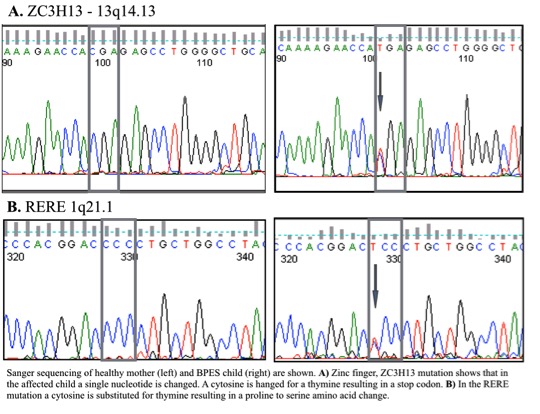
Phenotypic features of this disease were found in 2 generations of living relatives (father, 2 male children and female child) As well as documented in the grandparents as well. The inheritance demonstrated a Mendelian autosomal dominant pattern with 100% penetrance. Genetic analysis confirmed that a conserved mutation was responsible for the progression of disease, while whole exome sequencing identified candidate genes ZC3H13, and RERE with a nonsense and missense mutation, respectively.

We have identified a patient with orbitoblepharophimosis and, together with the father, the subjects were found to have a normal FOXL2 gene sequence, which was originally thought to manifest the disease. Whole exome sequencing and Sanger sequencing confirmed that FOXL2 was normal. The point mutation in ZC3H13 results in a premature stop codon of a gene which is known to be a strong transcription factor for FOXL2. Addition the single point mutation in RERE changes a cytosine for a thymine resulting in a proline to serine amino acid change.

Conclusion:

We have identified a missense and a nonsense mutation that together result in the BPES phenotype. Furthermore we have shown that FOXL2, a gene initially thought to be responsible for the mutation, to be completely normal in these patients.

74.13 Tumor Inhibition by Excisional Wounds in an Immune Competent Mouse Allograft Model

M. S. Hu1, T. Leavitt1, J. Gonzalez1, C. Marshall1, S. Malhotra1, L. Barnes1, A. T. Cheung1, G. G. Walmsley1, G. Gurtner1, A. J. Giaccia1, P. Lorenz1, M. T. Longaker1 1Stanford University,School Of Medicine,Palo Alto, CA, USA

Introduction:
We previously demonstrated that the placement of an adjacent splinted full-thickness excisional wound will inhibit human xenograft tumor growth in an immunocompromised mouse by outcompeting for neovascularization. Herein, we further explore this fascinating observation by placement of a full-thickness excisional wound both adjacent and directly over a mouse allograft tumor

Methods:
Mouse breast cancer 4T1 cells (5 x 10⁴) were injected into the left mid-dorsum of BALB/c mice. After engraftment was confirmed at 7 days, either a splinted full-thickness excisional wound was created adjacent to the tumor on the right mid-dorsum or an unsplinted full-thickness excisional wound was created directly over the engrafted tumor. Wounds were 6 mm in diameter. Tumor growth was assessed via 3-way caliper measurements taken every other day.

Results:
Tumors with an adjacent splinted full-thickness excisional wound were smaller than control tumors without a wound at 28 days post-wounding with a volume of 4639.14 mm3 versus 3742.32 mm3 and a weight of 3.27 g versus 2.47 g, respectively (*p<0.05). In addition, mice that had tumors and unsplinted full-thickness excisional wounds created directly over the tumor had inhibited growth and prolonged survival compared to mice with tumors alone (*p<0.05).

Conclusion:
These data show that placement of a full-thickness excisional wound adjacent to or directly over a tumor inhibits growth in an immune competent mouse allograft tumor model. These data have novel implications for ulcerated tumors and tumors that undergo an incomplete surgical resection. Further research promises to identify the mechanism for this inhibition.

74.14 Analysis of Gene Expression in E14 versus E18 Fetal Fibroblasts

M. S. Hu1, S. Malhotra1, W. Hong1, M. Januszyk1, G. G. Walmsley1, A. Luan1, D. Duscher1, D. Wan1, G. C. Gurtner1, M. T. Longaker1, P. Lorenz1 1Stanford University,School Of Medicine,Palo Alto, CA, USA

Introduction:
Early fetuses heal without scar formation, yet the biological mechanism behind this process is largely unknown. We aimed to examine fetal fibroblasts, which are intimately involved with the wound healing and scar formation process, in different stages of development to characterize differences that may contribute to the switch from wound regeneration to repair.

Methods:
Fetal fibroblasts were harvested and cultured from the dorsal skin of time-dated BALB/c embryos. Total RNA was isolated and microarray analysis was performed using chips with 42,000 genes. Significance analysis of microarrays (SAM) was utilized to select genes with greater than 2-fold expression differences between the wounds with a false discovery rate (FDR) of less than 2. Enrichment analysis was performed on significant genes to identify differentially expressed pathways.

Results:
Comparison of gene expression profiles revealed 275 genes that were differentially expressed between E14 and E18 fetal fibroblasts, with 30 genes significantly downregulated and 245 genes upregulated at the E18 time point compared to the E14 time point. Ingenuity pathway analysis (IPA) identified the top 20 signaling pathways that were differentially regulated between E14 and E18 fetal fibroblasts.

Conclusion:
This work represents the first instance where differentially expressed genes and signaling pathways between fetal fibroblasts at E14 and E18 have been identified. These genes and pathways drive the mechanism behind the transition from scarless fetal wound regeneration to scarring adult wound repair and may prove to be key targets for future therapeutics aimed to promote regeneration.

74.11 Influence of Topical Vasodilator-Induced Pharmacological Delay on Flap Viability

Z. Wu3, M. M. Ibrahim1, R. Schweller2, B. Phillips1, B. Klitzman1,2,3 1Duke University Medical Center,Division Of Plastic, Maxillofacial And Reconstructive Surgery,Durham, NC, USA 2Duke University Medical Center,Biomedical Engineering,Durham, NC, USA 3Duke University Medical Center,School Of Medicine,Durham, NC, USA

Introduction: Surgical delay is a well-known technique that improves perfusion of random and pedicle cutaneous flaps. The aim of this study was to create a model of pharmacological delay that would induce vascular remodeling and decrease overall flap necrosis.

Methods: A modified caudally based McFarlane flap was created using a rat model. Seven groups of random flaps were created (n=8) that included application of topical minoxidil and iloprost for various durations beginning 2 weeks prior to flap elevation. A standard surgical delay group was performed for a positive control. Surgical flaps were elevated, re-inset and observed at various time points until postoperative day 7. Gross viability, histology, perfusion analysis, tissue oxygenation and vascular casting were performed for analysis.

Results:Pharmacologic delay with preoperative application of topical minoxidil and iloprost were found to have equivalent flap viability when compared to standard surgical delay. A significant increase in viability was observed when comparing these groups to a negative control using a topical vehicle. Pharmacologic delay was found to increase blood flow during the preoperative period through vascular remodeling rather than acute vasodilation. These changes were not observed in flaps that were only treated in the postoperative period.

Conclusion:Preoperative topical application of vasodilatory agents, minoxidil and iloprost, yield equivalent viability in a random cutaneous flap model compared to the gold standard surgical delay. We have created a model of non-invasive pharmacological delay that improves tissue viability and potential postoperative complications without an additional surgical procedure.

74.09 Investigation of Visceral Skin Graft Revascularization and Separation from Peritoneal Contents

K. E. Caldwell1, R. M. Clark1, B. B. Coffman1, T. R. Howdieshell1 1University Of New Mexico HSC,Department Of Surgery,Albuquerque, NM, USA

Introduction:
Inability to close the abdominal wall after laparotomy for trauma may occur as a result of visceral edema, retroperitoneal hematoma, use of packing, and loss of tissue. When direct fascial closure is not feasible at initial admission, skin grafting of visceral granulation tissue provides temporary cutaneous coverage. Definitive abdominal wall reconstruction is planned when palpation yields graft separation from underlying viscera suggesting resolution of peritoneal inflammation.

Methods:
Following laparotomy for trauma, patients with persistent visceral distension or abdominal wall tissue loss precluding fascial closure underwent index admission visceral split thickness skin grafting and readmission graft excision and reconstruction. Real-time visceral skin graft perfusion and functional revascularization was determined by serial laser speckle contrast imaging (LSCI). Image analysis of CD-31 and α-SM actin immunostained histologic sections of harvested skin (nongrafted control) and excised visceral skin grafts was utilized to determine microvascular density and morphology. Quantitative RT-PCR arrays were performed on nongrafted control skin and excised visceral grafts to analyze the expression of a focused panel of genes involved in tissue injury and repair.

Results:
Five patients (3 male, 2 female) ranging in age from 26 to 45 years (mean 36 years) underwent visceral skin grafting for cutaneous coverage of an open abdomen. Time to graft excision ranged from 5 months to 1.5 years (mean 6.5 months). LSCI documented mean perfusion of pre-graft visceral granulation tissue of 1100 PU (range 900-1200 PU), and immediate skin graft application perfusion of 150 PU (range 120-190 PU), primarily a result of background perfusion through meshed interstices. By POD 5, skin graft perfusion doubled to a mean value of 300 PU, peaked at a mean of 350 PU by POD 14 concomitant with closure of meshed interstices, and remained unchanged until excision. Histologic examination revealed a 5-fold increase in excised graft thickness (mean control 400 µm vs. mean excised 2100 µm). Immunostaining documented a 2-fold increase in vessel number, a 3-fold increase in vascular surface area, and a dramatic increase in percentage of vessels covered by smooth muscle in excised grafts compared to control skin. RT-PCR demonstrated statistically significant up-regulation of genes involved in matrix structure and remodeling, cytoskeleton regulation, and WNT signaling; and down-regulation of key inflammatory cytokine and chemokine genes.

Conclusion:
Our preliminary data documents early visceral skin graft revascularization with a plateau in perfusion over the study interval. Histology reveals a marked increase in the mass of graft subdermal matrix composed of a complex supporting vascular network. The excised graft transcriptome is rich in extracellular matrix remodeling enzyme gene activity which may be important in graft separation from peritoneal contents.

74.10 The CD248+ Subpopulation of Adipose Derived Stromal Cells Posesses Enhanced Angiogenic Potential

E. R. Zielins1, M. Januszyk1, C. Blackshear1, E. A. Brett1, M. Chin1, S. Vistnes1, S. Menon1, S. Shailendra1, G. C. Gurtner1, M. T. Longaker1, D. C. Wan1 1Stanford University,Plastic And Reconstructive Surgery,Palo Alto, CA, USA

Introduction: Adipose-derived stromal cells (ASCs), the stem cell containing population derived from the stromal vascular fraction (SVF) of adipose tissue, has shown much promise as a technique to improve fat graft retention. As ASCs are a significantly heterogenous cell population, identification of cell subpopulations with enhanced secretion of pro-angiogenic growth factors would facilitate their use in strategies to further improve fat graft take.

Methods: Human lipoaspirate was enzymatically digested in order to obtain SVF cells. Individual ASCs were isolated via flow cytometry based on an established surface marker profile. As we have previously described, single cell transcriptional profiling of select angiogenic and adipogenic genes, as well as multiple cell surface markers, was employed. Applying a Fuzzy C-Means algorithm to this data allowed for partitioning of ASCs into multiple, functionally distinct clusters. Linear discriminant analysis was then performed to correlate surface marker expression with cluster definition. We then utilized flow cytometry, a cell proliferation assay, qRT-PCR, and an in vitro endothelial tube formation assay, to evaluate CD248, the most promising of these markers, for the potential to isolate cells with enhanced angiogenic potential.

Results: Using this strategy, we identified multiple markers with the potential capacity to delineate functional subgroups of ASCs based on angiogenic gene expression. Analysis of freshly harvested SVF cells by flow cytometry using CD248, the strongest correlating surface marker, showed 16% of cells were CD248+ and 84% were CD248. Proliferation, gene analysis, and endothelial tube formation assays were performed, showing differences between CD248+, CD248-, and unsorted cell populations. qRT-PCR showed CD248+ cells to have significantly higher VEGFa secretion (**p<0.01) compared to both unsorted and CD248- cells. CD248+ cells additionally were found to promote increased endothelial tube formation in vitro in comparison to unsorted and CD248- cells.

Conclusion: Our methodology has identified multiple cell surface markers associated with ASC angiogenic capacity. The most highly correlated marker, CD248, characterizes a cell population with significantly enhanced angiogenic potential, suggesting that it may be used in in vivo applications for improvement of fat graft retention.

71.22 Using IntegriSense 750 for Fluorescence-Guided Surgery in a Head and Neck Cancer Model

T. Ramesh1, L. Moore1, S. Bevans1, M. Limdi1, E. Rosenthal2, J. Warram1 1University Of Alabama,School Of Medicine,Birmingham, Alabama, USA 2Stanford University,School Of Medicine,Palo Alto, CA, USA

Introduction:
The integrin avb3 is overexpressed in pathologic processes associated with tumor growth and invasion, including in tumor cells and angiogenic blood vessels. IntegriSense 750 (Perkin Elmer) is a targeted fluorescent imaging agent composed of a selective av?b3 antagonist and a near-infrared fluorochrome. We hypothesize IntegriSense 750 can effectively produce contrast for applications in fluorescence-guided surgery as demonstrated in flank xenograft, orthotopic tongue, and lymph node metastatic disease models of head and neck cancer. A dose escalation study was performed at three doses (1.25μg/mouse, 2.5μg/mouse, and 5μg/mouse) in mice bearing luc+ SCC1 flank xenografts (n=5). Mice bearing orthotopic tongue (n=3) and lymph node (n=3) models received 2.5μg of IntegriSense 750 per mouse.

Methods:
Fluorescence imaging was performed twice daily after infusion using commercially available open-field NIR and closed-field NIR imaging systems. NIR renderings of resected specimens were compared to bioluminescence images to confirm localization of probe to tumor. Tissue fluorescence was correlated with histology. Progressively smaller tumor fragments were re-introduced into the wound bed in order to determine the smallest cancerous mass detectable using this agent. Exploratory surgeries were performed to demonstrate agent localization to orthotopic tumors and lymph node metastases. A biodistribution study was performed four days after dosing.

Results:
Intraoperative imaging successfully differentiated tumor from normal tissue with mean tumor-to-background ratios of 3.73, 5.35, and 9.62 on day 4 for the 1.25μg, 2.5μg, and 5μg doses, respectively. Tumor-to-background ratios for all three groups peaked at four days post-infusion. Tumor fragments as small as 0.5mg were visualized in all three dosing cohorts. 3/3 diseased lymph nodes were fluorescently visible in situ in mice during exploratory surgeries. The 5μg dose was found to be the optimal dose for applications in fluorescence-guided surgical oncology in this study.

Conclusion:
IntegriSense 750 appeared to be effective for use in fluorescence-guided surgical applications as demonstrated in flank xenograft, orthotopic, and lymph node metastatic disease models of head and neck cancer.

70.21 Murine Breast cancer cells eliminated in non-derived strain mice; using an improved breast cancer model

E. Katsuta1, S. DeMasi1, K. P. Terracina1, H. Aoki1, M. Aoki1, P. Mukhopadhyay1, K. Takabe1 1Virginia Commonwealth University School Of Medicine And Massey Cancer Center,Division Of Surgical Oncology, Department Of Surgery,Richmond, VA, USA

Introduction: We have previously established a murine syngeneic breast cancer model utilizing cell implantation under direct vision technique, which mimic human cancer progression (Rashid, Takabe et al. Breast Cancer Research and Treatment 2014). Other groups have reported that cell implantation using Matrigel produced stable results in xenograft models. Here, we report the establishment of improved syngeneic orthotopic murine breast cancer model using Matrigel. In this study, we determined the maximum amount of Matrigel to be implanted without spillage, the tumor growth with various number of cells, and utilizing this new model, we investigated the growth of murine cancer cell derived from different strain mice.

Methods: Matrigel was injected to #2 and #4 mammary glands. Various number of murine breast cancer E0771cells in Matrigel were implanted into bilateral #2 and #4 mammary gland of C57Blk6 mice. Three weeks after inoculation, tumorigenesis were compared. 1 x 10⁴ of murine breast cancer 4T1-luc2 cells, derived from Balb/C mice, were implanted into the Right side #2 gland of Balb/C or C57Blk6 mice. Tumor growth was monitored by bioluminescence (IVIS) imaging.

Results: We found that implantation of the cells will be more efficient with less variability when the cells are suspended in Matrigel compared with PBS, which was the technique we reported previously. Maximum volume of Matrigel inoculated without spillage was 20 μl in #2 gland, 30 μl in #4 gland, respectively. Therefore, we implanted 20 μl of Matrigel in #2 gland, and 30 μl in #4 gland in the subsequent experiments. In order to determine the difference of tumor development, 504, 105, 505, 106 E0771 cells suspended in 20µl Matrigel were inoculated. Three weeks after inoculation, ‘the take rates’ (tumorigenesis) were 0%, 12.5%, 75%, 75%, 100%, respectively. Utilizing 4T1-luc2 cells in Matrigel suspended cell implantation method, we investigated how long the mouse-derived cancer cells survive in mice from a different background. The fold increase in tumor growth in both backgrounds were nearly identical 24 h after inoculation at 5-fold increase measured by bioluminescence imaging. By 7 days after inoculation, tumor in C57Blk6 reached a maximum increase of approximately 720-fold their Day 0 size, whereas the tumor in Balb/C had almost a 2000-fold increase in tumor size. The Balb/C tumor continued to increase rapidly to reach an almost 3000-fold increase in size, while the C57Blk/6 mice tumors swiftly decreased from Day 7 and was eliminated by Day 14.

Conclusion: We identified the maximum amount of Matrigel that can be implanted into #2 or #4 mammary gland without spillage, and the difference in take rates with various number of cells for murine orthotopic breast cancer model. Utilizing Matrigel implantation method, we found that cancer cells will continue to grow until one week, then it will eliminated by 2 weeks when implanted into different background strain mice.

61.12 Finite element analysis: Assessing the optimal patient-specific mitral valve repair.

A. E. Morgan1, L. Ge4, W. G. Morrel5, J. L. Pantoja5, E. A. Grossi3, M. B. Ratcliffe1,2 1University Of California – San Francisco,Surgery,San Francisco, CA, USA 2San Francisco VA Medical Center,Surgery,San Francisco, CA, USA 3New York University School Of Medicine,Cardiothoracic Surgery,New York, NY, USA 4University Of California – San Francisco,Bioengineering,San Francisco, CA, USA 5University Of California – San Francisco,School Of Medicine,San Francisco, CA, USA

Introduction: Mitral valve repair is superior to replacement in terms of long-term survival, but recurrent mitral regurgitation after repair for degenerative disease occurs at a rate of ~2.6% per year, with a 20% reoperation rate at 20 years. We hypothesize that durability of repair is related to geometry of initial repair, as well as stress distribution over the mitral valve and left ventricle. Previous studies demonstrate that repair by posterior leaflet resection increases stress across the posterior leaflet; we tested the hypothesis that repair by placement of prosthetic chordae tendinae resulted in reduced overall stress as compared to leaflet resection.

Methods: Magnetic resonance imaging and intra-operative 3D trans-esophageal echocardiography were performed before surgical repair of isolated P2 prolapse in a single patient. A finite element model of the left ventricle and mitral valve was created. Stress was examined in the preoperative state for the mitral valve and left ventricle, and for the following repairs: Triangular resection; Placement of one PTFE chord; Placement of two PTFE chords.

Results: The principal findings of this study are the following: 1) Placement of prosthetic chordae resulted in stable or decreased overall posterior leaflet stress for all phases of the cardiac cycle; 2) In contrast, triangular resection resulted in increased posterior leaflet stress, most prominently in diastole; 3) All repair techniques reduced stress on the anterior leaflet; 4) All repair techniques restored normal leaflet coaptation. An example of pre- and post-repair leaflet geometry is shown in the attached figure, for placement of two prosthetic chordae.

Conclusion: All repair techniques eliminated mitral regurgitation and restored normal leaflet coaptation. Finite element analysis revealed that mitral valve repair with prosthetic chordae restored normal leaflet geometry without creation of excessive stress on the valvular tissues, in contrast to leaflet resection, which significantly increased stress across the posterior leaflet. Future studies will examine this effect across a broad range of patients with the aim of developing a patient-specific tool for mitral valve repair preoperative planning and surgical education.

61.01 Effects of Diabetes and CPB on Adherens-Junction-Protein Expression of Human Peripheral Tissue

J. Feng1, A. K. Singh1, J. Liang1, Y. Liu1, F. W. Sellke1 1Brown University School Of Medicine,Cardiothoracic/Surgery,Providence, RI, USA

Introduction: Cardiac surgery with cardiopulmonary bypass (CPB) is often associated with the increased vascular permeability/tissue edema, microvascular endothelial cell injury/dysfunction. Especially, these disturbances are more pronounced in patients with poorly controlled diabetes. Recent study has demonstrated that the increased permeability after cardiopleogic arrest/CPB is associated with changes in the expression/phosphorylation of adherence-junction-proteins in the coronary vasculature in patients with type-2 diabetes (T2DM). We hypothesized that T2DM may be associated with altered adherence-junction-protein expression/phosphorylation of skeletal muscle/vessel in the setting of CPB . The aim of the current study was to investigate the changes in adherens-junction-proteins, such as VE-cadherin, and β-catenin of skeletal muscle and vessels in patients with or without T2DM in the setting of CPB and coronary artery bypass graft (CABG) surgery.

Methods: Chest wall skeletal muscle tissue was harvested pre- and post-CPB from the controlled diabetic (HbA1c: 6.3 ± 0.1), uncontrolled diabetic (HbA1c: 9.6 ± 0.3) and non-diabetic patients (HbA1c: 5.4 ± 0.1) undergoing CABG surgery (n = 6/group). The expression/phosphorylation of adherence-junction-proteins such as, VE-cadherin and β-catenin were assessed by immunoblotting and immuno-histochemistry. Skeletal-muscle-arteriolar endothelial function was assessed by videomicroscopy in response to the endothelium-dependent vasodilator substance P.

Results:There were no significant differences in basal protein expression of VE-cadherin between UDM and CDM or ND patients or between pre- and post-CPB among groups. The level of pre-CPB phosphorylated VE-cadherin tends to be 25% higher in the UDM group compared with ND (P = 0.03). CPB induced more phosphorylation of VE-cadherin (21% in ND; 30% in CDM and 43% vs. pre-CPB; P<0.05, respectively) and this effect was more pronounced in the UDM group (P<0.05 vs. ND or CDM). The post-CPB β-catenin was significantly decreased as compared with pre-CPB in all three groups (P<0.05) and the decrease was more pronounced in the UDM group (P<0.05). There were significantly decreases in vasodilatory response of skeletal muscle arterioles to substance P after CPB in all three groups (P<0.05). This alteration was more pronounced in the UDM patients (P<0.05).

Conclusion: These findings suggest that poorly controlled diabetes down-regulates endothelial adherence-junction-protein expression of skeletal muscle/vessel tissues in the setting of CPB. The enhanced tyrosine phosphorylation of VE-cadherin and degradation of β-catenin indicates deterioration of these proteins and the damage of the cell-cell endothelial junctions, specifically in the diabetic vessels. These alterations may lead to the increases in periphral vascular permeability and endothelial dysfunction and affect the outcomes in diabetic patients after CPB/cardiac surgery.

06.11 Gender Differences in Mortality Following Ischemia-Reperfusion Injury in Diabetic Animals

N. N. Rizk1, E. Abbott1, M. Rizk2, A. Fischer1,2, J. Dunbar2 1Beaumont Health System Research Institute,General Surgery,Royal Oak, MI, USA 2Wayne State University,Physiology,Detroit, MI, USA

Introduction: The physiologic differences between men and women are well characterized so their clinical manifestations in pathologic states should potentially exhibit differences. Much of preclinical research utilizes male animal models to develop treatment modalities for clinical applications assumed to extrapolate to both genders. In this study we evaluated normal and diabetic female rats response to stroke and reperfusion injury and compared to our previously characterized outcomes in male rats.

Methods: We utilized middle cerebral artery occlusion (MCAO) and reperfusion model in normal and diabetic Wistar female animals. Following 24-hour MCAO and 2-hour MCAO followed by 24-hour reperfusion interval, several CNS parameters were examined, lesion volume (Triphenyltetrazolium Chloride), up regulation of apoptosis [TUNEL immunoreactivity in the sensori-motor cortex (layers-5 and 6) and the hippocampal pyramidal cell layers (CA1 and CA3 sectors)], as well as the percent survival.

Results: The two parameters in which the female animals differed significantly from their male counter parts were i) in lesion volume following restoration of blood flow with 2-hour MCAO and ii) in mortality following 24-hour MCAO and in following restoration of blood flow with 2-hour MCAO. Restoration of blood flow following 2-hour MCAO significantly decreased lesion volume in the normal and diabetic female animals (13.95 ± 8.16* and 20.32 ± 6.85*, respectively). In comparison to the male counterparts, lesion volumes following reperfusion were significantly less for normal male rats and significantly more for the diabetic male animals (3.8 ± 2.36# and 31.25 ± 2.5#, respectively). Twenty-hour MCAO and restoration of blood flow with 2-hour MCAO proved to be significantly more detrimental to the female animals with lower survival (normal 65%, diabetic 55% survival, p<0.05) in comparison to the male animals (normal 90%, diabetic 65% survival, p<0.05). There was a significant increase in TUNEL immunoreactive cells in the diabetic animals following reperfusion injury compared with the control counterpart. There were no appreciable differences in TUNEL immunoreactivity between male and female diabetic animals following reperfusion.

Conclusion:This paper highlights the dramatically different outcomes and metrics in different genders in animal models. Thus one gender alone may not translate into clinically relevant data applicable to both genders. We have demonstrated that both male and female diabetic animals have an exaggerated response to stroke and to restoration of blood flow but differ in their outcome with a significantly more detrimental outcome for the female sex. Understanding the exaggerated vascular response to injury in the female animal model is important in developing gender specific treatment modalities.

06.12 Non-Platelet Length Polyphosphates Inhibits tPA Mediated Fibrinolysis

P. J. Lawson1,2, H. B. Moore1,2, A. W. Bacon1,2, E. Gonzalez1,2, A. P. Morton1,2, A. Banerjee1, J. H. Morrissey3, E. E. Moore1,2 1University Of Colorado Denver,Surgery,Aurora, CO, USA 2Denver Health Medical Center,Surgery,Denver, CO, USA 3University Of Illinois,Surgery,Urbana, IL, USA

Introduction:
Fibrinolysis shutdown is the dominant phenotype in severely injured patients, but the mechanisms driving post-injury tPA resistance remain unknown. Polyphosphates are ubiquitous inorganic compounds that have recently been linked to coagulation. Polyphosphates are stored in platelets, and platelets are known to inhibit fibrinolysis. We hypothesize that polyphosphate polymers that are similar in size to the length of chains stored in platelets will inhibit tPA mediated fibrinolysis.

Methods:
Polyphosphates of four different polymer lengths were evaluated. Monophosphate, platelet length polyphosphates, medium size polyphosphates found in tissue, and large polyphosphates of the size produced by microbes. Polymers of different sizes were added at a concentration of 20 micromolar in whole blood from eight healthy volunteers and assessed with thrombelastography (TEG) for changes in clotting parameters with and without tissue plasminogen activator (tPA) to quantify inhibition in fibrinolytic activity.

Results:
Large polyphosphates reduced R time and increased angle in the presence of tPA compared control blood (p<0.001, p=0.008) and trended towards an increase in clot strength (MA p=0.052). Large, medium, and mono polyphosphates reduced LY30 compared to control (p=0.034, Figure 1). The other polymer lengths in the presence of tPA did not significantly alter other clotting parameters. Without tPA, the same relationship of shortened clotting time and increased angle were appreciated with the large polyphosphates (p<0.001, p=0.015). None of the other polymer lengths affected clot parameters.

Conclusion:
Large chain polyphosphates are associated with a rapid forming strong clot. Polyphosphates polymers in the length stored in platelets, however, did not impact clotting parameters. But all other polymer lengths inhibited tPA mediated fibrinolysis. These experiments indicate that platelet derived polyphosphates are either modified after release from stored granules, or the polyphosphates regulating coagulation are from another source.

06.13 High Glucose Does Not Inhibit Aortic Endothelial Cell Sprouting and Stimulates Proliferation

A. D. Morris1, H. Li1, K. Kuo1, S. Dalal1, L. P. Brewster1,2 1Emory University School Of Medicine,Division Of Vascular Surgery,Atlanta, GA, USA 2Atlanta VA Medical Center,Division of Vascular Surgery,Atlanta, GA, USA

Introduction:
Diabetes increases the risk of cardiovascular disease and major amputation, but the aggressive control of blood sugar levels in recent clinical trials did not correspond to better clinical outcomes. Endothelial cell regeneration is critical to preventing thrombotic and myointimal complications after vascular injuries and interventions, but the impact of hyperglycemia on this process is not well understood. Since hyperglycemia directly inhibits cellular homeostasis, we propose that it negatively impacts endothelial cell regeneration.

Methods:

Proliferation Protocol:

Human aortic endothelial cells(HAEC) were grown in standard culture conditions, and then plated in a 96-well plate at 5000 cells per well. After reaching ~70% confluence, the cells were quiesced with serum starvation for 24 hours. The cells were restimulated with growth media (positive control) or quiescent media (negative control) with 40mM glucose or 40mM mannose (oncotic control) added to both groups. After 72 hours, HAEC proliferation was collected by commercially available cell viability assay using absorbance.

3D Angiogenesis Assay:

HAECs were formed into pellets (40,000 cells) using mechanical rotation. The pellets were suspended in a fibrin hydrogel. The cell pellets were exposed to either 40mM glucose in growth media, 40mM mannose in growth media, or growth media for 3 days. HAEC invasion was captured by daily microscopy and analyzed as average sprout length at 10-degree intervals through 360 degrees by Matlab programming.

Results:

Contrary to our hypothesis, HAECs had greater proliferation in a 72-hour high glucose and high mannose environment compared to normal growth media (p<0.0001, p=0.030). Glucose media had additional proliferation potential compared to mannose media (p=0.012). High glucose and mannose conditions did not inhibit HAEC sprout length at any time point in the 3D angiogenesis assay.

Conclusion:

While there is literature supporting an inhibitory effect of high glucose on endothelial cells, it is related to a combination of concentration and time of exposure to high glucose conditions. In this work, we are surprised to find not only a lack of inhibition of EC sprout formation, but also an increase in EC proliferation with both high glucose and high mannose conditions. Perhaps, the 24 hours of quiescence positively affects proliferation in functionally starving cells. We are actively testing this by including the high glucose levels from the beginning of the experiment. The lack of inhibition on EC sprouting suggests that acute hyperglycemia may not inhibit EC angiogenic activity in a meaningful manner. The duration and severity of hyperglycemia that must be avoided remains a clinically important idea that must be further pursued.

06.09 Leukadherins: A Novel Agonist Approach To Treat Atherosclerosis Via Mac-1 Activation

A. Mesa1, S. Rahimpour2, N. Fernandez1, L. Song1, S. M. Pham2, K. A. Webster1, V. Gupta3, R. I. Vazquez-Padron1 1University Of Miami,Miami, FL, USA 2University Of Maryland,Baltimore, MD, USA 3Rush University Medical Center,Chicago, IL, USA

Introduction:
Atherosclerosis is a chronic systemic disease characterized by the accumulation of lipids and leukocytes in the arterial intima which eventually lead to myocardial infarction and other detrimental vascular events. New investigations revealed the role of integrin Mac-1 in leukocyte adhesion, migration and recruitment during the development of vascular proliferative diseases.

Methods:
In this study, a novel Mac-1 agonist drug named leukadherins (LA-1) is used as an innovative therapeutic tool to control inflammation with the ultimate goal to diminish atherogenesis. To test our hypothesis, daily intraperitoneal LA-1 or vehicle (control) injections (10 mg/ml) were administered to 20 hypercholesterolemic mice (ApoE null mice) kept in high fat diet for 16 weeks.

Results:
Mice treated with LA-1 exhibited a significantly reduction of monocytes and pro-inflammatory molecules in blood (p ≤ 0.001). Assessment of aortic atherosclerosis burden of treated and control mice indicated that LA-1 treatment significantly decreased atherosclerotic development (p = 0.03). Additionally, the results suggested that LA-1 reduces inflammation by retaining monocytes in bone marrow and spleen given that treated mice showed a significant reclusion of monocytes in these primary hematopoietic organs (p ≤ 0.001).

Conclusion:
These results suggest LA-1 treatment via Mac-1 activation represents a novel agonist approach for treating vascular inflammation and atherosclerosis.

06.10 Radiographic Kidney Volume Perfusion Correlates with Aortic Anatomic Severity Grade

C. I. Aghaie1, L. E. Trakimas1, D. S. Mix1, K. Rasheed1, M. Seaman1, J. L. Ellis1, R. J. Glocker1, A. J. Doyle1, M. C. Stoner1 1University Of Rochester,Vascular Surgery,Rochester, NY, USA

Introduction:
Anatomic Severity Grade (ASG) represents a quantitative mechanism for assessing anatomical suitability for endovascular aortic repair. ASG has been correlated with perioperative and mid-term outcomes, and resource utilization. The purpose of this study was to identify the correlation of a novel renal perfusion metric and ASG.

Methods:
Elective infrarenal aortic repair cases were identified in a retrospective database, describing patient operative and anatomical factors. Anatomic grading was undertaken by independent reviewers. Using volumetric software, kidney volume and a novel measure of kidney functional volume – radiographic kidney volume perfusion (RKVP) – was recorded. Systematic evaluation of the relationship of kidney volume and RKVP to CKD (glomerular filtration rate (GFR) < 60 ml/min) and ASG was undertaken using linear regression and receiver-operator statistical tools.

Results:
A total of 386 cases with patient and anatomic data were identified and graded. Mean age was 72.9 ± 0.4 years. Renal volume < 281 mL was correlated with CKD (AUC = 0.708, p = <0.0001). RKVP < 22.5 HU*L was correlated with CKD (AUC = 0.764, p = <0.0001). High (≥15) ASG scores correlated with both renal volume (AUC = 0.628, p = < 0.0001) and RKVP (AUC = 0.628, p = < 0.0001). Regression analysis demonstrated a strong, inverse relationship between ASG and RKVP (Figure).

Conclusion:
These data demonstrate that the novel metric of RKVP correlates with both CKD and ASG in a large database of patients undergoing elective aneurysm repair. The inverse relationship between renal function (renal volume, RKVP or GFR) and ASG has not been described in the literature before. Outcomes in patients with poor renal function may be related to anatomical issues in addition to well-described systemic ramifications.

06.06 Novel Link Between Cholesterol & Hydrogen Sulfide Production Potential in Vascular Disease Patients

W. W. King1, A. Longchamp1, M. Tao1, R. Kulkarni1, J. R. Mitchell2, C. K. Ozaki1, G. Sharma1 1Brigham And Women’s Hospital,Vascular And Endovascular Surgery,Boston, MA, USA 2Harvard School Of Public Health,Department Of Genetics And Complex Diseases,Boston, MA, USA

Introduction: Hydrogen sulfide (H2S) is a vasoactive gasotransmitter that is endogenously produced in the vasculature. In animal models, H2S is required for the maintenance of normal vascular function and protects from surgical stress including visceral and muscular ischemia-reperfusion injury, cardiac overload, and stroke. However, data regarding H2S biology in humans remains sparse. We hypothesized that there are specific clinical factors and circulating biomarkers that link with H2S generative capacity.

Methods: After obtaining institutional review board approval, we prospectively collected demographic information, clinical history, peripheral blood, and subcutaneous and perivascular adipose tissues from consented patients undergoing carotid endarterectomy (CEA), major leg amputation (AMP), or open lower extremity (arterial) revascularization (LER). Standard laboratory data were extracted from the medical record. Nine adipose-associated biologic mediators (adiponectin, IL-1β , IL-6, IL-8, leptin, MCP-1, PAI-1, resistin, and TNF) were assayed in the adipose tissues and plasma. In addition, serum H2S production capacity was determined utilizing the lead sulfide method. For categorical clinical and demographic characteristics, the continuous H2S and mediator data were analyzed using either Wilcoxon rank-sum or Student’s t-test, while for continuous clinical and demographic variables, adipose mediators, and H2S Pearson or Spearman correlation testing was utilized depending upon the normality of distribution.

Results: The cohort included 78 patients (49 CEA, 12 AMP, 17 LER). While several weak associations were identified between adipose related mediators and H2S production potential, the most powerful association among the variety of factors examined was total serum cholesterol: regression analysis revealed a negative correlation between total cholesterol and H2S (r=-0.53, p=<0.001) (Figure 1).

Conclusions: In human patients with vascular disease, there is a strong negative correlation between total serum cholesterol and H2S generative capacity. This unexpected relationship likely holds important mechanistic implications for the genesis and management of arterial vascular disease.

06.07 Model of Hemodynamic Changes after Arteriovenous Fistula Creation

L. Laquian1, Y. He1, S. Berceli1,2 1University Of Florida,Division Of Vascular And Endovascular Surgery,Gainesville, FL, USA 2Malcom Randall Veterans Affairs Medical Center,Gainesville, FL, USA

Introduction: Chronic inflammation and hemodynamic changes are major components of the altered physiology in hemodialysis (HD) patients. Parallel studies in our laboratory have identified significant changes in leukocyte biology and their genomic profile in association with arteriovenous fistula (AVF) creation. The purpose of this study is to characterize the changes in blood flow and wall shear stress (WSS) associated with AVFs. We hypothesize that increased WSS and turbulence associated with AVFs act as a driving force for the altered biology in HD patients.

Methods: Utilizing vessel dimension and blood flow data compiled from the literature, normal systemic circulation was modeled and then adapted for left brachiocephalic AVF creation using postoperative values. For each vascular segment, transit time, peak WSS, and peak Reynolds number (as an indicator of turbulent flow) were calculated. Fraction of circulation time spent at high WSS (>40 dyn/cm2) and turbulent flow (Reynolds number >4000) was computed in normal and AVF models.

Results: Increased flow after AVF creation results in elevated peak WSS and Reynolds number in the subclavian and brachial arteries. Peak WSS in the left subclavian artery increases by more than seven fold from 32 dyn/cm2 preoperatively to 232 dyn/cm2 after AVF creation. Left brachial artery peak WSS increases from 46.0 dyn/cm2 to 209.5 dyn/cm2 after AVF placement. Overall, high peak WSS in normal upper extremity circulation occurs for 9.5% of circulation time but does not exceed 50 dyn/cm2 (Fig 1A). After AVF creation, 13.5% of circulation time is spent at high WSS, with 7.4% of circulation time spent at over 200 dyn/cm2. With an AVF, subclavian artery flow turns turbulent as Reynolds number increases from 1309 in normal circulation to 9366. Peak Reynolds number in the brachial artery increases from 660 to 3008 after AVF, reflecting more disordered and chaotic flow. In normal circulation, 2.5% circulation time is spent at turbulent flow, while an AVF increases circulation time at turbulent flow to 13.9% (Fig 1B).

Conclusion: AVF creation leads to elevated WSS and more turbulent flow compared to normal circulation. Furthermore, the time spent at high WSS and turbulent flow increases after AVF placement. Ongoing studies have been initiated to investigate the influence of this increased exposure time to higher WSS and turbulent flow on circulating immune cell phenotype and their role in the chronic inflammatory state that characterizes the typical HD patient.

06.08 Development of a Novel Murine Carotid Artery Neointimal Hyperplasia Model

A. M. Steely1,2, T. Mann-Gow2, B. S. Childs2, M. Kida2,3, P. Zvara1,2 1The University Of Vermont Medical Center,Department Of Surgery,Burlington, VT, USA 2The University Of Vermont,College Of Medicine,Burlington, VT, USA 3The University Of Vermont Medical Center,Department Of Pathology And Laboratory Medicine,Burlington, VT, USA

Introduction: Neointimal hyperplasia remains a common problem following vascular surgery. Current murine mechanical injury models, which denude the endothelium in a retrograde fashion and require suture ligation of some portion of the arterial tree, have been criticized for the small proliferative response and high inter-animal variability. Our aim was to develop a simple, reproducible, mechanical, antegrade murine common carotid artery (CCA) neointimal hyperplasia model that does not require suture ligation of any portion of the cervical arterial tree.

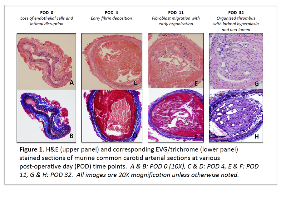
Methods: After creating an arteriotomy in the CCA with a 30G needle, a 0.010-inch hydrophilic wire was used to cannulate the CCA via the arteriotomy and denude the endothelium in an antegrade fashion. Hemostasis at the arteriotomy site was achieved by placing a topical cellulose matrix (Surgicel, Ethicon, Inc. Somerville, New Jersey) over the arteriotomy site. Mice were euthanized 0, 4, 11, and 32 days following CCA injury after systemic formaldehyde perfusion fixation. CCA sections were stained with hematoxylin and eosin (H&E), trichrome, and elastin van Gieson (EVG).

Results: Loss of endothelial cells and intimal disruption was observed at POD 0. Early fibrin deposition was seen in the injured arterial segment at POD 4, however, fibroblast migration with early organization was not observed until POD 11. Organized thrombus with intimal hyperplasia and neo-lumen formation was evident on POD 32.

Conclusion: Antegrade mechanical injury with a hydrophilic wire effectively and reproducibly denudes the endothelium of the murine CCA with subsequent neointima formation throughout the injured arterial segment. This novel, physiologic, and reproducible CCA injury model may aid in the development of innovative pharmaceuticals to prevent and treat neointimal hyperplasia.

06.03 The Effect of Autophagy on Wound Healing in the Murine Model

H. Albadawi1,2, M. W. Koulopoulos1, L. M. Crowley1, H. Yoo1, M. T. Watkins1,2 1Massachusetts General Hospital,Division Of Vascular And Endovascular Surgery, Department Of Surgery,Boston, MA, USA 2Harvard School Of Medicine,Brookline, MA, USA

Introduction: Delayed wound healing is a significant comorbidity associated with peripheral vascular disease. Autophagy is an important cellular process for homeostasis during tissue repair. The role of autophagy in the wound healing process has not been investigated. This study evaluated the effect of the autophagy inhibitor, chloroquine, on cutaneous wound healing in mice.

Methods: Two groups of mice were subjected to excisional full-thickness splinted wounds created on the upper dorsum using a 5mm biopsy punch. The first group of mice were treated daily with an intraperitoneal injection of 50mg/kg chloroquine (CQ, n=8) starting 24hrs before wounding. The second group received saline as an untreated control (CON, n=10). Wound closure was assessed using serial photography and image analysis at days 0, 3, 7, 11 and 14, and expressed as a percentage of the original wound size. On day 14, the mice were euthanized and the wounds were harvested and analyzed by western blotting for markers of autophagy, the ratio of the lipidated autophagosome localized isoform of the microtubule-associated protein Light Chain-3 (LC3-II) to its non-lipidated cytosolic localized isoform (LC3-I) and the expression of Sequestosome-1 (p62). Western blot data was expressed as arbitrary units (AU). Statistical analysis was performed using student-t test and Pearson correlation test.

Results: Wound healing was significantly delayed at days 11 (CQ: 78.0±4.9, CON: 55.7±6.4 percent, p<0.0001) and 14 (CQ: 37±6.8; CON: 18.1±4.8 percent, p=0.028) in the CQ group. Western blot analysis showed significant increase in p62 protein levels (CQ: 0.61±0.06; CON: 0.44±0.04 AU, p=0.02) and LC3-II/I ratio (CQ: 0.61±0.04; CON: 0.37±0.08, p=0.024) in the CQ group. There was a positive correlation between the wound size at day 14 and the p62 protein levels in the same wound (Pearson r=0.83, p=0.02).

Conclusion: These data suggest that autophagy plays a significant role in the wound healing process. The significant increase in the LC3-II/I and p62 protein accumulation demonstrated chloroquine’s inhibitory effect on autophagy. This finding was supported by the positive correlation between wound size at day 14 and the corresponding p62 levels.