06.04 Thrombin and Laminar Flow have Synergistic Effects on YAP Activation in Endothelial Cells

J. Kurita1,2, G. Chitragari1,2, B. E. Sumpio1,2 1Yale University School Of Medicine,Vascular Surgery,New Haven, CT, USA 2VA Connecticut Healthcare System,Vascular Surgery,West Haven, CT, USA

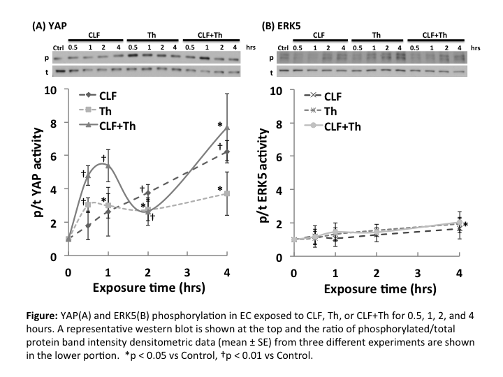
Introduction: The endothelium can be activated not only by mechanical forces of the circulation but also by circulating chemicals such as thrombin (Th). However, the effect of combined mechanical and chemical stimulation of endothelial cells (EC) is poorly understood. The purpose of this study was to investigate the effect of steady laminar flow (CLF), as a mechanical stimuli, and Th, as a chemical stimuli, on yes-associated protein (YAP) and extracellular signal-regulated kinase 5 (ERK5) activities in EC. YAP is a mechano-signaling protein that controls growth and proliferation depending on cell shape. ERK5 is an intracellular protein that maintains EC integrity.

Methods: Bovine aortic ECs seeded on fibronectin coated glass slides were grown to confluence in culture medium containing 10% fetal bovine serum. Thereafter, they were serum starved for 16 hours and were subjected to 0.5, 1, 2, or 4 hours of CLF utilizing a parallel plate flow chamber system controlled by a computerized pump for 4 hours at 37? in the presence or absence of Th (4U/ml). Western blot analysis of total and phospho-YAP and ERK5 was performed. The activities of YAP and ERK5 were determined based on the ratio of immunoblot intensity of phosphorylated to total protein. Fold change compared to static control (Ctrl) at 0 hour was calculated and compared for significant difference using t-test. P value of <0.05 was considered statistically significant.

Results: ERK5 activity rose slightly through 4 h for all experimental groups but was only significantly increased in the Th group at 4 hours (1.95±0.39; p<0.03 vs Ctrl) (Figure B). YAP was phosphorylated by Th and the combination of CLF+Th (2.96±0.73; p<0.01 and 2.44±1.16; p<0.05 vs Ctrl, respectively). YAP phosph/total ratio was significantly increased with time through 4 h in the CLF group (p<0.05 vs Ctrl), while the ratio in the CLF+Th group showed the similar time course pattern as in the Th group within 2 hours. However, at 4 h it rose to the level of the CLF groups (Figure A).

Conclusion: Thrombin and flow did not activate ERK5. YAP was phosphorylated in EC exposed to both thrombin and flow although flow stimulation was stronger. The combination of flow and thrombin had an additive effect on YAP phosphorylation. These results indicate selective activation of intracellular proteins by mechano-chemical signals. Further studies have to be done to elucidate the interactions of these signaling pathways and to understand their roles in endothelial response to stimuli.

06.05 Baseline Adipose Phenotype Predicts Vascular Surgery Wound Complications

R. Kulkarni1, W. W. King1, S. Shah1, A. Longchamp1, M. Tao1, K. Ding1, C. K. Ozaki1, G. Sharma1 1Brigham And Women’s Hospital,Vascular And Endovascular Surgery,Boston, MA, USA

Introduction: Wound complication rates after vascular surgery may be as high as 30%, and represent a major cause of morbidity, mortality, and cost. The authors have previously demonstrated that local adipose tissue can exhibit an exacerbated inflammatory response to local surgical trauma. To date, however, links between human adipose phenotype and procedural outcomes such as wound complications have not been reported. We hypothesized that specific adipose-related biomarkers uniquely link to 30-day wound complication rates in patients undergoing open vascular surgical procedures.

Methods: Clinical history, peripheral blood, and subcutaneous and perivascular adipose tissue were prospectively collected from patients undergoing carotid endarterectomy (CEA), lower extremity revascularization (LER), and lower extremity amputations (AMP) at the time of surgery. Nine adipose-associated biologic mediators (adiponectin, IL-1β, IL-6, IL-8, leptin, MCP-1, PAI-1, resistin, and TNF) were assayed in the adipose tissues and plasma. Wound complications were classified according to a previously published grading system. Logarithmic transformation of mediator levels was performed based on positively skewed, non-Gaussian distribution and data were compared using the Student’s t-test. All statistical analyses were conducted using SAS software,v9.3 (SAS Institute, Inc., Cary, NC.)

Results: The cohort included 115 patients (49 CEA, 44 LER, 22 AMP). Median follow-up was 14 months (SD 9.97 months) and 30-day follow-up was 94.8%. At 30 days, 22 (19.1%) patients had wound complications—namely, superficial surgical site infections (14/22), hematoma (4/22), dehiscence (4/22), seroma or lymph leak (3/22), and deep surgical site infection (1/22). There were several statistically significant, plasma/perivascular/subcutaneous compartment-specific relationships between logarithmically transformed mediator levels and wound complications. Most notably, mean plasma TNF levels were higher in patients with superficial surgical site infections (figure, panel A), whereas plasma (figure, panel A), and perivascular (figure, panel B) TNF levels were lower in patients with wound dehiscence. There was also a trend relating elevated subcutaneous TNF levels and any wound complication (p=0.05.)

Conclusions: Adipose-associated mediator levels at the time of operation demonstrate a compartment-specific relationship to wound outcomes in patients undergoing vascular surgical procedures. These associations likely have implications for mechanisms underlying the pathogenesis of wound complications, and suggest novel interventional strategies to reduce wound complications based on the plasticity of the adipose organ’s phenotype.

06.01 Development of a Co-Culture Injury Model for Studying Vascular Smooth Muscle Cell Migration In Vitro

F. Galambo1, H. Bass1,2, R. Beard2, B. J. Cha2, P. R. Nelson1,2 2University Of South Florida College Of Medicine,Department Of Molecular Pharmacology And Physiology,Tampa, FL, USA 1University Of South Florida College Of Medicine,Division Of Vascular And Endovascular Surgery/Department Of Surgery/Morsani College Of Medicine,Tampa, FL, USA

Introduction: Vascular injury disrupts normal vessel wall architecture causing de-endothelialization, and SMC dedifferentiation. Unregulated SMC migration leads to neointimal hyperplasia and eventually restenosis. We sought to develop an in vitro co-culture model to study SMC physiology under conditions of injury. We hypothesized that disrupting quiescent co-cultures would lead to stimulation of SMC chemotaxis.

Methods: SMC/EC co-cultures were established by growing human aortic EC to confluence onto the bottom surface of 0.45µm porous polycarbonate membranes in 6-well Transwell® inserts. Human aortic SMC were then grown to confluence on the top/inner side of the membranes and the co-cultures were then incubated for 48-72 hours to reach equilibrium. The porous membrane allows both chemical and physical communication between the cells, but maintains the layered architecture found in the vessel wall in vivo. To confirm this, the model was characterized using multiphoton fluorescent microscopy. Migrating SMCs were grown to confluence, serum starved for 48 to 72 hours, and seeded onto a second 24-well Transwell® insert with 8.0 µm pores. These inserts were then suspended into SMC and EC monocultures, as well as our co-culture model, both uninjured and injured. Injury of co-culture was created using a cell scraper. Migration was measured using a 4 hour modified Boyden chemotaxis assay. Comparisons were performed using a Student’s T-test.

Results: Using a Z-stacked technique, 3-Dimensional renderings and transverse sections of co-culture membranes demonstrated not only the establishment of healthy co-culture, but also the presence of cell-cell contact through the pores. Under serum-free stimulant-free conditions, SMC demonstrated a low baseline level of migration. SMC exposed to either EC or SMC monoculture alone demonstrated significantly increased migration (P< .0001). SMC exposed to uninjured SMC/EC co-culture demonstrated migration that returned to unstimulated control levels (* P< .0001). SMC exposed to injured SMC/EC co-culture exhibited significantly increased migration levels compared to uninjured conditions (** P< .0001). Migrations results are summarized in Figure 1.

Conclusion: Confocal microscopy demonstrated the viability and utility of our co-culture model in studying vascular injury physiology in vitro. SMC and EC grown in co-culture induce a quiescence compared to either cell type alone, and as such had no influence on SMC chemotaxis. In contrast, disrupting this quiescence, by injuring the co-culture lead to a significant stimulation of SMC migration. This model holds promise to more accurately study the mechanism of restenosis.

06.02 Urotensin’s Mechanistic Role for Development of Intimal Hyperplasia in Androgen Deficient Males

J. Univers1, D. J. Mountain1, B. M. Freeman1, R. T. Fisher1, S. S. Kirkpatrick1, F. A. Klein1, M. B. Freeman1, O. H. Grandas1 1University Of Tennessee Graduate School Of Medicine,Department Of Surgery,Knoxville, TN, USA

Introduction: Androgen deficiency (AD) is associated with increased risk of vascular disease, though the underlying mechanisms remain unclear. We have previously demonstrated testosterone (TST) and dihydrotestosterone (DHT) inhibit vascular smooth muscle cell (VSMC) migration and proliferation in a dose dependent manner in vitro. Furthermore, we have shown that AD increases intimal hyperplasia (IH) in vivo in a rodent model of vascular injury, while physiological TST replacement attenuated this effect. Urotensin II (UTS) is a potent vasoconstrictor and can stimulate cellular proliferation. Activation of the UTS/UTS receptor mechanism has been shown to exacerbate vascular pathologies. Here we investigated the role of UTS in AD-induced IH.

Methods: Three groups of aged orchiectomized (AO) male rats underwent TST supplementation via controlled release pellet (0.5-5 mg). Young and aged intact (YI, AI) and AO placebo (Plac) groups served as controls. All groups underwent balloon angioplasty of the left common carotid following 14d TST therapy. Carotid tissue was collected 14d post-injury, and stained for UTS quantification. Human male VSMCs were treated with TST or DHT (0-3000nM) for 24h then subjected to qPCR for UTS/UTS receptor expression or PCNA/Ki67 proliferation marker analyses. Cells were stained with Phalloidin-FITC for visualization of cytoskeletal organization.

Results:UTS receptor staining was low in injured vessels of YI, AI, and Plac controls but was significantly upregulated in all AO groups receiving TST supplementation, irrespective of dose (Fig1A/B). UTS peptide in the tissue was not affected by TST exposure. In vitro exposure to DHT increased the expression of UTS receptor in VSMCs in a dose-dependent manner, with 3000nM increasing expression by 55±13% vs. 0nM; n=3; P<0.05. However, this did not correlate with any change in proliferative markers PCNA or Ki67. Phalloidin staining of filamentous-actin revealed that DHT induced cytoskeletal organization in a dose–dependent manner (Fig1C).

Conclusion:AD alone does not affect the UTS/UTS receptor mechanism. However TST and DHT increase the expression of UTS receptor, in vivo and in vitro, respectively. This regulation has no effect on proliferative markers but does correlate to a shift in filamentous-actin organization. Analyses of this organization in the presence of UTS receptor agonist/antagonists, and its downstream effect on vasotone, are ongoing. Future studies will examine the potential for exogenous TST therapy to exacerbate dysfunctional vasoconstriction via the upregulation of UTS receptor. These studies are needed in determining if TST replacement in AD men should be evaluated for attenuation of vascular pathogenesis.

47.08 Absence of Platelet-Derived TLR4 Improves Perfusion Recovery in Ischemic Skeletal Muscle

B. Xie1, M. Xie1, X. Cui1, J. Xu1, E. Tzeng1, T. Billiar1, U. Sachdev1 1University Of Pittsburgh,Surgery,Pittsburgh, PA, USA

Introduction: We have previously shown that lack of Toll-like Receptor 4 (TLR4), a pattern recognition receptor in the innate immune system, promotes perfusion recovery, angiogenesis and muscle fiber regeneration in a murine hindlimb ischemia model. However, the cell-type responsible for this protective effect has not been elucidated. In other experimental models, platelet-derived TLR4 has been shown to a mediator of damage, possibly through expression of the platelet activation marker P-selectin. We therefore hypothesized that platelet-specific TLR4 similarly mediates inflammation from muscle hypoxia, and that targeted deletion of TLR4 in platelets is protective in limb ischemia.

Methods: Targeted deletion of TLR4 was performed using Cre-Lox site-specific recombinase technology. To generate endothelial cell, platelet and myeloid specific TLR4 knockout mice, TLR4-floxed mice were bred with VE-cadherein-Cre (VEC), platelet-factor 4-Cre (PF4) and LysM-Cre(LysM) mice, respectively. Homozygous mutation was confirmed using PCR. In cell-specific TLR4KO as well as TLR4-floxed (control) mice, femoral artery ligation (FAL) was performed on the right hindlimb, while vessels were exposed without ligation on the left. Perfusion was assessed at baseline, 1, 7 and 14 days after FAL using Laser Doppler perfusion imaging (LDPI). IL-6 ELISA levels 6 hours after FAL were measured from control and PF4-TLR4KO mice. Platelets from control and global TLR4KO mice were isolated from whole blood and activated with buffer, LPS (TLR4 agonist), Pam3CSK4 (TLR2 agonist), HMGB1 (TLR2 and TLR4 agonist) or thrombin (0.25U), and subjected to flow cytometry to assess P-selectin expression.

Results: FAL uniformly resulted in significant, unilateral ischemia. Two weeks after FAL, platelet-specific TLR4KO mice (PF4) demonstrated more perfusion recovery compared to endothelial cell specific (VEC) or myeloid specific (LysM) TLR4KO mice (Figure 1, p<0.02; N=5-8/group; ANOVA). PF4-TLR4KO mice had less serum IL-6 levels compared with controls six hours after FAL (p<0.03, N=3-5/group;t-test). Platelets from control and global TLR4KO mice responded to treatment with thrombin and Pam3CSK4 with increased P-selectin expression. However, when exposed to either LPS or HMGB1, platelet P-Selectin expression did not increase significantly over baseline.

Conclusion: As opposed to myeloid and endothelial cells, platelet sources of TLR4 negatively mediated perfusion recovery following FAL, promoting release of IL-6 after injury. However, platelets with functional TLR4 did not respond to traditional TLR4 agonists with P-selectin expression. Thus, the mechanism for a protective effect of platelet –specific TLR4 deletion in muscle ischemia may be P-selectin independent.

47.09 Cytokines and Neuropeptide Receptors in a Neuroischemic Rabbit Model of Wound Healing

J. M. Johnson1, M. Orrgren1, M. Auster1, F. W. LoGerfo1, L. K. Pradhan-Nabzdyk1 1Beth Israel Deaconess Medical Center,Vascular And Endovascular Surgery/Surgery,Boston, MA, USA

Introduction: The role of inflammatory cytokines and neuropeptides in peripheral neuropathy and ischemia is not completely understood. These complications of diabetes are known to be major factors in the development of chronic diabetic foot ulcers and non-traumatic limb amputation. This study investigates gene expression of inflammatory cytokines (IL-8, its receptor CXCR1 and IL-6) and neuropeptide receptors (NPY receptors, NPY2R and NPY5R and Substance P receptor, NK1R) in a diabetic rabbit neuroischemic wound healing model.

Methods: New Zealand White rabbits received saline or alloxan monohydrate for the induction of diabetes. 30 days post-alloxan or saline all rabbits underwent wound procedure. In all rabbits, the central and rostral, arteries and nerves in one ear were ligated and resected to induce neuroischemia while in the other ear, arteries and nerves were left intact (sham). Four 6mm full thickness wounds were created in both ears. Rabbits were euthanized at two or ten days post-surgery and wounds were harvested. To confirm diabetes, blood glucose (BG) levels and glycated hemoglobin (HbA1c) were monitored. Rabbits with BG over 250mg/dL and HbA1c levels above 6.5 were considered diabetic. Wound size (% of original wound) was monitored in the 10-day group using computerized planimetry. Using Q-RT-PCR, gene expression was compared between the skin of non-diabetic and diabetic rabbits. Change in gene expression within the sham and neuroischemic wounds (D2 or D10) relative to skin (D0) were also measured and comparisons were made between non-diabetic and diabetic rabbits. All measurements are presented as mean±SEM. N= 3-6 rabbits.

Results: Compared to non-diabetic rabbits, rabbits treated with alloxan had higher BG and HbA1C levels on the day of surgery (112±5.16 mg/dL and 4.65±0.18 vs. 289±22.6 mg/dL and 7.3±0.36). Ten days post-surgery, compared to non-diabetic sham wounds, healing was significantly impaired in diabetic sham wounds (51.74±5.66% vs.62.97±4.65%). Within the neuroischemic wounds, there was no difference in healing between non-diabetic (83.52±3.34) and diabetic (86.49±1.73) rabbits. Gene expression of inflammatory markers, IL-8, CXCR1 and IL-6 was different between non-diabetic and diabetic sham wounds but not neuroischemic wounds, and that of neuropeptide markers, NPY2R, NPY5R and NK1R was significantly different between non-diabetic and diabetic sham and neuroischemic wounds (Figure).

Conclusion: Inflammatory cytokines and neuropeptide markers may play an important role in diabetic wound healing. The rabbit neuroischemic model can be used to study chronic wound-healing impairment in diabetes.

47.10 Resistin Promotes Inflammation by Upregulating Expression of Inflammatory Markers in Macrophages

M. C. Zuniga1,2, G. Raghuraman1,2, E. Hitchner1,2, W. Zhou1,2 1VA Palo Alto Healthcare Systems,Vascular Surgery,Palo Alto, CA, USA 2Stanford University,Vascular Surgery,Palo Alto, CA, USA

Introduction: Monocytes and macrophages (MΦ) are key players in atherosclerotic plaque inflammation. Resistin has been associated with increased risk for cardiovascular complications but its mechanisms of action remain to be elucidated. We have previously observed higher levels of inflammatory cytokines in patients with ‘high’ resistin levels (4.97 – 13.3 ng/mL, AVE = 7.29), implicating that resistin may act through inflammatory pathways. In this study, we evaluated the direct effects of high levels of resistin on the monocyte/macrophage inflammatory state.

Methods: Monocytes were isolated from buffy coats of healthy controls, and some monocytes were differentiated into MΦ. Monocytes and MΦ were treated with resistin at a ‘high’ physiological level (10 ng/mL) with or without a selective PKC-ε inhibitor (1 µM). Monocytes/MΦ profiles were evaluated using immunocytochemistry (ICC) and verified with Western blot. CD80 and CD40 were used as markers for the M1 (pro-inflammatory) phenotype, and CD206 (mannose receptor) and CD163 were used for the M2 (anti-inflammatory) macrophage phenotype; CD14 and CD16 were used to assess monocyte states. Levels of resistin post-treatment were measured with ELISA. Data was analyzed with ANOVA, and a p-value of <0.05 was considered significant.

Results: Despite a very low basal expression of resistin (0.15 ng/mL), MΦ treated with resistin (10 ng/mL) showed significantly higher than expected production of resistin up to 18.2 ng/mL ± 1.17 (AVE ± SEM), suggesting a potential positive feedback mechanism in MΦ. This phenomenon was not observed in monocytes. ICC revealed that monocytes treated with resistin did not exhibit a change in CD14 or CD16 expression compared to the controls. Similarly, resistin-treated MΦ did not show significant changes in CD80 or CD163 expression. However, decreased CD206 and a markedly increased CD40 expression were observed (p<0.01). Western blot results confirmed these observations. Moreover, inhibition of PKC-ε mitigated resistin-induced CD40 upregulation (Figure 1 a, b, c).

Conclusion: Our study suggests that resistin stimulates MΦ toward a pro-inflammatory subtype through a positive feedback mechanism. PKCε-specific inhibition decreased CD40 up-regulation, highlighting a possible approach to prevent resistin-mediated M1 polarization and associated cardiovascular complications.

47.05 CD44 Increases Matrix Production By Promoting Inflammatory Responses During Fistula Maturation

J. J. Hanisch1,2, G. Kuwahara1, K. Yamamoto1, T. Hashimoto1, C. D. Protack1,2, T. Foster1,2, H. Bai1, S. M. Jay3, A. Dardik1,2 1Yale University School Of Medicine,Department Of Surgery,New Haven, CT, USA 2VA Connecticut Healthcare System,Department Of Surgery,West Haven, CT, USA 3University Of Maryland,Department Of Bioengineering,College Park, MD, USA

Introduction:
Arteriovenous fistulae (AVF) remain the optimal conduit for hemodialysis access but continue to demonstrate poor patency and with poor rates of primary maturation. We have previously shown that there is a distinct temporal regulation of extracellular matrix (ECM) components during AVF maturation. CD44 is a widely expressed cellular adhesion molecule that serves as a major receptor for ECM components such as hyaluronic acid as well as promoting adhesion of leukocytes to endothelial cells and stimulating smooth muscle cell proliferation and migration. We hypothesized that CD44 promotes wall thickening and ECM deposition during AVF maturation by promoting inflammation in the maturing vein.

Methods:
Aortocaval fistulae were performed by needle puncture in wild-type (WT) C57BL/6J and CD44 knockout (KO) mice. AVF diameter was serially assessed weekly by duplex ultrasound. AVF were harvested at days 7 or 21 and histology analyzed using computerized morphometry, as well as qRT-PCR and Western blot. In both WT and CD44 KO mice, after AVF formation, microspheres containing MCP-1 were placed over the adventitia using a pluronic gel.

Results:
CD44 expression was increased 2.1-fold in AVF (day 7). CD44 KO mice showed reduced AVF wall thickness (8.9 μm vs. 26.8 μm; p = 0.0114, ANOVA), collagen density (1.7 fold; p = 0.0186, ANOVA), and hyaluronic acid density (2.6 fold; p = 0.0004, ANOVA), but similar elastin density (p = 0.9315) when compared to control AVF (day 21). CD44 KO mice also showed reduced VCAM-1 expression (12.0 fold; p = 0.0092, ANOVA), ICAM-1 expression (30.5 fold; p = 0.0002, ANOVA ), and MCP-1 expression (11.4 fold; p = 0.0006, ANOVA) in the AVF compared to control AVF; there were also reduced M2 macrophage markers (TGM2: 81.5 fold; p = 0.0001, ANOVA and IL-10: 7.6 fold; p = 0.0228, ANOVA) in CD44 KO mice. Delivery of MCP-1 to the AVF in WT mice resulted in thicker AVF walls (32.0 μm vs. 24.5 μm; p = 0.0383, t-test), increased collagen density (1.6 fold; p = 0.0184, t-test), and increased number of M2 macrophages (1.5 fold; p = 0.0241, t-test).

Conclusion:
CD44 promotes accumulation of M2 macrophages, ECM deposition and inflammation, enhancing AVF maturation. These data suggest that promoting CD44 activity may be a strategy to enhance AVF maturation, and also show the importance of inflammation to enable wall thickening during AVF maturation.

47.06 Eph-B4 Mediates Arteriovenous Fistula Maturation via Akt-1

T. Foster1,2, C. Protack1,2, T. Hashimoto1, K. Yamamoto1, H. Bai1, J. Hanisch1,2, A. Dardik1,2 1Yale University School Of Medicine,New Haven, CT, USA 2VA Connecticut Healthcare System,West Haven, CT, USA

Introduction:
Although the arteriovenous fistula (AVF) is the gold standard for dialysis access, low rates of fistula maturation prevent optimal fistula use. We have previously shown that stimulation of Eph-B4 in vein grafts prevents wall thickening during venous remodeling. Since Akt-1 is a downstream mediator of Eph-B4 signaling in endothelial cells, we hypothesized that Akt-1 mediates Eph-B4 function during AVF maturation.

Methods:
The infrarenal aorto-caval AVF model was created in wild type C57BL/6 mice and Akt-1 knockout mice as previously described. To stimulate Eph-B4 activity, mice were treated with either EphrinB2/Fc (20 μg IP) or a control vehicle at 48 hr intervals over a 21 day study period. Fistula maturation was monitored with weekly ultrasound measurements; AVF were harvested at day 21 and infrarenal IVC wall thickness was measured using computerized morphometry. Eph-B4 and Akt-1 expression were measured with immunofluorescence.

Results:
In WT mice, Eph-B4 expression increased 4.6-fold in the vein after AVF surgery compared to veins that had a sham operation (p=0.007, t-test). Stimulation of Eph-B4 in WT mice resulted in increased merged Eph-B4 and phospho-tyrosine signal (60% vs 92%; p=0.04, t-test) as well as an 8.2-fold increase in phospho-Akt-1 signal (p=0.04, t-test). Eph-B4 stimulation in WT mice was associated with reduced fistula diameter (45% vs. 98.6%; p<0.05, ANOVA) and reduced fistula wall thickness (8.9μm vs. 20.0μm; p=0.0261, t-test). However, in Akt-1 KO mice, Eph-B4 stimulation resulted in no change in fistula diameter (38% vs 22%; p=0.09, ANOVA) and no change in fistula wall thickness (15.3 vs 17μm; p=0.46, t-test).

Conclusion:
Although Eph-B4 expression is increased in an AVF, Eph-B4 in the AVF is not phosphorylated. However, stimulation of Eph-B4 increases the percentage of phosphorylated Eph-B4 that appears to be active with reduced fistula diameter and wall thickness. Akt-1 knockout abolishes the effects of Eph-B4 stimulation on wall thickness and diameter suggesting that Akt-1 mediates the effect of Eph-B4 on venous adaptation to the arterial environment.

47.07 Limb Demand Ischemia Modulates Revascularization and Adipocyte Differentiation in Obese Mice

H. Albadawi1,2, L. M. Crowley1, M. W. Koulopoulos1, H. Yoo1, M. T. Watkins1,2 2Harvard School Of Medicine,Brookline, MA, USA 1Massachusetts General Hospital,Department Of Surgery, Division Of Vascular And Endovascular Surgery,Boston, MA, USA

Introduction: Claudication, a form of demand ischemia (DI) in patients with peripheral arterial disease (PAD) has been associated with altered skeletal muscle fiber morphology and fatty degeneration. Metabolic syndrome exacerbates exercise intolerance in PAD due to impaired circulation and metabolism. The diet-induced obese (DIO) mouse provides a robust model of humans at risk for PAD and diabetes. This study evaluated the expression of growth factors, cytokine signaling pathways, and regulators of adipocyte differentiation in DIO mice in the setting of DI.

Methods: DIO C57BL6 mice underwent unilateral femoral artery ligation followed by two weeks recovery. Mice were then randomized into two groups. The first group (n=8) was subjected to 1hr of daily treadmill exercise (12m/min, 10° incline) for 4 weeks to induce DI, and a parallel group (n=7) remained sedentary (SI) for 4 weeks. Hindlimb muscles were examined histologically for capillary density (CD31 immunohistochemistry) and muscle cross sectional area (CSA). Hindlimb skeletal muscles were analyzed for the levels of the pro-angiogenic cytokines G-CSF and VEGF using multiplex assay while the phosphorylation of the signaling transduction molecules, pSTAT3 and pERK1/2 and the brown adipocyte marker uncoupling protein-1 (UCP-1) levels were measured by western blotting. Real-time qPCR analysis was used to assess gene expression of the energy metabolism transcription factor PGC1-α. Statistical analysis was performed with student t-test.

Results: (see table) Exercise significantly enhanced capillary density (p=0.0086) and markedly lowered CSA (p=0.0012) in DI muscle compared to SI. These findings were paralleled by a significant increase in G-CSF (p= 0.0006) and pSTAT3 (p=0.0114) protein levels as well as enhanced PGC1-α (p= 0.014) gene expression. There was no difference in the pERK1/2 (p=0.23) or VEGF (p=0.2) protein levels between the two groups. Furthermore, DI muscle had significantly diminished UCP-1 protein levels compared to SI muscle.

Conclusions: These data suggest that DI enhances capillary density but lowers muscle CSA. Since VEGF and ERK1/2 levels were not different between DI and SI muscles, the enhanced capillary density may be primarily dependent on G-CSF and STAT3 signaling pathway after 4 weeks of DI. Exercise also appears to modulate adipogenic differentiation and muscle energy metabolism, evident by the decrease in UCP-1 protein and increase in PGC1-α gene expression. Ongoing regular exercise in patients with PAD is important to augment microvascular perfusion and decrease fatty degeneration, thereby enhancing muscle adaptation to ameliorate claudication and improve limb function.

47.02 atRA Polymeric ePTFE Grafts Inhibit Intimal Formation in a Rat Aortic Interposition Graft Model

E. K. Gregory4, A. R. Webb2,3, J. M. Vercammen4, M. E. Flynn4, W. Jiang4, R. van Lith2, G. A. Ameer2,4, M. R. Kibbe4 2Northwestern University,Biomedical Engineering/ McCormickSchool Of Engineering,Chicago, IL, USA 3University Of Florida,Department Of Materials And Science Engineering,Gainesville, FL, USA 4Feinberg School Of Medicine – Northwestern University,Surgery/ Vascular,Chicago, IL, USA

Introduction: The poor performance of expanded polytetrafluoroethylene (ePTFE) vascular grafts demonstrates the need for alternative prosthetic graft materials. All-trans retinoic acid (atRA) has many beneficial effects on the vasculature, including inhibition of proliferation. We previously demonstrated that localized delivery of atRA through a poly (1,8) octamethylene citrate (POC) periadventitial membrane significantly reduced neointimal hyperplasia after carotid balloon injury in rats. The objective of this study was to develop an atRA-POC coated ePTFE graft and evaluate the biocompatibility and efficacy of this graft in the vasculature. We hypothesize that atRA-POC ePTFE grafts will be biocompatible and inhibit intimal formation after aortic interposition grafting in rats as compared to standard ePTFE grafts.

Methods: To evaluate biocompatibility ex vivo, compliance was evaluated via compression testing, platelet aggregation was evaluated using plasma from Sprague Dawley rats (n=3), and complement activation was assessed using human serum (n=3). To establish efficacy and biocompatibility in vivo, an aortic interposition bypass was performed on 12-week-old Sprague Dawley rats. Treatment groups included ePTFE (n=6), POC ePTFE (n=7), atRA ePTFE (n=6) and atRA-POC ePTFE (n=6). At 28 days the grafts with 1 cm of native aorta and blood were collected. Morphometric analysis was performed on H&E stained cross sections using ImageJ software. Analysis was performed on collected blood samples to evaluate renal and liver function.

Results: All 3 grafts (POC, atRA, and atRA-POC) ePTFE exhibited similar compliance as compared to standard ePTFE grafts. There were no differences in platelet aggregation or complement activation between the treatment groups. Rats who received the atRA-POC ePTFE grafts were found to have 40% less intimal formation at the proximal graft anastomosis and 56% less intimal formation at the distal graft anastomosis compared to control ePTFE grafts (P<0.03). atRA ePTFE grafts resulted in a 53% decrease in intimal formation at the distal graft anastomosis compared to ePTFE control (P< 0.01); however, atRA ePTFE grafts did not reduce intimal formation at the proximal graft anastomosis. There were no clinically significant differences in the liver function tests and renal chemistries between treatment groups.

Conclusion: atRA-POC ePTFE grafts resulted in less intimal formation at both the proximal and distal graft anastomoses compared to ePTFE grafts. Assessment of biocompatibility revealed no alteration of graft compliance, platelet aggregation, complement activation, or hepatic or renal toxicity. This novel drug-eluting prosthetic vascular graft has great potential to impact prosthetic graft patency rates. Further study in a large animal model is warranted.

47.03 Targeted Therapeutics for the Prevention of Restenosis Following Vascular Interventions.

M. A. Wasserman1,2,3, J. S. Rink1, C. S. Thaxton1,4, M. R. Kibbe1,2,3 1Simpson Querrey Institute For BioNanotechnology,Chicago, IL, USA 2Northwestern University,Department Of General Surgery,Chicago, IL, USA 3Northwestern University,Department Of Vascular Surgery,Chicago, IL, USA 4Northwestern University,Department Of Urology,Chicago, IL, USA

Introduction:
Current surgical and endovascular therapies for severe atherosclerotic disease often fail due to the development of neointimal hyperplasia with resultant arterial restenosis and occlusion. The objective of this study is to evaluate the targeting specificity, cellular binding, and internalization patterns of a novel synthetically engineered targeted delivery vehicle that can subsequently be designed to deliver a therapeutic agent to prevent arterial restenosis. Our hypothesis is that the targeted gold nanoparticle will bind with specificity to the site of arterial injury and demonstrate cellular binding and internalization to the cells that comprise the vascular wall.

Methods:
A citrate-stabilized 13 nm gold nanoparticle (AuNP) was surface functionalized with a molecular fluorophore and a collagen-binding peptide (CBP) or a scrambled peptide (SCR). In vivo assessment of targeting specificity was accomplished utilizing the rat carotid artery balloon injury model in 10-week-old male Sprague-Dawley rats. After balloon angioplasty of the left carotid artery, systemic injection of the AuNP into the inferior vena cava was performed with either the CBP-AuNP (15-60 nM, n=14) or the SCR-AuNP (30-60 nM, n=7). Arteries were harvested 20 minutes post injection. Binding was detected using fluorescent microscopy. The right carotid artery served as the control (n=21). In vitro determination of cellular binding and internalization was performed on rat aortic endothelial cells (EC), adventitial fibroblasts (AF), and smooth muscle cells (SMC) exposed to 50 nM of the AuNP. Fluorescent uptake was quantified at 4 and 24 hours using the Cytation 3 Cell Imaging Multi-Mode Reader. Statistical analysis was performed using ANOVA on Ranks.

Results:
In vivo, the collagen-targeted CBP-AuNP bound to the site of arterial injury and brightly fluoresced, with the most prominent signal at 45 nM. No binding was noted to the contralateral, uninjured right carotid artery. Rats injected with the SCR-AuNP expressed no fluorescent signal in either the right or left carotid arteries. In vitro, the AuNP bound to all three cell types and fluorescence significantly increased over time (EC p<0.001, AF p<0.001, SMC p<0.001), suggesting internalization within the cell. Among the three cell types, the greatest fluorescence was noted with EC (2.0-fold increase vs. 1.6-fold AF and 1.3-fold SMC at 24 hours).

Conclusion:
Our collagen-targeted AuNP binds with specificity to the site of vascular injury and demonstrates binding to and internalization within the cells that comprise the vascular wall. These studies serve as the foundation for further evaluation and optimization of our delivery vehicle for the vasculature. Ultimately, our goal is to systemically administer a therapeutic agent in a targeted manner to prevent neointimal hyperplasia and arterial restenosis following vascular interventions.

47.04 Apicidin Inhibits Major Vascular Smooth Muscle Cell Pathogenic Phenotypes

C. J. Little1, A. Kent1, B. Wang1, M. A. Chaudhary1, K. Kent1, L. Guo1 1University Of Wisconsin School Of Medicine And Public Health,Department Of Surgery,Madison, WI, USA

Introduction: Intimal hyperplasia leads to failure of 20-50% of vascular reconstructions. The underlying cause is smooth muscle cell (SMC) pathophysiology characterized by proliferation, migration and dedifferentiation. Apicidin, a class I histone deacetylase (HDAC3) inhibitor, has been shown to have a potent anti-proliferative effect in a number of cancers. The mechanism of its effect appears to be through activation of the tumor suppressor factor P53. The purpose of this study is to evaluate the effectiveness of Apicidin in preventing recurrent stenosis after vascular reconstruction.

Methods: In vitro assays included proliferation (CellTiter-Glo), migration (scratch assay), and western blotting. In vivo analysis was conducted using a rat model of carotid balloon angioplasty.

Results: Rat SMCs were pretreated (2 hours) with Apicidin and then stimulated with 10% Fetal Bovine Serum (FBS) for 72 hours followed by measurement of proliferation. Based upon an initial dose response curve, we found the optimal concentration of Apicidin to be 500nM, which reduced proliferation by 72.9% ± 5.3% (p < 0.05). Pretreatment of SMCs with Apicidin also reduced SMC migration by 77.1% ± 2.0% (p < 0.01) at 24 hours in response to 10% FBS. We have previously shown that the combination of TGF-β/Smad3 is a potent stimulant of SMC dedifferentiation. To evaluate whether Apicidin can reverse this effect and promote differentiation, SMCs transfected with Smad3 were pretreated for 2 hours with Apicidin and then stimulated with TGF-β (10ng/mL) for 48 hours. Reduction of several proteins indicative of SMC differentiation by TGF-β/Smad3 was rescued by Apicidin by the following percentages: Calponin (22.3%), Myosin Heavy Chain (108.9%), and Smooth Muscle Actin (30.0%). Blots were re-probed for levels of acetylated histone, which were increased 14.3 ± 2.1 fold (p < 0.05), confirming the expected function of the HDAC inhibitor. Finally, adult rats underwent carotid balloon angioplasty with periadventitial application of Apicidin (500µg) in 23% pluronic gel. Cross-sectional carotid samples were harvested at 21 days, yielding preliminary results indicating that Apicidin reduces neointimal area by 55% while preserving luminal and vessel integrity.

Conclusion: The HDAC3 inhibitor, Apicidin, can reduce SMC proliferation and migration, promote SMC differentiation, and subsequently lead to a significant reduction in intimal hyperplasia. Inhibition of HDAC3 could be an effective therapeutic intervention for the prevention of recurrent stenosis leading to improved long-term outcomes in patients treated for vascular disease.

46.08 Bcl-2 induces paradoxical intrinsic and extrinsic enterocyte apoptosis in septic Bcl2xTAg mice

J. D. Lyons1, N. Klingensmith1, Z. Liang1, C. Coopersmith1 1Emory University,Surgery,Atlanta, GA, USA

Introduction: Changes in intestinal epithelial cell apoptosis and proliferation have been associated with mortality in animal models of sepsis, and preventing gut cell death by local overexpression of Bcl-2 has repeatedly been shown to increase survival. However, our unpublished observations indicate that when anti-apoptotic Bcl-2 is overexpressed in conjunction with the pro-proliferative transgene SV40 T-antigen, the epithelium is not protected from sepsis-induced apoptosis, but instead displays a paradoxical increase in cell death. We here attempt to shed light on this phenomenon by examining mechanisms of cell death in the gut of septic Bcl2xTAg animals.

Methods: Fabpi-Bcl2 mice were crossed to fabpl-TAg mice to produce bi-transgenic Bcl2xTAg mice with small intestinal enterocyte-specific overexpression of Bcl-2 and SV40 T-antigen. Mice aged 8-10 weeks old were subjected to cecal ligation and puncture (CLP) and sacrificed at 24 hours. Jejunal sections were collected at time of sacrifice for western blot analysis. Septic Bcl2xTAg mice were compared to septic TAg mice to determine how the additional expression of Bcl-2 might lead to the increased cell death seen in Bcl2xTAg enterocytes.

Results: Septic Bcl2xTAg mice expressed the Bcl-2 protein at a concentration 4.1-fold greater than septic TAg controls (p=0.001, n=9-10). Expression of the apoptosis effector caspase-3 was significantly increased in these animals (3.1-fold increase, p=0.017, n=5-6). Bcl2xTAg mice also displayed increased expression of caspase-9 (24.5-fold increase, p=0.005, n=7-9) and caspase-8 (1.8-fold increase, p=0.013, n=9-10), suggesting both intrinsic and extrinsic signaling upstream of caspase-3 activation. These changes were associated with trends in increased concentrations of the mitochondrial apoptosis mediators Bax (8.9-fold increase, p=0.051, n=9-11) and cytochrome C (2.0-fold increase, p=0.052, n-5-6), but not apoptosis inducing factor (1.06-fold increase, p=0.65, n=5-6).

Conclusion: The overexpression of Bcl-2 in the gut of Bcl2xTAg mice paradoxically results in increased sensitivity to sepsis-induced enterocyte apoptosis by both intrinsic and extrinsic mechanisms. The greatly increased caspase-9 expression suggests the intrinsic pathway may predominate. Overexpression of an anti-apoptotic factor, when coupled with overexpression of a pro-proliferative factor, may trigger a counter compensatory response that increases the likelihood of cell death rather than survival.

46.09 Cold-inducible RNA-binding Protein Promotes a Th1 T Cell Response via TLR4 in Sepsis

A. C. Bolognese1,2, A. Sharma3, W. Yang1,2,3, J. Nicastro1, G. F. Coppa1, P. Wang1,2,3 1Hofstra North Shore-LIJ School Of Medicine,Department Of Surgery,Manhasset, NY, USA 2Elmezzi Graduate School Of Molecular Medicine,Manhasset, NY, USA 3The Feinstein Institute For Medical Research,Center For Translational Research,Manhasset, NY, USA

Introduction: Cold-inducible RNA-binding protein (CIRP) was recently discovered as a proinflammatory mediator in sepsis that stimulates cytokine release from macrophages. In light of the immune dysregulation seen in sepsis, the effect of CIRP on the innate and adaptive immune response of T cells is an area of interest that has not yet been characterized. Here, we hypothesized that extracellular CIRP activates splenic T cells via a toll-like receptor 4 (TLR4)-dependent mechanism and promotes a proinflammatory Th1 response.

Methods: C57BL/6 wild type (WT) and TLR4 knockout (TLR4KO) mice received intravenous (IV) injection of recombinant murine CIRP (rmCIRP, 5 mg/kg BW) or PBS (vehicle). At 20 h after injection, splenocytes were isolated, labeled, and analyzed by flow cytometry. The early T cell activation marker CD25 was used to identify activated T cells. CD4 T cells isolated from the spleens of WT and TLR4KO mice were incubated on anti-CD3/anti-CD28-coated plates with or without rmCIRP (1 µg/ml) for 20 h, followed by flow cytometry or PCR array analysis.

Results: After rmCIRP injection in WT mice, the CD25+ CD4 T cell population was increased 4.1-fold compared to PBS; however, there was no significant change in the number of CD25+ CD4 T cells in TLR4KO mice (Table). Furthermore, incubation of WT CD4 T cells with rmCIRP and CD3/CD28 co-stimulation increased the population of CD25+ CD4 T cells by 11% compared to PBS control. In contrast, CD4 T cells isolated from TLR4KO mice showed a decreased percentage of CD25+ CD4 T cells with rmCIRP incubation (Table). Additionally, an 84-gene Th1 & Th2 response PCR array (QIAGEN) showed significant upregulation of seven genes in CD4 T cells treated with rmCIRP compared to PBS control (n=3, P < 0.05). Of these, four were Th1-related genes; interferon-γ, interleukin 12 receptor beta 2, T-bet and colony stimulating factor 2 were increased 7.1-, 5.3-, 3.7- and 2.3-fold, respectively. Two CD4 T cell markers, interleukin 6 and suppressor of cytokine signaling 3, were increased 3.9- and 2.9-fold, respectively. Lastly, the Th2 cytokine interleukin 13 was increased 3.7-fold. No genes were downregulated.

Conclusion: Our findings demonstrate that CIRP activates T cells via a TLR4-dependent mechanism. We have further shown that CIRP promotes a proinflammatory Th1 response profile in activated CD4 T cells. These findings demonstrate that CIRP plays an important role in T cell dysregulation in sepsis.

46.10 Protective Effect of Different HDAC Inhibitors on Hypoxia/Reoxygenation Induced Cardiomyocyte Injury

W. He1, Z. Chang1, B. Liu1, X. Cheng1, T. Bambakidis1, I. Halaweish1, Y. Li1, P. Patrick2, V. Nikolian2, H. B. Alam1 1University Of Michigan,General Surgery,Ann Arbor, MI, USA 2University Of Michigan Health System,Ann Arbor, MI, USA

Introduction: We have previously shown that histone deacetylase inhibitors (HDACIs) preserve organ function and improve survival in a rat model of hemorrhagic shock. It remains unknown whether the different types of HDACIs differ in their cytoprotective potential. Here we used an in vitro model of hypoxia/ reoxygenation (H/R) to assess whether different HDACIs, such as SAHA (pan), MS-275 (class I), MC-1568 (class IIa), Tubastatin-A (Tub-A, class IIb), and EX-527 (class III), could protect the cardiomyocytes from injury. The most effective agent was also subjected to a mechanistic study.

Methods: Experiment I: H9c2 cardiomyocytes from ATCC were subjected to an in vitro model of H/R (12/8 hours), and treated with or without SAHA, MS-275, MC-1568, Tub-A, or EX-527. Cell viability was measured by MTT assay and terminal deoxynucleotidyl transferase dUTP nick end labeling (TUNEL) staining. Lactate dehydrogenase (LDH), an indicator of cellular toxicity, in cell culture media was detected by a LDH Cytotoxicity Assay Kit. The levels of acetylated histone H3 and acetylated α-tubulin were measured by Western blots. Experiment II: Tub-A was chosen for further investigation to analyze the underlying protective mechanisms. Cultured H9c2 cells were randomly divided into Sham control, H/R, and H/R+Tub-A groups. Expression of phospho-Akt (p-Akt)/Akt, phospho-mTOR (p-mTOR)/mTOR, phospho-p70s6k (p-p70s6k), phospho-rpS6 (p-rpS6)/rpS6 proteins were determined by Western blots. In addition, the changes in expression of microtubule-associated protein 1 light chain 3 (LC3)-II, a marker of autophagy, were assessed.

Results: High levels of acetylated histone H3 were detected in all of the HDACI treated-groups, while acetylated α-tubulin were only seen in Tub-A or SAHA treated-groups. Moreover, MC-1568, Tub-A, or EX-527, but not SAHA, or MS-527, markedly improved cell viability and decreased LDH release (Fig 1, p<0.05). Pro-survival effect of Tub-A was further confirmed by TUNEL assay. In addition, Tub-A treatment stimulated expression of p-Akt, p-mTOR and p-p70S6 kinases, as well as p-rpS6, in H9c2 cells. Expression of LC3-II was also significantly decreased in the Tub-A group.

Conclusion: HDACIs improve cell viability in a model of cardiomyocyte hypoxia/reoxygenation, with class IIa, IIb, and III inhibitors showing superior efficacy. This effect, at least in part, is due to the activation of AKT- mTOR- rpS6 signaling pathway.

47.01 Utilization of I-domain of LFA-1 to Mediate Binding of Nanocarrier-coated EPC to Inflamed EC

Z. Liu1, L. Zhang1, Y. Li1, S. Joel1, S. Deo1, S. Daunert1, O. C. Velazquez1 1University Of Miami,Miami, FL, USA

Introduction: Intercellular adhesion molecule-1 (ICAM-1) is induced on inflamed endothelial cells (iEC) under several pathological conditions, such as atherosclerosis and inflammation. Integrin LFA-1 recognizes ICAM-1 as its ligand, and the I-domain of LFA-1 (idLFA-1) is responsible for bind to ICAM-1. idLFA-1/ICAM-1 pair is thus an attractive target for delivery of therapeutic cells to tissues where iEC are presented. We are aimed to develop a safe and targeted cell delivery method for therapeutic administration of stem/progenitor cells, which carry repair function, to atherosclerotic lesion to stave off the development of atherosclerosis. We have recently developed a novel cell delivery platform by coating the surface of the cells to be delivered with nanocarriers composed of nanoparticle–adhesion molecule complex. These nanocarriers guide the coated cells to their destination via recognition and association with their counterpart adhesion molecules, which are highly or selectively expressed on the diseased tissue, and execute their therapeutic roles. Here, we tested effectiveness of idLFA-1 complexed nanocarriers in mediating interaction of nanocarrier-coated endothelial progenitor cells (EPC) to iEC in vitro.

Methods: Recombinant human idLFA-1 protein was denatured, refolded, and purified by gel filtration. Binding activity of idLFA-1 to ICAM-1 was validated by testing association of FITC-conjugated idLFA-1 with human aorta endothelial cells (HAEC) pre-transduced with Notch1IC/lentivirus to induce high ICAM-1 (ICAM-1hi) expression. Nanocarriers were created by conjugation of idLFA-1 with dendrimers. Human EPC were labeled by transduction with DsRed/Lentivirus. DsRed+EPC were then coated with idLFA-1-dendrimers and BSA-dendrimers (control), respectively. The effectiveness of idLFA-1 complexed nanocarriers in mediating interaction of DsRed+EPC to ICAM-1hiHAEC (Notch1IC/lentivirus transduced) vs ICAM-1loHAEC (LacZ/lentivirus transduced) was tested using in vitro cell-cell binding assay. Interaction of DsRed+EPC-HAEC was quantified by measuring fluorescence intensity.

Results: Purified human idLFA-1 protein was characterized based on its size. HAEC transduced with Notch1IC/lentivirus are ‘inflamed’ since expression of ICAM-1 along with a panel of inflammatory citokines, including IL-1α, IL-6, IL-8 and RANTES, are elevated significantly. FITC-conjugated idLFA-1 preferentially bound to ‘inflamed’ HAEC compared to control HAEC (p<0.01). idLFA-1-complexed nanacarriers mediated increased interaction of DsRed+EPC to ICAM-1hi HAEC compared to ICAM-1lo HAEC (>1 fold, p<0.01).

Conclusion: We demonstrated effectiveness of idLFA-1 complexed nanocarriers in mediating interaction of nanocarrier-coated EPC to iEC in vitro. This work paves way to test feasibility of idLFA-1 complexed nanocarriers for targeted delivery of therapeutic cells to atherosclerotic lesion or other inflammation tissues in in vivo model.

46.05 Sepsis Enhances TH2 Cytokine Phenotype, but TH1 Function Remains Robust Post-Sepsis

B. N. Jacobs1, M. J. Delano1 1University Of Michigan,General Surgery,Ann Arbor, MI, USA

Introduction:
Sepsis is the leading cause of death in the critically ill.. Poor outcomes are attributed to immune dysregulation and susceptibility to nosocomial infection. Sepsis produces a TH1 to TH2 immune shift associated with poor outcomes. The specific impact of theTH1 to TH2 shift on immune function is unknown. We investigate the timing and impact of the TH1 to TH2 response on immune function.

Methods:
C57Bl/6 mice underwent cecal ligation and puncture (LD10) or sham procedure. Plasma cytokine measurements were taken over 10 days. On days 3 and 7 post sepsis, baseline antibody production was measured. Mice were immunized with NP-KLH. T-cell dependent B-cell class-switching and antigen-specific immunoglobulin production were measured. On day 3 or 7 TH1 and TH2 functions were tested by inoculating mice with a lethal dose of L. monocytogens or P. aeruginosa. Spleen and liver were evaluated for bacterial colonization. At days 3 and 7 post sepsis mice were euthanized, and spleen and bone marrow monocyte number and function assessed.

Results:
TH1 (IFN, GM-CSF, TNF, IL-2, IL-12p40, IL-3, IL12p70) and TH2 (IL-4, GM-CSF, IL-5, IL-10, IL-6, IL-13) cytokines were elevated in a biphasic pattern. T-cell dependent B-cell class-switching and antigen-specific production showed equivalent increases in both TH1 and TH2 antibody subtypes. Septic mice eradicated and survived lethal Listeriosis at both 3 and 7 days post sepsis while sham and control mice did not. Mice treated with lethal Pseudomonas on day 3 post sepsis died, whereas mice treated on day 7 preferentially survived. After sepsis mice were able to eradicate Listeria from the spleen and bone marrow, but not Pseudomonas form the lung. Monocyte phagocytic function was greatly increased in CLP vs sham mice.

Conclusion:
The data indicate that sepsis produces both functional TH1 and TH2 immune responses. There is a transient deficit in the TH2 immunity early after sepsis that may contribute to nosocomial infection development.

46.06 Urine Intestinal Fatty Acid Binding Protein (I-FABP) Predicts Acute Mesenteric Ischemia in Patients

S. Y. Salim1, P. Young1, Y. Li1, T. A. Churchill1, R. G. Khadaroo1,2 1University Of Alberta,Div. Gen Surgery, Dept. Surgery,Edmonton, AB, Canada 2University Of Alberta,Div. Critical Care Medicine/ Dept Surgery,Edmonton, AB, Canada

Introduction:
Acute mesenteric ischemia (AMI) has a high morbidity and mortality and often presents as a diagnostic challenge. Currently there are no blood, urine or radiological tests that provide a definitive diagnosis of AMI. The aim of this study was to evaluate if intestinal fatty acid binding protein (I-FABP), especially detected in urine, can predict AMI in patients.

Methods:
Eighteen patients referred to the Acute Care Surgery service at University of Alberta Hospital with suspected AMI taken to the operating room for definitive diagnosis, were recruited. Pathological findings from surgical specimens confirmed gold standard diagnosis for intestinal ischemia. The patients found to be non-ischemic became the internal controls. ELISAs for I-FABP and IL-6 were done on blood and urine samples collected at the time of surgery.

Results:
Ten patients were diagnosed with AMI while 8 patients were non-ischemic. There was no difference in age or gender between ischemic and non-ischemic patients (65+21 vs. 53+17 years old, respectively; 4 females with no ischemia and 5 females in the ischemic group). Additionally, there was no difference in serum lactate and creatinine between the 2 groups. Serum IL-6 levels in patients with AMI were significantly higher than non-ischemic controls (0.6+0.2 ng/mL vs. 0.08+0.04 ng/mL, respectively, p<0.006). There was an increase in serum I-FABP in AMI patients, though it was not statistically significant compared to internal controls (11+4 ng/mL vs. 1.9+0,6 ng/mL, respectively, p=0.06). Alternatively, urine I-FABP was significantly higher in patients diagnosed with AMI than in controls (8.7+1.4 ng/mL vs. 2.5+0.7 ng/mL, respectively, p<0.002). The receiver operating characteristic (ROC) illustrated that urine I-FABP does discriminate between patients with AMI and controls (area under ROC=0.9, p<0.003).

Conclusion:
The traditional clinical marker, lactate, was not able to differentiate AMI from non-ischemic bowel. However, we found that urine I-FABP can be used as a non-invasive biomarker with high specificity and sensitivity for accurately diagnosing AMI in patients. A non-invasive accurate tool for AMI would facilitate a rapid treatment, while preventing unnecessary surgical interventions in high-risk patient populations.

46.07 Diffuse Brain Injury Induced Circuit Reorganization Coincides With Thrombospondin-1 Expression

S. Ogle1,2,3, H. G. May2,4, P. Adelson1,2, J. Lifshitz2,3, T. Currier Thomas2,3, S. B. Johnson1 1Banner University Medical Center- Phoenix,Surgery,Phoenix, AZ, USA 2University Of Arizona,College Of Medicine, Child Health,Phoenix, AZ, USA 3Barrow Neurological Institute At Phoenix Children’s Hospital,Phoenix, AZ, USA 4University Of Bath,Bath, UK, United Kingdom

Introduction: Traumatic brain injury (TBI) disrupts neuronal processes and connections (circuits), which initiate reparative events to rebuild damaged circuits (synaptogenesis). Aberrant synaptogenesis, however, can lead to circuit reorganization which is thought to manifest as persistent neurological dysfunction. In rodents, circuit reorganization in the thalamocortical circuit leads to the development of late-onset sensory sensitivity to whisker stimulation; similar to light/sound hypersensitivity in TBI survivors. Developmental synaptogenesis is, in part, stimulated by astrocyte-secreted thrombospondin-1 and 2 (TSP) via interaction with the α2δ-1 subunit on neuronal voltage-gated calcium channels (α2δ-1). TSP levels are high during development, but diminish with age. After focal trauma in rodent models, TSP levels increase and experimental TSP antagonism decreases epileptiform activity. We, therefore, hypothesize that TBI-induced TSP expression will coincide with circuit reorganization in the thalamocortical circuit.

Methods: Adult male Sprague-Dawley rats underwent sham or moderate midline fluid percussion brain injury. At multiple time points over 2-months post-injury, expression of TSPs and synaptic markers were quantified from thalamocortical circuit biopsies using qPCR and automated capillary westerns, respectively. Tissue sections were stained for glial acidic fibrillary protein and processed using silver and Golgi staining. GFAP and silver pixel density were quantified over time in cortical and thalamic regions. Golgi stained neurons were 3D reconstructed and analyzed for neuronal process length, number and surface area with Neurolucida software.

Results: In the thalamus, TSP-1 gene and protein expression significantly increases 7 days post-injury (DPI). Gene and protein expression of α2δ-1, pre- and post-synaptic markers also significantly change over time. In the cortex, TSP-1 gene expression increases at 7 DPI which coincides with significant changes of expression of α2δ-1, pre- and post-synaptic markers. Protein analysis of the cortex is still on going. Histologically, there is increasing neuronal pathology and astrocytosis at 7 DPI. Neuronal process length, number, and complexity also differ significantly from shams in both relays over time.

Conclusion: Changes in synaptogenic and synaptic marker expression, pathology and neuronal morphology coincide with the development of late-onset of symptoms. This supports post-TBI reparative events leading to aberrant synaptogenesis and circuit reorganization resulting in neurological dysfunction. Persistent astrocytosis after TBI provides a source of the increased expression of TSP, which likely mediates synaptogenesis and ongoing events. Understanding the unique time course and mechanism of chronic neurological dysfunction after TBI is critical to development of treatment strategies.