42.02 Nafamostat mesilate enhances effect of gemcitabine + nab-paclitaxel therapy for pancreatic cancer.

T. Horiuchi1,2, H. Shiba1, N. Saito1,2, Y. Shirai1,2, R. Iwase1, K. Haruki1, Y. Fujiwara1, K. Furukawa1, T. Uwagawa1, T. Ohashi2, K. Yanaga1 1The Jikei University School Of Medicine,Department Of Surgery,,Tokyo, , Japan 2The Jikei University School Of Medicine,Department Of Gene Therapy, Research Center For Medical Science,Tokyo, , Japan

Introduction: Pancreatic cancer has a poor prognosis among visceral cancers, with an overall 5-year survival rate of around 5%. Recently, gemcitabine plus nab-paclitaxel therapy was reported to be superior to gemcitabine alone in patients with advanced pancreatic cancer. However, the tumor resistance to chemotherapy is common and activation of nuclear factor-kappa B (NF-κB) plays a key role in attenuation of anti-tumor effect of chemotherapy in pancreatic cancer. We previously reported that nafamostat mesilate, a synthetic serine protease inhibitor, inhibited NF-κB activation and induced antitumor effects for pancreatic cancer. We hypothesized that nafamostat mesilate downregulates the activity of NF-κB and improves therapeutic outcome of pancreatic cancer.

Methods: In vitro, we assessed NF-κB activity, cell viability, induction of caspase cascade, and quantification of apoptosis of human pancreatic cancer cell lines (PANC-1, MIA PaCa-2, ASPC-1) in the following five groups: 1) gemcitabine and nab-paclitaxel, 2) nafamostat mesilate alone, 3) triple combination (gemcitabine, nab-paclitaxel and nafamostat mesilate), or 4) vehicle as control. In vivo, we established orthotopic pancreatic cancer model in mice by injection of PANC-1 cells to the pancreas. At six weeks after injection, the animals were treated with nafamostat mesilate 3 times a week (Nafamostat mesilate group), with gemcitabine plus nab-paclitaxel once a week (Chemotherapy group), or with combination of nafamostat mesilate 3 times a week and gemcitabine plus nab-paclitaxel once a week (Triple combination group). In Control group, only vehicle of gemcitabine and nafamostat mesilate were injected at the same time course.

Results:In vitro, NF-κB activity in Chemotherapy group was higher than that in Control group, and NF-kB activity was significantly surpressed in Nafamostat mesilate group and Triple combination group as compared with Control group and Chemotherapy group, respectively (PANC-1: p<0.001, MIAPaCa-2: p<0.001, ASPC-1: p<0.001). Cell viability in Triple combination group was lower than that in Chemotherapy group. Cleaved caspase-3 and 8 level in Triple combination group was the greatest in the 4 groups. Apoptosis in Triple combination group was significantly more frequent than that in Chemotherapy group. In vivo, tumor size in Tripe combination group was smaller than those in other groups. NF-κB activation of the excised tumor in Triple combination group was significantly inhibited in comparison with that in Chemotherapy group (p<0.01). Cleaved caspase-3 level and the number of apoptotic cells in the Triple combination group were the greatest in the 4 groups.

Conclusion:Combination chemotherapy of gemcitabine plus nab-paclitaxel with nafamostat mesilate exerts enhanced anti-tumor effect against orthotopic pancreatic cancer model in mice.
42.03 A Novel Dual Chamber Stent to Maintain Organ Perfusion in a Porcine Model of the Cardiac Death Donor

B. W. Tillman1,4, Y. Chun3, T. Richards1, T. Maul3, N. L. Liang1, A. Tevar2 1University Of Pittsburgh,Division Of Vascular Surgery, Department Of Surgery,Pittsburgh, PA, USA 2University Of Pittsburgh,Starzl Transplant Institute, Department Of Surgery,Pittsburgh, PA, USA 3University Of Pittsburgh,Department Of Bioengineering,Pittsburgh, PA, USA 4University Of Pittsburgh,McGowan Institute For Regenerative Medicine,Pittsburgh, PA, USA

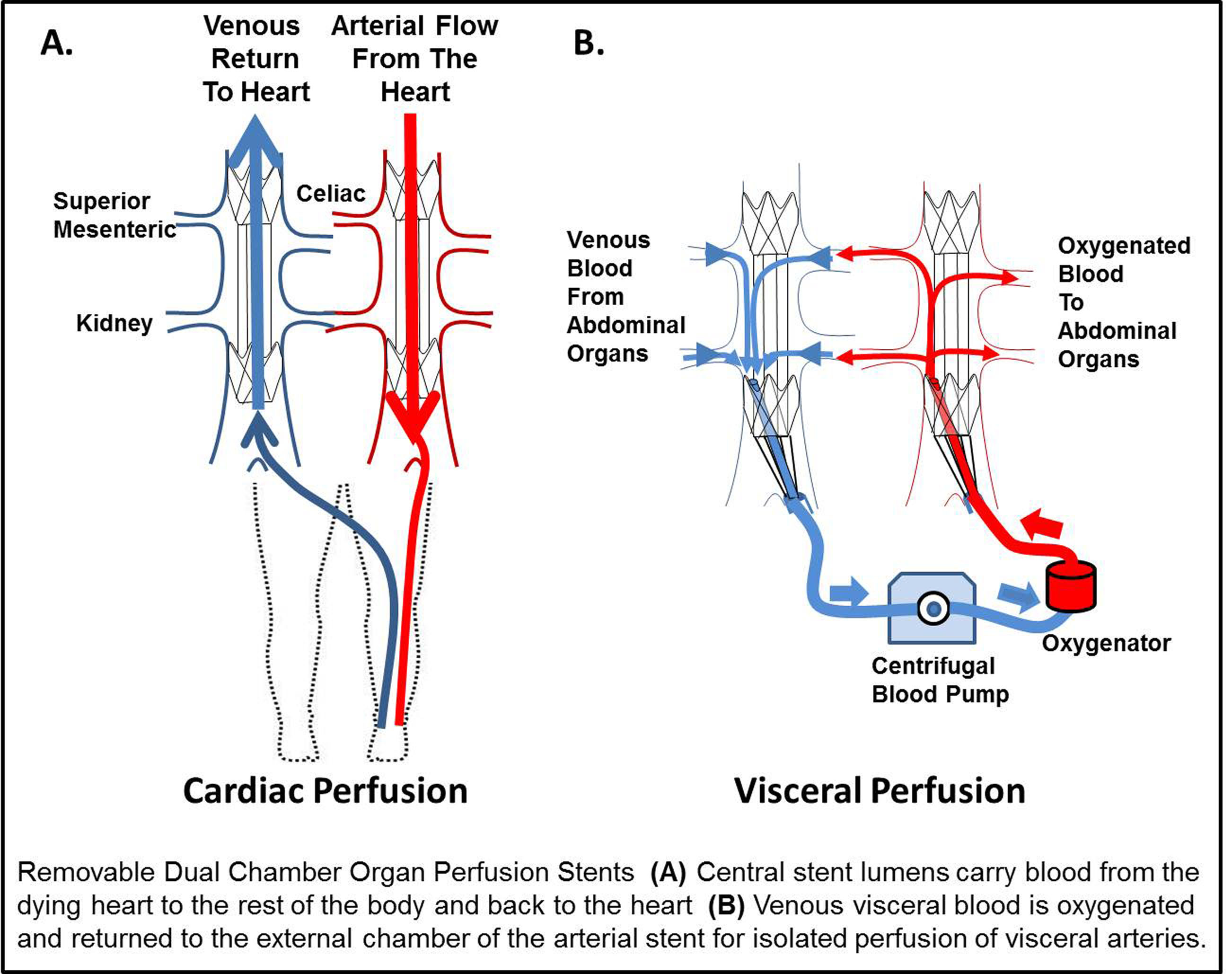
Introduction: The single most significant obstacle facing organ transplantation remains the critical shortage of donor organs. While donation after cardiac death (DCD) has demonstrated significant growth, these organs have inferior outcomes compared to other types of donation. During the ‘agonal period’, after life support is discontinued, malperfusion of organs leads to ischemic organ damage. As a result of the tenet that cardiac death should not be expedited, previous approaches could not be used until after cardiac death and after organ injury. In contrast to the global withdrawal of life support in DCD, among non-donors end of life care is determined by the family for individual organ systems. We have designed a dual chamber organ perfusion stent (OPS) to reduce ischemic injury by isolation of the visceral vessels and to perfuse only those organs with oxygenated blood while the heart fails on its own terms. This differs from all prior strategies because this removable stent would be placed by a needlestick femoral access and would allow the failing heart to continue to perfuse the rest of the body while protecting against ischemic injury of the visceral organs.

Methods: Prototype OPS were constructed from commercial stent components and final versions were custom made from nitinol and electrospun polyurethane. An OPS (n=5) was evaluated in a porcine model compared to non-stented controls. Paired venous and arterial OPS were deployed. Next, venous blood from the viscera in the external chamber was oxygenated and cycled to the external chamber of the arterial stent. Blood from the heart perfused the body through the large central lumen of the arterial OPS. DCD conditions were simulated for one hour with profound hypotension (SBP < 50 mm Hg).

Results: Invasive cardiac monitoring revealed no significant change in heart rate, mean arterial pressure, mixed venous oxygen, or venous pressure during stent deployment (P< 0.05). A small decrease was observed in the cardiac output consistent with exclusion of the visceral blood flow. Visceral blood flow of over 650 mL/min was achieved with the OPS and oxygen pressures were increased nearly 5 fold compared to agonal control animals. Control organs demonstrated severe malperfusion, contrasting to the well perfused OPS organs.

Conclusion: This pilot study suggests that an OPS offers: 1) rapid endovascular delivery 2) minimal impact on cardiac physiology 3) improved oxygen delivery to organs. Our early results suggest this novel stent design may increase availability of donor organs without impacting the natural cardiac death of the consented donor. Further development of this approach may ensure that nearly every consented donor successfully donates lifesaving organs.

42.04 Nanofiber Scaffold for Recreation of the Abdominal Wall in Rattus norvegicus II: Histopathology

L. Fluke1, R. Restrepo1, B. Hoagland1, J. Stephenson1 1Naval Medical Center Portsmouth,General Surgery,Portsmouth, VIRGINIA, USA

Introduction:

Full thickness soft tissue defects from congenital absence or traumatic loss are difficult to surgically manage. Healing requires cell migration, organization of an extracellular matrix (ECM), inflammation, and wound coverage.

We have previously shown that Poly(L-lactide) (PLLA) nanofibrous scaffolds enhance cell infiltration in vitro and in vivo in wound healing models, and we have demonstrated that the PLLA scaffold provides adequate strength to serve as a replacement for a full thickness abdominal wall defect in the rat model. Our current study compares aligned and unaligned nanofibrous scaffolds to a commonly used absorbable mesh, Vicryl, and permanent mesh, Gortex. We hypothesized that aligned electrospun PLLA nanofibrous grafts best facilitate native tissue ingrowth, providing physiologic strength necessary for repair of soft tissue defect while serving as a scaffold to guide tissue regeneration.

Methods:
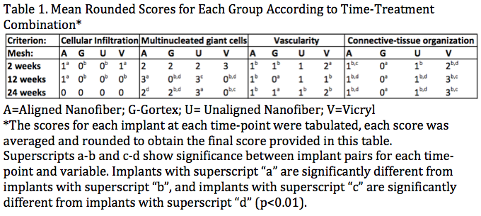
Abdominal wall defects were created in 72 rats, 6 rats per mesh per time frame, followed by underlay implantation of Vicryl, Gortex, aligned or unaligned PLLA Nanofiber mesh. Abdominal walls were harvested at 2, 12, and 24 weeks. Histopathologic semiquantative analysis was graded for cellular infiltration, multinucleated giant cells (MNG), vascularity, and tissue organization in a blinded fashion by an expert reviewer using a validated scoring system. Mean scores were compared and analyzed with non-parametric Mann-Whitney Wilcoxon testing.

Results:
Both aligned PLLA and Vicryl showed improved cellular infiltration at 2 weeks versus unaligned PLLA and Goretex. At 12 weeks, only the aligned PLLA had persistent cellular infiltration of the graft, while both aligned and unaligned PLLA grafts showed the presence of multinucleated giant cells (MNG). The presence of MNG’s decreases in the aligned PLLA graft by 24 weeks. There was no significant difference in graft vascularity at any timepoints. Vicryl graft had significant disruption at 2 and 12 weeks resulting in moderately organized connective tissue by the end of the study. Meanwhile, both aligned and unaligned PLLA grafts were broken down with less densely organized connective tissue, or scar.

Conclusion:
PLLA graft maintains structural integrity until at least 12 weeks and exhibits substantial tissue replacement at 24 weeks. Vicryl and PLLA grafts have similar tissue responses; however the Vicryl mesh has substantial degradation at 2 weeks and results in more dense scarring. These results demonstrate that PLLA Nanofiber mesh offers early strength comparable to Gortex but breaks down, replaced with cellular growth creating a favorable option in management of complex surgical wounds or native soft tissue defects.

04.21 Targeting HDM-2 Over-Expression in Multi-Drug Resistant Ovarian Cancer

E. Gleeson1, H. Glatthorn1, D. Zimmerman1, C. Carballo1, S. Mahmood4, P. Love1, M. F. Shaikh1, W. F. Morano1, S. D. Richard2, M. R. Pincus3, W. B. Bowne1 1Drexel University College Of Medicine,Surgery,Philadelphia, Pa, USA 2Drexel University College Of Medicine,Gynecologic Oncology,Philadelphia, Pa, USA 3New York Harbor Healthcare System VAMC,Pathology,New York, NY, USA 4University Of Pittsburg,Pittsburgh, PA, USA

Introduction: Of an estimated 22, 000 women diagnosed with ovarian cancer in the United States each year, 80% will develop lethal multi-drug resistance (MDR) to conventional chemotherapy. HDM-2 over-expression in the cancer cell membrane may be a potential anti-cancer target. We tested this target using a p53-derived, HDM2-binding peptide for anti-cancer activity in MDR ovarian cancer.

Methods: Western blot analysis was used to demonstrate expression of HDM-2 in MDR SKOV-3 human ovarian cancer cell membranes compared to the membranes of untransformed fibroblasts. 5×10⁴ cells were treated with HDM2-binding construct PNC–27 and control peptide. Anti-cancer activity and mechanism of PNC-27 were studied for cell viability (MTT), necrosis (LDH), apoptosis (Caspase-3) and co-localization of HDM-2 with PNC-27 (immunofluorescence).

Results: HDM-2 was strongly expressed in the cell membranes of MDR SKOV-3 by western blot analysis. Accordingly, PNC–27 was selectively cytotoxic to these cells, inducing nearly 100% reduction in cell proliferation compared to control PNC–29 and untreated cells (p<0.001; See Figure). We observed a rapid (4h) cellular necrosis with a 2.5 fold increase in LDH (p<0.001) and no caspase-3 activity. This was shown to occur in a dose-dependent manner. In contrast, PNC-27 had no effect on untransformed fibroblasts. This observed effect was due to over-expression of HDM-2 in MDR SKOV-3 cells which demonstrated co-localization of PNC–27 and HDM–2 along their membranes.

Conclusion: These findings demonstrate that HDM-2 over-expression in MDR ovarian cancer cells may be used as a potential target for anti-cancer therapy. Therapies that target HDM-2 in ovarian cancer cell membranes may increase our armamentarium for the treatment of this lethal disease.

04.17 MicroRNA dysregulation associated with function of the impaired monocyte

N. J. Galbraith1, S. Walker1, M. Cahill1, S. Gardner1, H. C. Polk1 1Price Institue Of Surgical Research,Department Of Surgery,Louisville, KY, USA

Introduction:
Major trauma often leads to impaired host defenses. Specifically, subnormal monocyte function (seen in approximately a sixth of all such patients) has repeatedly been shown to be associated with infection-related complications. However, identifying the ‘at risk’ patient, as well as understanding the pathophysiology behind this disturbance, continues to elude the investigators. MicroRNAs hold the potential as stable biomarkers that fine tune host defenses. We aim to identify novel microRNA (miRNA) associated with the impaired monocyte response.

Methods:
We produced ‘impaired’ human monocytes by exposure to LPS 10ng/mL (each containing 2.5 x 10⁵ monocytes) and compared them to similarly incubated naïve monocytes. Both preparations were then challenged with LPS 100ng/mL and the production of TNFα, IL-6 and IL-10 (ELISA) and HLA-DR expression (flow cytometry) were determined. MiRNA profiling was undertaken using microfluidic array technology (n = 4 for each determination).

Results:
Monocytes exposed to LPS 10ng/mL were found to have lower TNFα, IL-6 and IL-10 production in response to an LPS 100ng/mL challenge, when compared to the naïve monocyte (p < 0.05, paired T-test). Furthermore, these impaired cells had lower HLA-DR expression (p<0.05). MiRNA profiling revealed that when compared to the naïve monocyte, the LPS-impaired cells had upregulated miRNA- 487a & miRNA-655, yet downregulated miRNA-433 & miRNA-450a (p = <0.05).

Conclusion:
The precise processes behind immune dysfunction that appear following major trauma remain unclear. We have identified four dysregulated miRNAs associated with the impaired monocyte inflammatory response. Further study of these, and approximately 20 more differentially expressed miRNAs based on our preliminary data and literature consultation, may identify other markers, some of which may be associated with important signaling mechanisms present in the high risk patient.

04.18 Hm-Chitosan Gauze: A New And Effective Topical Hemostat

A. Chaturvedi2, M. B. Dowling4, J. P. Gustin3, T. M. Scalea2, S. R. Raghavan3, M. Narayan2 2University Of Maryland,R Adams Cowley Shock Trauma Center,Baltimore, MD, USA 3University Of Maryland,Department Of Chemical & Biomolecular Engineering,College Park, MD, USA 4University Of Maryland,Fischell Department Of Bioengineering,College Park, MD, USA

Introduction:
Currently, the standard of care for treating severe hemorrhage in a military setting is Combat GauzeTM(CG). Previous work has shown that hydrophobically modified (hm) chitosan has great hemostatic potential. This work aims to create a hm-chitosan coated gauze to directly compare to CG as well as ChitoGauze® (ChG) in a lethal in vivo hemorrhage model.

Methods:
Twelve Yorkshire swine were randomized to receive either hm-chitosan gauze (n = 4), ChG (n = 4), or CG (n = 4). A standard hemorrhage model was used in which animals underwent a splenectomy prior to a 6 mm punch arterial puncture of the femoral artery. Thirty seconds of free bleeding was allowed before dressings were applied and compressed for 3 minutes. Baseline mean arterial pressure was preserved via fluid resuscitation. Experiments were conducted for three hours after which any surviving animal was euthanized.

Results:
Hm-chitosan gauze was found to be at least equivalent to both CG and ChG in terms of overall survival(100% v 75%), number of dressing used(6 v 7), and duration of hemostasis (3 hrs v 2.25 hrs). Total post-treatment blood loss was significantly lower in the hm-chitosan gauze treatment group (4.7 mL/kg) when compared to CG(13.4 mL/kg) and ChG(12.1 mL/kg) groups (p =< 0.0001)

Conclusion:
Hm-chitosan gauze appears to have outperformed both CG and ChG in a lethal hemorrhage model. However given the small treatment group size, the only measured outcome that was significantly different was total post-treatment blood loss. Future comparison of hm-chitosan gauze to CG and ChG will be performed on a hypothermic and coagulapathic model that should allow for outcome significance to be differentiated under small treatment groups.

04.19 DPR Increasd sICAM-1 But Not Lung Integrin-aL or Integrin-aM in Resuscitated Hemorrhagic Shock

S. C. Jones1, P. J. Matheson1,2, C. D. Downard1,2, J. C. Frimodig1, C. J. McClain1,2, R. N. Garrison1,2, J. W. Smith1,2 1University Of Louisville,Department Of Surgery,Louisville, KENTUCKY, USA 2Robley Rex VAMC,Department Of Surgery,Louisville, KENTUCKY, USA

Introduction: ~~Hemorrhagic shock (HS) in trauma patients can result in gut and liver hypoperfusion and a pro-inflammatory systemic condition, which often initiates acute lung injury (ALI) or the more severe acute respiratory distress syndrome (ARDS). The pathophysiology of ALI/ARDS is multifactorial, but can involve alarmins, activated immune cells, and inflammatory chemokines/cytokines. DPR improves intestinal and hepatic blood flow to prevent gut-associated inflammatory changes and thus might also prevent or mitigate ALI/ARDS. We hypothesized that soluble intracellular adhesion molecule-1 (sICAM-1), which is sloughed from endothelial cells in inflammatory conditions, might be elevated in resuscitated HS, and that DPR might prevent that event.

Methods: ~~Male Sprague-Dawley rats (225-250g) were anesthetized and randomized to groups (n=8/group): 1) Sham (no HS, no conventional resuscitation or CR); 2) HS/CR; or 3) HS/CR+DPR. HS was 40% of mean arterial pressure for 60 minutes. CR was shed blood plus two equal volumes of lactated Ringers over 30 minutes total. DPR was 25mL of pre-warmed 2.5% glucose peritoneal dialysis solution at the time of blood infusion. Serum and lung tissue were collected at 4 hours post-CR. Adhesion molecule levels were measured by ELISA: serum sICAM-1, lung integrin-alpha L (ITGAL or LFA-1), and lung integrin-alpha M (ITGAM or MAC-1).

Results:~~HS/CR increased sICAM-1 levels compared to Sham, and DPR increased sICAM-1 further compared to Sham or HS/CR (see Figure). There were no differences in lung LFA-1 or MAC-1 levels in HS/CR or HS/CR+DPR compared to Sham.

Conclusion:~~These data suggest that circulating levels of sICAM-1 are not mediated by gut blood flow and/or gut-associated inflammatory mediators in this model of resuscitated shock at this 4-hour post-CR time point. Other post-resuscitation time points might reveal a benefit to DPR, which has been shown to improve the number of lungs suitable for organ transplant in human organ donors compared to donors who did not receive DPR treatment.

04.14 Monocyte Differentiation Alters CHRFAM7A: Implications for a Human-Specific Inflammatory Response

S. M. Langness1, B. Eliceiri1, R. Coimbra1, A. Baird1, T. W. Costantini1 1University Of California – San Diego,Trauma, Surgical Critical Care, Burns And Acute Care Surgery,San Diego, CA, USA

Introduction: In humans, the alpha-7 nicotinic acetylcholine receptor (a7nAChR) is encoded by the CHRNA7 gene and mediates an anti-inflammatory response to injury and infection. However, the human genome also encodes a uniquely human gene called CHRFAM7A that is an antagonist to a7nAChR-mediated signaling. Several studies have suggested that in humans, the ratio of CHRNA7 to CHRFAM7A modulates receptor activity. We have previously shown that: a) CHRFAM7A is highly expressed in human leukocytes; b) the ratio of CHRNA7 to CHRFAM7A gene expression in leukocytes varies widely amongst individuals; and c) CHRFAM7A is biologically active in the human THP1 pre-monocyte cell line. Here, we postulated that PMA differentiation of these monocyte precursors to monocytes might alter the CHRNA7 to CHRFAM7A ratio and alter monocyte gene expression. The effects of preventing a change in differentiation-induced CHRFAM7A gene expression was evaluated by its constitutive expression using gene delivery.

Methods: THP-1 cells were used in all experiments and monocyte differentiation was induced by incubation with PMA for 3 days in culture. Cell appearance and viability was assessed by light microscopy and flow cytometry respectively. CHRNA7 and CHRFAM7 gene expression was measured by quantitative RT-PCR. Constitutive expression of CHRFAM7A was achieved with lentiviral transfection prior to PMA-induced differentiation. Gene expression in transfected CHRFAM7A and GFP-transduced differentiated monocytes were compared using RNA-seq with subsequent pathway analyses.

Results: CHRFAM7A expression was decreased by 70% in PMA-differentiated monocytes compared to untreated THP-1 cells. In contrast, there was no difference in CHRNA7 expression resulting in a net 5-fold increase in the expression of CHRNA7 compared to CHRFAM7A (see Figure). Over-expression of CHRFAM7A in differentiated monocytes produced viable cells with monocytic phenotype based on microscopic appearance and the presence of monocyte cell surface markers like CD14, CD22 and CD163. In contrast, the constitutive expression of CHRFAM7A in differentiated monocytes changed the expression of genes in pathways traditionally associated with regulating inflammatory response signaling and antigen presentation.

Conclusions: The ratio of CHRNA7 to CHRFAM7A gene expression in THP1 cells changes after differentiation to monocytes. Preventing this change by gene transfection, CHRFAM7A has no effect on differentiation but alters the expression of genes that mediate the inflammatory response. Because CHRFAM7A is unique to the human genome, these data support the existence of a human-specific mechanism that regulates a7nAChR anti-inflammatory signaling and hence, the human immune response to injury and infection.

04.15 Effect of Swine Leukocyte Antigen Class II on Human PBMCs in a Mixed Cell Reaction

J. M. Ladowski1, J. Tchervenkov3, J. Butler1, G. Martens1, M. Tector1, J. Blum2, J. Tector1 1Indiana University School Of Medicine,Transplant Surgery,Indianapolis, IN, USA 2Indiana University School Of Medicine,Microbiology/Immunology,Indianapolis, IN, USA 3Royal College Of Surgeons,Dublin, DUBLIN COUNTY, Ireland

Introduction: As the antibody barrier to clinical xenotransplantation is reduced, attention within the community will turn to cellular-mediated rejection. The direct recognition of swine leukocyte antigen (SLA) class II by PBMC may play a significant role in this rejection. To characterize the proliferative effect of SLA class II, human bulk PBMCs and isolated CD4+ T-cells were incubated with a SLA class II positive and negative adherent fibroblast cell line in a mixed lymphocyte reaction (MLR) variant. The proliferative response was observed through CFSE MLR staining. This novel adherent cell MLR assay allows for faster analysis of candidate antigens and therapeutics compared with studies using cells isolated from animal models for xenotranaplantation.

Methods: An aGal, SLA class I deficient cell line was transfected with a human version of class II transactivator (CIITA) and sorted via flow cytometry based on SLA class II expression. A positive and negative clone were selected as stimulators for a MLR containing human whole or CD4+ isolated PBMCs as the responder population. Responders were stained with carboxyfluorescein succinimidyl ester (CFSE) and monitored for proliferation via flow cytometry.

Results: The human transcription factor CIITA functions as an up-regulator of SLA class II, producing cells with constitutive and stable class II expression. Both whole and CD4+ isolated PBMCs incubated with fibroblasts lacking expression of SLA class II demonstrated considerably less proliferation (0.39 and 0.15% respectively) compared to responders incubated with SLA class II positive cells (1.73 and 1.22%) given the near identical proliferation seen in whole PBMC versus isolated CD4+ population.

Conclusion: This novel swine adherent cell MLR assay facilitates the analysis of cross-species reactivity using human PBMCs, providing for a new assay for xenotransplant study. The proliferation stimulated by SLA class II positive cells appears to predominately occur through direct recognition by CD4+ cells. This information will help focus immunosuppressant therapy in future clinical xenotransplantation.

04.16 Histone Deacetylase-2 Gene Deletion In Mice Extends Tolerance of Renal Ischemia/Reperfusion Injury

D. Murken1, D. Aufhauser1, Z. Wang1, G. Ge1, T. Bhatti3, W. Hancock3, M. Levine1,2 1University Of Pennsylvania,Department Of Surgery,Philadelphia, PA, USA 2Children’s Hospital Of Philadelphia,Department Of Surgery,Philadelphia, PA, USA 3Children’s Hospital Of Philadelphia,Department Of Pathology And Laboratory Medicine,Philadelphia, PA, USA

Introduction: Ischemia Reperfusion Injury (IRI) causes significant morbidity in renal transplantation and other surgical scenarios. A better understanding of the molecular mechanisms leading to IRI is required so that new strategies for prevention and treatment can be developed. Histone deacetylases (HDACs) regulate diverse cellular processes. We have previously shown the benefit in renal IRI of non-selective pharmacologic HDAC inhibitors, and now report on the benefit of targeting one specific HDAC isoform, HDAC2, via inducible whole body gene deletion in order to extend renal IRI tolerance.

Methods: Female wild type C57BL/6 (WT) or female whole body HDAC2-deficient C57BL/6 (HDAC2-/-) mice were subjected to a unilateral warm ischemia model with contralateral nephrectomy under strict temperature control in order to yield maximum reproducibility. Plasma concentrations of BUN were assessed for four days post-operatively.

Results: WT mice survived 28 minutes of warm ischemic time, but developed substantial injury (Figure 1). When subjected to longer periods of warm ischemia, these mice all developed renal insufficiency requiring sacrifice within 48 hours of ischemic insult. HDAC2-/- mice were subjected to longer periods of warm ischemia than survivable by WT mice. HDAC2-/- mice tolerated 35 minutes of ischemia with comparable renal injury to that observed in WT mice after 28 minutes of IRI (p=0.48, Figure 1). Prolongation of the ischemic time by a further two additional minutes (37 min) resulted in non-survivable injury in HDAC2-/- mice.

Conclusion: Murine HDAC2 gene deletion leads to significant extension of renal IRI tolerance. Pharmacologic HDAC2 inhibition may offer a novel therapeutic strategy to avoid organ injury in kidney transplantation and other surgeries requiring renal ischemia.

04.10 A Role For RGS5 In MYCN Amplified Neuroblastoma

M. J. Metzner1, J. Mazar2, A. Rosado1, T. J. Westmoreland1 1Nemour’s Children’s Hospital; University Of Central Florida,Pediatric General Surgery,Orlando, FL, USA 2Sanford Burnham Presbys Medical Discovery Institute,Orlando, FL, USA

Introduction:

Neuroblastoma is a childhood predominant cancer, and its prognosis is greatly affected by MYCN overexpression. MYCN amplification is directly related to poor tumor prognosis and has been previously shown to attenuate RGS5, a regulator of G-protein signaling involved in tumor growth, metastasis, and angiogenesis. It has been previously shown that a decrease in RGS5 expression has been associated with increased tumor aggressiveness. Next Generation sequencing has enabled many advancements in cancer research and has enabled researchers to look at potential cancer markers to help further stratify how cell types react to treatments. We hypothesize that MYCN amplification correlates with RGS5 expression.

Methods:
To test this hypothesis we performed Next-Generation Sequencing on IMR-32 (MYCN amplified) cell line as well as MYCN knockdown in the same cell line. Next, total cellular RNA was isolated from the human neuroblastoma pre-treatment cell lines (SK-N-Be(1), SMS-KAN, IMR32) and post-treatment cell lines (LA-N-6, SK-N-As, SK-N-FI). The pre-treatment cell lines have overexpression of MYCN. The expression of RGS5 was measured using real-time reverse transcriptase polymerase chain reaction (qPCR).

Results:
Utilizing Next Generation sequencing, we demonstrated that the expression of RGS5 in MYCN amplified cell lines showed attenuated expression when compared to post-treatment cell lines where MYCN was non-amplified. The qPCR data using both pre and post-treatment cell lines followed the same pattern and showed that RGS5 expression was decreased in the MYCN amplified pre-treatment lines.

Conclusion:
RGS5 is differentially expressed in neuroblastoma cell lines with and without MYCN amplification. There is an inverse correlation of MYCN and RGS5, which follows the theory that low levels of RGS5 and high MYCN expression lead to a worse prognosis. It has been shown that RGS5 works in many cell growth, metastatic, and neovascularization pathways. This work further correlates the connection between expression of MYCN and RGS5. Because RGS5 expression levels are increased in post-treatment cell lines, RGS5 could serve in the future as a possible marker to further stratify and characterize the aggressiveness of recurrent neuroblastoma.

04.11 Continuous Cold Perfusion vs. Static Cold Preservation for Intestinal Graft Preservation in Rats.

A. Munoz-Abraham2, A. Alkukhun2, S. Judeeba2, R. Patron-lozano2, T. Alfadda2, A. Bertacco1,2, M. I. Rodriguez-Davalos1,2, J. P. Geibel2 1Yale University School Of Medicine,Transplantation/Surgery,New Haven, CT, USA 2Yale University School Of Medicine,Surgery,New Haven, CT, USA

Introduction: In the current transplantation era, new preservation techniques have been developed to further preserve the organs in an optimal status. Continuous perfusion for organs such as kidney, liver, heart and lung have already proven to have a value in comparison to static preservation. However few attempts have been done to develop or find the ideal technique to preserve intestinal grafts. The aim of this study was to assess the optimal preservation technique for intestinal grafts intended for transplantation by comparing the static cold preservation against continuous cold perfusion in a rat model.

Materials and

Methods:

Comparison between static cold preservation (Control Group) with University of Wisconsin (UW) Preservation Solution versus Continuous Cold Perfusion with UW for perfusion (UW Perfusion Group) or a Modified Preservation Solution (MPS) containing UW with L-Arginine (MPS Perfusion Group) with rat intestinal grafts.

30 cms. of Male Sprague-Dawley rats’s small intestine were procured. The intestinal lumen was flushed with HEPES solution (pH 7.40) to remove intestinal debris. Two intestinal loops (10 cm each) were then connected to two custom perfusion chambers of an intestinal perfusion device. One chamber received a constant flow of UW solution and the other MPS solution and both submerged in a bath of deionized water at 4°C. A third intestinal loop was placed in cold static preservation with UW solution. Intestinal tissue samples were taken at T0, T1 (1 hr), T3 (3 hrs.), and T6 (6 hrs.), fixed in Formalin at 10%, and sent to pathology for processing, analysis, and grading according to Park/Chiu Scoring System for Grading Intestinal Ischemia.

Results: Both the UW and the MPS Perfusion groups better preserved the intestine in comparison to the static cold preservation after T6.

Conclusion: Continuous cold perfusion can better preserve intestinal grafts when compared to static cold preservation. These early results might open the door for new techniques for intestine preservation for transplantation that can eventually be applied in the clinical setting. Based on this findings, the next goal of this study is to determine the ideal preservation solution for continuous cold perfusion of the intestinal graft.

04.12 Normothermic Perfusion Using UW compared to HTK Solution on a Rat Intestinal Model

A. Bertacco1, A. Alkukhun1, R. Agarwal1, C. P. LeBlanc1, A. Munoz-Abraham1, F. D’Amico1, M. Rodriguez-Davalos1, J. Geibel1 1Yale School Of Medicine,Surgery/Transplantation,New Haven, CT, USA

Introduction: Prevention of intestinal ischemia during small intestine transplantation still remains a challenge . A safe and effective preservation solution is a precondition for successful small intestine transplantation. The most commonly used hypothermic preservation solutions for intestinal transplantation are the University of Wisconsin solution (UW) and histidine-tryptophan-ketoglutarate (HTK). We have previously shown that intestinal ischemia results in an increased fluid shift towards the lumen of the intestine. In the present study we demonstrate that as the temperature increases (4-37 C) the HTK becomes more acidic. Using this knowledge, our aim was to evaluate the effect of the UW and HTK solution on the viability of an intestinal preparation at normothermic temperature in a rat perfusion model.

Methods: Male Sprague Dawley rats (average weight 440g) were anesthetized and euthanized in accordance with the Animal Care and Use Committee at Yale University. Two sequential small intestinal segments were procured and flushed with regular HEPES solution (with a pH 7.40, mOsm 300 at 37 C) to remove any remaining intestinal debris. The intestines were then connected to our intestinal chambers, which allowed both luminal and extraluminal perfusion. The first intestine was filled intraluminally with 1.5ml of UW Solution including 50µM of FITC-Inulin, and was surrounded by standard UW solution on the vascular side. The second intestine was filled with 1.5ml of HTK Solution including 50µM of FITC-Inulin, and was surrounded by HTK solution on the vascular side. Both chambers were maintained at 37o C. Sample measurements of intraluminal perfusate were made to measure FITC-Inulin concentration, using the Nanodrop 3300™and were taken at times 0,45,90 and 120 minutes.

Results: When comparing the FITC-Inulin concentration in both intestines, there was a mean reduction of 43.25% of FITC-Inulin concentration in the UW arm vs a 9.5% decrease in the HTK arm, indicating more fluid secretion in the intestine exposed to UW.

Conclusion: We have shown that UW solution increases the fluid shift towards the lumen at normothermic temperature compared to HTK solution. Surprisingly, we did not observe the expected amount of secretion from the acidic HTK. With that response, we recognized that UW is a better preservation solution at 37 C due to either it’s buffering capacity in the intestinal lumen, or starch component present in it’s composition. We hypothesize that UW results in decreased cellular swelling in the small intestine cells at normothermic conditions, and thus could leed to a a better outcome for intestinal preservation at normothermic temperatures.

04.13 IL-l Induces Endothelial Hyperpermeability via Activation of p38 MAPK and JNK and Production of MMP-9

Z. Wang1, L. Kong1, J. Kang1, D. K. Nakayama2, P. S. Dale1, D. W. Ashley1 1The Medical Center Navicent Health, Mercer University School Of Medicine,Division Of Trauma, Division Of Basic Sciences, Division Of Surgical Oncology, Department Of Surgery,Macon, GA, USA 2West Virginia University School Of Medicine,Department Of Surgery,Morgantown, WV, USA

Introduction: Endothelial hypermeability mediates edema formation, and contributes to morbidity and death in trauma and the systemic inflammatory response syndrome. Inflammatory cytokines including interleukin-1beta (IL-l) are known to activate matrix metalloproteinases (MMPs), a potential mechanism by which cytokines may disrupt endothelial barrier. The mechanism by which proinflammatory cytokines disrupt endothelial permeability is poorly understood. We investigated if IL-1 would induce endothelial hypermeability through activation of p38 mitogen-activated protein kinase (MAPK) and Jun N-terminal kinase (JNK) and induction of MMP-9.

Methods: Vascular endothelial cells (EC) were incubated with and without IL-1, a MMP-9 inhibitor, and inhibitors of p38 MAPK (SB202190) and JNK (SP600125). RT-PCR and Western blotting measured MMP-9 mRNA and protein, respectively. Gelatin zymography quantitated MMP-9 release. Horseradish peroxidase diffusion through MVEC monolayer assessed endothelial permeability. The kinases were assessed with immunoblotting and activity assay.

Results: IL-l activated p38 MAPK and JNK, promoted expression of MMP-9 mRNA and protein and release of MMP-9 activity, and increased endothelial permeability. IL-1-induced endothelial hypermeability was significantly attenuated by pharmacological inhibitors of p38 MAPK, JNK and MMP-9, respectively. Moreover, pharmacological inhibition of p38 MAPK and JNK reduced MMP-9 release in response to IL-1 stimulation.

Conclusion: The proinflammatory IL-1 induces endothelial hypermeability via activation of p38 MAPK and JNK, and subsequent release of MMP-9. Endothelial MMP-9 expression and activity may play a role in the pathogenesis of vascular hyperpermeability in microcirculation in the setting of trauma and cytokine activation syndromes. JNK, p38 MAPK and MMP-9 may be therapeutic targets for endothelial protection from inflammatory injury.

04.08 Evaluation of apoptosis in carcinoid cells via concomitant treatment of siRNA and Thailandepsin-A

S. Odorico1, R. Jaskula-Sztul1,2, A. Harrison1, S. Clarkson1, S. Golden1, H. Jin1,2, H. Chen1,2 1University Of Wisconsin,Department Of Surgery,Madison, WI, USA 2University Of Alabama,Surgery,Birmingham, Alabama, USA

Introduction: Carcinoids are neuroendocrine tumors that often metastasize and other than surgery, no curative options are available. We have shown that Thailandepsin-A (TDP-A), a novel histone deacetylase inhibitor, can induce apoptosis in carcinoid tumor cells in vitro and reduce tumor size in vivo. However, treatment with TDP-A alone achieved only cytostatic effect. The suppression of anti-apoptotic proteins with concomitant TDP-A treatment could induce cytotoxic effect of both drugs. Therefore, our goal was to investigate a potential synergistic effect on apoptosis in carcinoid cells in vitro with dual treatment of TDP-A and siRNA silencing of three different anti-apoptotic markers, cyclin D1, MCL1 and BCL2.

Method: siRNA of three anti-apoptotic oncogenes, cyclin D1, MCL1 and BCL2, were transfected into gastrointestinal carcinoid cells (BON) using Lipofectamine. Treatment with TDP-A 7 nM (previously tested IC50 value of the drug) occurred 4 hours following siRNA transfection. Cells were incubated for 48 hours before harvesting. qRT-PCR analysis was completed to assess the efficiency of siRNA silencing and flow cytometry was analyzed to determine the apoptotic induction.

Results: In flow cytometric analysis, we assessed pre-apoptotic and apoptotic percentages of total cells after 48 hours of treatment with either siRNA alone, TDP-A alone or siRNA and TDP-A. Figure 1A represents a summary of pre-apoptotic and apoptotic percentages of total cells observed in all samples. We observed a synergistic induction of apoptosis when MCL1 siRNA and TDP-A were applied concurrently. Figure 1B depicts flow cytometric images visualizing the synergistic induction of apoptosis. For qRT-PCR, we observed reduction in relative gene expression in all target genes following transfection of siRNA as compared to scrambled siRNA transfection.

Conclusion: While BCL2 and cyclin D1 siRNA did indeed knock down the expression of their target genes, no synergistic induction of apoptosis occurred with the treatment of both the siRNA and TDP-A. However, combined MCL1 siRNA and TDP-A treatment resulted in a synergistic induction of apoptosis in BON carcinoid cells. These results suggest selective gene silencing of MCL1 enhanced TDP-A induced apoptosis in carcinoid cells in vitro, and warrants further investigation into the relationship between MCL1 knockdown and concomitant treatment of TDP-A.

04.09 Molecular Mechanisms of the Inflammatory Response in Different Aged Fibroblasts

T. S. Lefcourt1, C. Zgheib1, J. Hu1, J. Deacon1, J. Zuk1, J. Xu1, K. W. Liecthy1 1Laboratory For Fetal And Regenerative Biology, Department Of Surgery, School Of Medicine, University Of Colorado Denver Anschutz Medical Campus And Childrens Hospital Colorado,Surgery,Aurora, CO, USA

Introduction: Compared to the adult response to injury, the inflammatory response and scar formation in the neonates is decreased. We have previously shown that increased fibroblast age is associated with increased production of proinflammatory cytokines. Recent evidence suggests that one of the most important regulatory factors of protein production are microRNAs. One such microRNA, microRNA-146a (miR-146a), regulates the inflammatory response by suppressing interleukin-8 (IL-8). Epigenetic regulation such as histone modification has been implicated as a modulatory factor in microRNA expression. We hypothesize that differentially expressed miR-146a in different age groups is under epigenetic control, and that the increased production of proinflammatory cytokines by fibroblasts with increasing age may be due to decreased expression of miR-146a.

Methods: With IRB approval, we isolated fibroblasts from the discarded foreskin of young (<1 month of age) or old (>6 months) males undergoing elective circumcision. Fibroblasts were isolated and expanded no more than 5 passages. Young or old fibroblasts were then cultured for 24 hours with or without two different pan Histone Deacetylase inhibitor (HDACi) conditions: suberanilohydroxamic acid (SAHA), or Apicidin. Total cellular RNA was isolated. Expression of IL-8 and miR-146a was analyzed by quantitative RT-PCR analysis. Expression at baseline and following HDACi treatment was compared to vehicle (DMSO) control.

Results: Young fibroblasts had significantly decreased IL-8 gene expression compared to the older fibroblasts. Young fibroblasts also had significantly increased miR-146a expression compared to older fibroblasts. Treatment with an HDACi resulted in an increase in miR-146a expression in both young and old fibroblasts. In addition, expression of IL-8 was significantly decreased in the older fibroblasts under both HDACi treatment conditions (SAHA p < 0.01, Apicidin p < 0.05).

Conclusion: These findings identify age specific molecular mechanism(s) that modulate the inflammatory response. Increased inflammation and subsequent scar tissue formation with increasing age may be due to decreased expression of miR-146a. Our results also indicated that anti-inflammatory miR-146a is differentially expressed with age, and that the mechanism governing this differential expression may be under epigenetic control. Further studies are warranted to examine miR-146a as a potential therapeutic target to modulate the inflammatory response, and decrease the complications of fibrosis and scar formation.

04.04 Thrombin Stimulated Endothelium Downregulates Fibrinolysis Through Prestored PAI-1 Release

B. R. Huebner1,3, C. C. Silliman1,2,4, E. Gonzalez1,3, S. Mitra1, A. Banerjee1, E. E. Moore1,3 1University Of Colorado Denver,Department Of Surgery,Aurora, CO, USA 2University Of Colorado Denver,Department Of Pediatrics,Aurora, CO, USA 3Denver Health Medical Center,Aurora, CO, USA 4Bonfils Blood Center,Denver, CO, USA

Introduction:
Recently, there’s evidence that injured patients exhibit diverse phenotypes ranging from hyperfibrinolysis coagulopathy to fibrinolysis-shutdown resulting in microvascular occlusion and multi-organ failure. The primary mediators of these phenotypes are tissue plasminogen activator (tPA), a driving force behind hyperfibrinolysis, and plasminogen activator inhibitor-1 (PAI-1), a serine protease inhibitor that couples tPA nullifying its physiologic effect. Endothelial cells activate during stress and are capable of releasing tPA and PAI-1, but their role in critically injured patients remains unclear. Thrombin is a key agent which can stimulate shutdown or enhancement of fibrinolysis. While it is known that tPA is stored in Weibel-Palade bodies in endothelium, the timing and source of PAI-1 remains unclear. We hypothesize that thrombin causes PAI-1 release from preformed vesicles in endothelium thus serving as a rapid counter-regulation of tPA release.

Methods:
Human liver sinusoidal endothelial cells (HLSECs) were grown to an 80-90% confluence in wells after passage 3. HLSECs were treated with thrombin at a concentration of 5units/ml known to be present in the plasma of trauma patients. Supernatant samples were collected at 15 and 30 minute timepoints. Each timepoint was run in duplicate. The supernatants were analyzed with ELISAs for total (free) tPA, total (free) PAI-1, and tPA-PAI-1 complex.

Results:
Thrombin-stimulated endothelial cells released more PAI-1 than the control (>600ng/ml for thrombin 15 min and 30 min vs 441ng/ml control). Free tPA was lower in the thrombin treated cells (0.781ng/ml at 15m, 0.617ng/ml at 30m, 0.977ng/ml control). tPA-PAI-1 complex values were slightly higher from the control at 15min and lower at 30min for the thrombin-treated cells (10.95ng/ml for 15m, 8.56ng/ml for 30m, and 10.2ng/ml control).

Conclusion:
Thrombin provokes the rapid release of PAI-1 from hepatic endothelium with the subsequent formation of tPA-PAI-1 complexes causing a decrease in free tPA and an increase in PAI-1 resulting in fibrinolysis shutdown. Endothelial cells are important in contributing to the fibrinolysis phenotype seen in severely injured patients.

04.05 Post-Shock Accumulation Of Succinate Accelerates Fibrinolysis Via Platelet-Dependent Mechanism

A. L. Slaughter2, H. B. Moore2, A. Bacon2, N. Butler1, A. Banerjee2, C. Silliman2,3, A. D’Alessandro2, E. Peltz2, E. Moore1,2 1Denver Health Medical Center,Aurora, CO, USA 2University Of Colorado Denver,Aurora, CO, USA 3Children’s Hospital Colorado,Aurora, CO, USA

Introduction:
Previous data demonstrate that the plasma concentration of succinate can increase 20-fold within minutes of hemorrhagic shock. Succinate receptors are highly expressed on platelets and dysfunctional platelets play a pivotal role in trauma-induced coagulopathy (TIC). However, the contribution of post-shock succinate accumulation to platelet-mediated TIC is unknown. Fibrinolysis is an important cause of early post-traumatic death due to hemorrhage, yet the mechanism driving acute platelet/fibrin clot failure remains elusive. We hypothesize that succinate alters platelet function, accelerating fibrinolysis, at plasma concentrations observed during hemorrhagic shock.

Methods:
Venous whole blood (WB) from healthy individuals (n=8) was mixed with 5μl of succinate solution to achieve 250-1000μM concentrations per 500μl of WB. Tissue plasminogen activator (75ng/mL, +tPA) was added at each concentration to simulate post-shock endogenous tPA release. Samples were analyzed using native thromboelastography (TEG) and functional fibrinogen (measurement of clot formation independent of platelets, FF-TEG) assays. Repeated measures ANOVA with Bonferroni adjustment was used to determine statistically significant change from baseline TEG and FF-TEG parameters at increasing succinate concentration ± tPA.

Results:
Elevated succinate concentrations (±tPA) decreased time to clot initiation (R), while increasing clot potentiation (angle) and the maximum amplitude of clot formation (MA) (R 250μM+tPA vs. R WB+tPA, p=0.026; angle 500μM+tPA vs. angle WB+tPA, p=0.004; MA 500μM+tPA vs. MA WB+tPA, p=0.026, Figure 1). tPA independently decreased R and increased angle (R WB+tPA vs. R WB, p=0.016; angle WB+tPA vs. angle WB, p=0.016); however these effects were more profound in the setting of succinate (R +tPA vs. R succinate+tPA, -33% vs. -51%, p=0.042; angle +tPA vs. angle succinate+tPA, +37% vs. +67%, p=0.001). Functional fibrinogen levels (FLEV) were unchanged by elevated tPA and succinate concentration ± tPA.

Conclusion:
Pathophysiologic concentrations of succinate in whole blood accelerate clot initiation and potentiation, precipitating earlier tPA-mediated fibrinolysis. Functional fibrinogen levels, measuring clot formation independent of platelets, are unchanged by increasing succinate (± tPA), implicating platelets as culprit mediators. These data support succinate’s role in a platelet-dependent mechanism that drives early post-shock hyperfibrinolysis.

04.06 Examining Virulence Activation of Acinetobacter in Soft Tissue Injuries with an Agent Based Model

A. J. Benjamin1, G. An1 1University Of Chicago,Surgery,Chicago, IL, USA

Introduction: Acinetobacter baumannii is a microbe of growing importance in trauma related soft tissue infections (STIs), and is currently one of the most common causes of late mortality following combat injuries. It has also become a significant cause of multi-drug resistant infection in the intensive care setting. Injury and ischemia have been shown to enhance A. baumannii virulence, both directly and by altering the fitness function of any microbes present in the wound. Characterizing for the ecological dynamics of host-pathogen interactions (HPIs) in the wound milieu will provide insight into tipping points that lead to A. baumannii STIs (AbSTIs). Towards this end we have adapted a prior agent-based model (ABM) of STI to incorporate virulence activation mechanisms of A. baumannii in order to characterize the dynamics of wound HPI and identify conditions that promote AbSTI.

Methods: Our prior muscle wound ABM (MWABM) was extended by adding known A. baumannii virulence mechanisms and host tissue responses to hypoxia and inflammation. The resulting ABM incorporates muscles cells, neutrophils, macrophages, avirulent commensal bacteria, and the detailed representation of A. baumannii. Parameter sweeps were performed to evaluate dynamic patterns of of injury, microbial community composition, virulence activation and host mediator levels in relation to the generation of non-healing and the development of AbSTI. Simulation interventions included differing levels of wound debridement and topical antibiotic therapy.

Results: Simulation experiments identified a series of interconnected thresholds of microbial population composition, degree of initial injury and residual damaged tissue leading to AbSTIs. We demonstrated the role of free iron in the activation of virulence in relation to A. baumannii’s ability to sequester iron via siderophore production. Decreased free iron increased accumulation of HIF1α and decreased iNOS dimerization, which improved with greater debridement. Paradoxically, early topical antibiotic therapy demonstrated decreased microbial populations but increased relative rates of AbSTIs, suggesting the importance of microbial ecological suppression of A. baumannii virulence.

Conclusion: The modified MWABM effectively provides dynamic knowledge representation of A. baumannii virulence in the pathogenesis of AbSTI, demonstrating the effect of debridement, and also the unexpected result that topical antibiotic therapy may have adverse microbial ecological effects on the development of AbSTIs. These findings suggest the importance of incorporating intact microbial communities in future experimental wound healing STI studies in order to make them more clinically relevant. This type of model can serve as a framework to add more detailed mechanistic knowledge as well as investigate novel anti-virulence strategies.

04.01 The Use of FITC-Inulin as a Marker for Intestinal Ischemic Injury

A. O. AlKukhun1, A. Muñoz Abraham1, S. Judeeba1, C. Jasinski1, R. Patron-Lozano1, M. I. Rodriguez-Davalos1, J. P. Geibel1 1Yale University,Department Of Surgery,New Haven, CT, USA

Introduction:

Intestinal ischemia can be seen in conditions such as mesenteric ischemia, or in a challenging situation like intestinal transplantation. Intestinal ischemia leads to pathophysiological disruption that typically presents as increased fluid secretion into the lumen of the intestine with increased levels of ischemia. The aim of this study was to understand the fluid alterations that occur with intestinal ischemia, and determine if FITC-Inulin measurement is sensitive to measure the changes in fluid movement across an intact rat intestinal graft as a real-time measurement of ischemic injury.

Methods:

Male Sprague Dawley rats (average weight of 400g) were operated on in accordance with the Animal Care and Use Committee at Yale University. Two 10 cm small intestine segments were procured from the rats. The intestinal segments were cleansed with HEPES (pH: 7.40, Osmolarity 300±5 at 37°C) solution to clear remaining intestinal contents. The intestinal segments were connected to customized intestinal chambers that are part of a perfusion device. The chambers were kept in a water bath maintained at 37°C. Both intestines were perfused on the vascular side with HEPES buffer solution (pH: 7.40, Osmolarity 300±5 at 37°C). The control HEPES reservoir was open to air, while the experimental reservoir was sealed and bubbled with 100% Nitrogen (N2) to mimic an ischemic environment. Inside both lumens, 3ml of HEPES containing 50 µM FITC-Inulin was continuously perfused at a rate of 1ml/min. Intraluminal samples of the perfusates were collected at three 30 minutes’ intervals to measure FITC-Inulin concentration using a nanospectrofluorimeter.

Results:

When comparing measurements of FITC-Inulin in both intestines, there was a mean 37% reduction of FITC-Inulin concentration in the control versus a mean 57% reduction in the experimental arm, thus indicating increased fluid secretion that was stimulated by the ischemic environment. After examining and comparing the ischemic with the control tissue, an approximate doubling of the amount of secretion was observed over the same time period.

Conclusion:

FITC-Inulin can be used effectively as a volume marker that equates to the ischemic state of the intestinal tissue. This is a practical method to measure real time fluid changes inside a small intestine graft. This model can now be used to assess other intestinal ischemia models: assess viability of intestinal grafts prior to transplant, and monitor drug candidates against ischemia.