04.02 A Pilot Study On The Use of Mass Cytometry to Phenotype the Cellular Response to Trauma

G. Brat3, B. Yorkgitis3, A. Seshadri3, J. Keegan3, J. Dolan3, C. Hauser2, A. Salim3, R. Askari3, J. Lederer3 2Beth Israel Deaconess Medical Center,Trauma Surgery,Boston, MA, USA 3Brigham And Women’s Hospital,Department Of Surgery,Boston, MA, USA

Introduction: Trauma induces a complex physiological and immune response that requires a computational and systems biology research approach. Here, we used a novel technology, CyTOF mass cytometry (CyTOF), to characterize the multi-cellular immune response to severe trauma. By characterizing a massive number of circulating immune cell markers at 1 day after severe trauma, we are able to construct, for the first time, an immune phenotype of trauma. In this pilot study, we focused on myeloid and lymphoid markers that comprise a subset of our comprehensive immunophenotyping assay.

Methods: Blood mononuclear cells from severe trauma patients with injury severity score >20 and from normal controls were collected within 24 hours of initial injury. Cells were stained with lymphoid- or myeloid-cell centric CyTOF staining panels consisting of a combination of 35 cell-surface and intracellular subset identifying marker antibodies each. CyTOF staining data was analyzed using multi-dimensional visualization and statistical methods to reveal trauma-associated changes in circulating immune cell subsets using normal blood cells for comparison.

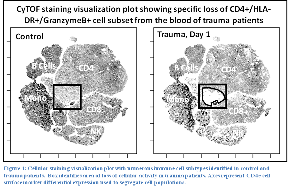
Results: Staining profiles of mononuclear cells using our lymphoid CyTOF panel revealed a specific loss of a unique subset of CD4 T cells that express MHC class II, GranzymeB, PD-1, and Perforin (Figure 1). Staining total leukocyte cells with a myeloid panel showed a complete loss of cells showing eosinophil/basophil staining phenotype and marked reduction in NK cells. As expected, neutrophils were dramatically increased in trauma patients. Other lymphoid populations were unchanged.

Conclusion: These preliminary results demonstrate the feasibility of CyTOF for large-scale profiling of circulating immune cells in trauma patients. Our pilot study of myeloid and lymphoid markers identified specific trauma-induced cellular phenotypic changes that are consistent with movement of certain immune cell types out the circulatory system and possibly towards affected tissues or organs.

04.03 Effect of Factor XIII in an ex vivo assay of hemostatic bandage adhesion

D. J. Zhou1,2, R. Spretz1,3, G. Larsen3,4, W. H. Velander4, M. A. Carlson1,2 1University Of Nebraska Medical Center,Department Of Surgery,Omaha, NE, USA 2VA Nebraska-Western Iowa Healthcare System,Omaha, NE, USA 3LNK Chemsolutions, LLC,Lincoln, NE, USA 4University Of Nebraska-Lincoln,Department Of Chemical & Biomolecular Engineering,Lincoln, NE, USA

Introduction:
Bandage adhesion to bleeding tissue in the setting of traumatic coagulopathy can be improved with fibrin glue (FG). The objective of this study was first to establish an ex vivo assay of bandage adhesion to liver and then to test our hypothesis that use of FG containing recombinant Factor XIII (rFXIII) would improve the adhesion strength (AS) of bandage glued to liver compared to FG without rFXIII.

Methods:
Customized FG (0.2 mL; 9 mg/mL plasma-derived fibrinogen, pdFI, + 106 U/mL r-thrombin + 0.36 mg/mL rFXIII) or commercial FG (0.2 mL Tisseel; Baxter), an FG that contains ~75 mg/mL pdFI and only trace pdFXIII, was applied to a 1×2 cm interface between custom electrospun polycaprolactone (PCL) mesh and a fresh porcine liver strip, and the interface was compressed with a 170 g weight for 5 min at 37°C (default setup). A T-peel adhesion test was performed with an Instron 5943 tensiometer with a 10 N load cell. Force vs. displacement data were used to calculate AS (N/cm), defined as average force during the peel divided by the interface width. AS data were compared with ANOVA (α<0.05) and unpaired t-tests (p<0.05).

Results:
Using the default setup, AS of custom FG was ~2-fold greater by gluing the PCL mesh to the capsular surface of the liver vs. raw parenchyma (Fig. 1A), so use of the liver capsule was incorporated into the default setup. Neither capsular surface wetness (patted dry vs. pre-wet with PBS) nor prolonged compression time (5 vs. 10 min) affected AS (Fig. 1B). There appeared to be decreased AS with lower temperature during compression (25 vs. 37?C), but this was not significant (Fig. 1B). Decreasing FG volume by 50% (0.05 vs. 0.1 mL/cm2) resulted in a lower AS (Fig. 1B). Increasing FG volume beyond 0.1 mL/cm2 was ineffective secondary to glue spillage during compression. Removing rFXIII from the default setup decreased AS by ~50%, but doubling the [rFXIII] did not increase AS (Fig. 1C). AS of customized FG vs. commercial FG was not different (Fig. 1C).

Conclusion:
An ex vivo adhesion test of synthetic resorbable mesh applied to porcine liver with customized FG was optimized with respect to liver surface qualities, adhesion compression time and temperature, and FG quantity. AS was augmented by rFXIII in the FG. The customized FG produced AS similar to that of commercial FG, despite the former having only ~1/8 the pdFI. The AS equivalence between these two FGs likely was a result of the added rFXIII to the customized FG, suggesting that efficacy testing of rFXIII addition to biologic hemostatic devices may be warranted.

31.08 Double Stranded RNA induced TLR-3 activation Drives inflammatory response in bacterial pneumonia

M. Deshmukh1, M. V. Suresh1, B. Thomas1, V. Dolgachev1, K. Kondapi1, D. Machado-aranda1, M. R. Hemmila1, K. Raghavendran1 1University Of Michigan,Surgery,Ann Arbor, MI, USA

Introduction: Lung contusion (LC) is an independent risk for the development of ARDS and Ventilator associated pneumonia. Our findings suggest that dsRNA released from injured cells drive the acute inflammatory response in LC through TLR3. These experiments were performed in order to evaluate the role of TLR3 in primary and secondary (following LC) Klebsiella pneumonia.

Methods: WT, TLR-3-/- mice were subjected to a lethal model of primary and secondary (concurrently and 6 hours following LC) insults with Klebsiella pneumonia. Survival and bacteriological analyses of the blood, lung was studied. DsRNA-TLR3 complex inhibitor was used to assess the effect on survival. Additional experiments were performed to evaluate the effect on lung injury and inflammation by cellular and cytokine analyses of the lung/Broncho-alveolar lavage. In separate experiments alveolar macrophages were isolated and markers of M1/M2 phenotype and apoptosis were assessed by Flow cytometry. Finally flow cytometry was performed to measure the amount of dsRNA staining (J2 ab) of the BAL cells. Standard ANOVA was used to measure statistical difference with an attributed significance of p<0.05.

Results:Survival and bacteriology. Compared to WT mice, TLR3 (-/-) mice that received the bacteria inoculation concurrently or 6 hours following LC had significantly lower mortality. The TLR3 (-/-) mice had reduced bacteria in the lungs with the TLR3 (-/-) mice with no bacterial colonies in the blood at 24 and 48 hours (WT-115-120 colonies), after bacterial challenge. The mortality in WT mice was partially reversed (50%) by the administration of dsRNA/TLR3 complex inhibitor.

The WT mice show increased lung injury and inflammation following lung contusion and pneumonia. Lung morphology of TLR3 (-/-) mice revealed reductions in lung inflammation following pneumonia. TLR3 (-/-) mice had reduced BAL albumin (permeability injury) and cytokine (IL1 β, KC) levels following LC + pneumonia and pneumonia alone. The levels of lung IL-1β and myeloperoxidase were lower in the TLR3 (-/-) mice subjected to LC + pneumonia and pneumonia alone group compared to corresponding WT .

The WT mice show increased M1 expression than TLR3 (-/-) with more of the M1 and M2 alveolar macrophage undergoing apoptosis following LC+pneumonia.

WT mice show increased staining than TLR3 (-/-) for dsRNA (necrosis) following LC+pneumonia

Conclusion:The interaction of dsRNA released from injured cells with TLR3 drives the acute inflammatory response following primary and secondary bacterial pneumonia. This finding has significant therapeutic implications. Agents to inhibit TLR3 have the potential to inhibit the inflammatory response following the primary insult (LC) as well the secondary bacterial infection.

31.09 Murine JAM-A-/- Causes Intestinal Hyperpermeability But Improves Survival Following Sepsis

N. J. Klingensmith1, Z. Liang1, J. D. Lyons1, C. M. Coopersmith1 1Emory University,Surgery/Critical Care,Atlanta, GA, USA

Introduction:

Sepsis has been shown to increase intestinal permeability and increase mortality. Intestinal epithelial cells and their associated tight junction proteins are crucial for regulating intestinal permeability. Junctional Adhesion Molecule A (JAM-A) is a protein found in the intercellular tight junction that has been implicated in regulating gut barrier function. Previous studies have shown that JAM-A-/- mice have increased gut mucosal permeability at baseline. However, how intestinal permeability and overall survival are altered in JAM-A deficient mice during sepsis has yet to be determined.

Methods:

6-8 week old male and female C57Bl/6 (WT) or JAM-A-/- mice underwent cecal ligation and puncture (CLP) to induce sepsis. Following sepsis, mice were followed for seven days for survival or sacrificed at 24 hours for functional studies. Intestinal permeability was assessed by quantifying serum fluorescent dextran (FD-4, 4.4kDa) following oral gavage 5 hours prior to sacrifice. Blood and peritoneal cultures were taken via sterile cardiac puncture and peritoneal lavage, respectively, and incubated on sheep blood agar plates for 48 hours before quantification. A Log-Rank test was performed for survival studies. For two-way comparisons, a Student’s t-test or Mann-Whitney test was performed depending on Gaussian distribution and a p-value of <0.05 was considered significant.

Results:

Intestinal permeability was increased in JAM-A-/- mice compared to WT mice (mean, 1031±516.3 ng/ml vs. 699±295.1 ng/ml, p=0.04, n=13-15). However, JAM-A-/- mice displayed a decrease in systemic bacterial burden compared to controls (log transform mean, 0.9731±1.819 CFU/ml vs. 2.751±2.013 CFU/ml, p=0.007, n=18-20) suggesting sepsis induced intestinal hyperpermeability does not correlate with greater bacteremia in JAM-A deficient mice. There were no statistical differences in peritoneal cultures in JAM-A-/- mice compared to controls (log transform mean, 3.495±2.069 CFU/ml vs. 4.646±2.305 CFU/ml, p=0.11, n=18-20). Importantly, JAM-A-/- mice had improved survival compared to WT mice (95% vs. 55%, p=0.003, n=20/group).

Conclusion:

Though JAM-A-/- mice have an increase in intestinal permeability during sepsis, they display less bacteremia and overall improvement in survival compared to WT mice. These opposing findings suggest that deleting JAM-A, while increasing intestinal permeability, alters other physiologic parameters which appear to predominate allowing for the improved survival.

31.10 Electroporation-mediated delivery of the FER Gene in the Resolution of Trauma-related Fatal Pneumonia

V. A. Dolgachev1, S. V. Madathilparambil1, B. Thomas1, M. R. Hemmila1, K. Raghavendran1, D. Machado-Aranda1 1University Of Michigan,Acute Care Surgery,Ann Arbor, MI, USA

Introduction: Lung Contusion (LC) and Bacterial Pneumonia (PNA) are common in the critically ill trauma patients and in combination can be fatal. The emergence of multidrug resistant organisms has mitigated the efficacy of many antibiotics. Recent genomic studies have shown FER, a cytosolic non-receptor tyrosine kinase of unknown role in the lung, with increased gene expression among survivors from sepsis and pneumonia. Our hypothesis is that using electroporation-mediated (EP) delivery of the FER gene we can improve survival in a murine model of combined LC and PNA.

Methods: C57Bl/6 mice were subject to a lethal model of unilateral LC followed by intratracheal inoculation of 500 CFU of Klebsiella sp. at 6 hrs. In the treatment group, plasmid DNA encoding FER (pFER) was provided via hypopharyngeal instillation right after LC injury (time 0.2 hr). External electrodes were placed under each forelimb and 8 square wave electroporation pulses were delivered. Two control groups were used: one receiving Na+/K+-ATPase genes (shown to be benefitial in LC injury model) in similar LC+PNA injury conditions followed by EP (pPump); and a second with no gene delivery (LC + PNA/no EP). 7-day survival curves were recorded. In a parallel experiment, animals were euthanized at 24 hrs post-LC, lung mechanics were measured and bronchial alveolar lavage fluid (BAL), blood and lung tissue were collected. BAL cellular subpopulations were immunotypified by flow cytometry. BAL cytokines were assayed using ELISA kits for IL-1α, IL-6, IL-10, IL-33, MCP-1 and MIP-2. Quantitative bacterial counts were determined by serial dilution and plating of blood and lung homogenates.

Results: 7-day survival was markedly improved by pFER treatment compared to positive (pPump) and negative-controls (LC+PNA/no EP). Better lung compliance was found in pFER animals at 24 hr post-LC. At this same timepoint, pFER treatment group showed a robust inflammatory response with absolute increases in BAL monocytes, neutrophils and macrophages. In contrast, BAL of pPump treated animals was macrophage-predominant while negative-controls had a relative lower total of inflammatory cells, especially of macrophages, compared to the other groups. pFER treatment diminished levels of early response pro-inflammatory cytokines IL-1β, IL-6, MCP-1 and MIP-2, whereas increases were seen in modulation-cytokines IL-10, IL-33 and TNF-α. Lung bacterial counts were significantly lower in pFER (ANOVA p=0.023) with a trend towards better systemic containment.

Conclusion: EP-mediated delivery of the FER gene significantly improved mouse survival in a combination model of lung contusion and pneumonia. Better containment of bacteria within the lungs was possibly mediated by alterations in the immune cell and cytokine response.

31.05 Plasma from Coagulopathic Trauma Patients Inhibits Donor Platelets: A Functional Proteomic Analysis

M. P. Chapman1,2, H. B. Moore1,2, E. Gonzalez1,2, A. P. Morton1,2, C. D. Fleming1,2, K. C. Hansen1, C. C. Silliman1,3,4, A. Sauaia1,2, A. Banerjee1, E. E. Moore1,2 1University Of Colorado Denver,Surgery,Aurora, CO, USA 2Denver Health Medical Center,Surgery,Aurora, CO, USA 3Children’s Hospital Colorado,Haematology,Aurora, CO, USA 4Bonfils Blood Center,Denver, CO, USA

Introduction: To answer the question of when to transfuse platelets to coagulopathic trauma patients with platelet dysfunction, it is ultimately necessary to answer the question of whether the observed platelet dysfunction is intrinsic to the platelets, or is extrinisic, i.e.: a function of a toxic circulatory milieu. We therefore endeavored to test a hypothesis analogous to the familiar Koch’s third postulate, wherein the causal agent of disease will transfer that disease state to a healthy organism. In our case, the causal agent in question was not infectious, but rather was plasma from coagulopathic trauma patients with platelet dysfunction. We hypothesized that plasma from coagulopathic trauma patients would cause inhibition of healthy donor platelets, and that this toxic effect would be associated with a shift in the plasma proteome.

Methods: We isolated platelet free plasma from 171 trauma activation patients, of which 8 were severely coagulopathic with severe platelet dysfunction. This plasma was mixed 1:1 with whole blood from healthy donors. Comparison was made to plasma from non-coagulopathic trauma patients and healthy donors (control). Platelet inhibition was measured using whole blood impedance aggregometry with TRAP and ADP agonists (ROTEM Platelet, Tem GmbH). Proteomic analysis of plasma samples was conducted using the QconCat platform (PolyQuant, GmbH).

Results: Plasma from severely coagulopathic trauma patients with platelet dysfunction, conveyed significant inhibition in platelet function to donor platelets (p=0.049); see figure. Plasma from non-coagulopathic trauma patients did not significantly decrease healthy donor platelet function, compared to exposure to self plasma. A subset of plasma samples, which inhibited healthy donor platelets ≥50% (p=0.0025), demonstrated significant proteomic shifts, including massive upregulation of alpha-enolase , GAPDH, fibronectin, von Willebrand factor, and alcohol dehydrogenase 1B, along with loss of coagulation factor V.

Conclusion: Plasma from coagulopathic trauma patients with platelet dysfunction is capable of conveying this dysfunction to healthy donor platelets. This pathogenic transfer is likely the result of a soluble toxic factor, as it is associated with an upregulatory proteomic shift, that is not buffered by healthy whole blood. These findings have two implications: (1) that it is critical to correct the platelet-inhibitory circulatory milieu of a coagulopathic trauma patient (e.g. with plasma resuscitation) before transfusing patients and (2) that the unidentified toxic soluble factor may represent a drug target for preserving platelet function in trauma patients.

31.06 Fibrinolysis Shutdown Initiated Through Inflammatory Degradation of Plasminogen

H. B. Moore1,2, E. Gonzalez1,2, A. P. Morton1,2, M. P. Chapman1, A. L. Slaughter1,2, C. C. Silliman1,3, K. C. Hansen1, E. E. Moore1,2 1University Of Colorado Denver,Surgery,Aurora, CO, USA 2Denver Health Medical Center,Surgery,Denver, CO, USA 3Bonfils Blood Center,Hematology,Denver, C), USA

Introduction: Plasminogen (PLG) is a complex multi-domain protein with diverse functions. PLG is cleaved by tissue plasminogen activator (tPA)and yields plasmin, which degrages fibrin. Neutrophil proteases, such as elastase, can also modify PLG and produce angiostatin, which retains the fibrin-binding site of plasmin without the proteolytic domain. Previous animal work has demonstrated that hemorrhagic shock increases fibrinolytic activity while tissue injury reduces fibrinolytic activity while both causing a depletion of PLG. We hypothesize trauma patients with hyperfibrinolysis and fibrinolysis shutdown will have decreased PLG compared to trauma patients with normal fibrinolytic activity and healthy controls, and that angiostatin is a potent inhibitor of fibrinolysis.

Methods: 150 trauma activations were screened and, patients(n=10) with the lowest fibrinolytic activity(shutdown) were contrasted patients with the highest fibrinlytic activity(hyperfibrinolysis n=10), normal fibrinolytic activity(physiologic n=10) and healthy controls(n=10). PLG levels and firbrinolytic regulator proteins were measured in each of these cohorts. Exogenous angiostatin and elastase were mixed ex vivo in healthy volunteer blood (n=6) and challenged with tPA to promote fibrinolysis. Fibrinolysis was quantified by the LY30 variable using thrombelastography (TEG).

Results:PLG levels were decreased in shutdown and hyperfibrinolytic patients compared to trauma patients with physiologic activity and healthy controls (p<0.001). Fibriolytic regulator protein were also depleted in these phenotypes (figure1). Exogenous angiostatin reduced LY30 by 62% (p=0.046) in tPA challenged blood of healthy volunteers. Elastase also reduced LY30 by 96% (p=0.046). Additional clotting parameters were not effected by the addition of angiostatin in whole blood with and without tPA. However, elastase reduced R time by 30% (p=0.028) with and without tPA (-34% p=0.028) and increased angle by 33% with tPA (p=0.075).

Conclusion: Depletion of plasminogen post injury is associated with the hyperfibrinolytic and fibrinolysis shutdown phenotype. The breakdown of PLG may be the result of different mechanisms. Exogenous elastase promotes a hypercoaguable clot that is resistant to fibrinolysis, which is a common hemostatic derangement of patients in the surgical intensive care unit. These data support inflammation initiates an alternative utilization of PLG promoting post injury fibrinolysis resistance despite depletion of other fibrinolytic regulatory proteins.

31.07 Supercomputing Sepsis Simulations for In Silico Outcome Prediction

R. C. Cockrell1, G. An1 1University Of Chicago,Surgery,Chicago, IL, USA

Introduction: Current predictive models for sepsis generally use correlative methods, and as such are limited in their individual precision due to patient heterogeneity and data sparseness. In particular, the inability of these methods to capture the clinical dynamics and mechanisms of sepsis limits achieving true prediction. Other fields of science have used computational modeling and simulation to help contextualize multi-dimensional data arising from complex systems in order to describe their behavior. Advances in supercomputer-aided modeling can provide this same capability to biomedical research. Herein we begin this process by defining the behavioral possibility space for sepsis across a range of physiological, microbial and environmental parameters, and apply advanced mathematical analysis to identify predictive metrics for system outcome.

Methods: 80 million microbial sepsis patients were simulated representing a 28-day hospital course using a previously validated agent-based model (ABM) of sepsis implemented on a Cray XE6 supercomputer. Parameter space was examined regarding system response with and without antibiotics regarding the following parameters: cardio-respiratory-metabolic resilience; two properties of microbial virulence, invasiveness and toxigenesis; and degree of contamination from the environment. Simulation data was analyzed using varied tiers of multi-dimensional space, which was then subjected to mathematical analysis using deterministic chaos and bifurcation theory.

Results: The identified parameter space had a clear structure with plausible boundaries reflecting the range of possible human behavior in response to microbial sepsis (Fig 1). The central region of highest outcome uncertainty corresponds to the critically ill population. Analysis of this region using chaos and bifurcation theory identified basins of attraction leading to three outcomes – complete recovery, hyperinflammatory system failure, and overwhelming infection – in a 32-dimensional phase space defined by various chemokine concentrations and their conjugate generalized momenta. These basins of attraction were used to develop metrics for prediction of individual outcomes based on networks of mediator and cell population levels.

Conclusion: Supercomputing simulations of sepsis can play a vital role in the contextualization of both big data output and mechanistic basic science research. The representation of the aggregate set of individual trajectories facilitates formal mathematical analysis and is a necessary step towards developing truly predictive models and precision therapies.

31.02 Hemorrhagic Shock Induces Pulmonary Metabolopathy Mediated by Gut Ischemia

A. W. Bacon1, A. D’Alessandro1, A. L. Slaughter1, H. B. Moore1, M. Fragoso1, C. Silliman1,3, A. Banerjee1, E. E. Moore1,2 1University Of Colorado Denver,Dept Of Surgery,Aurora, CO, USA 2Denver Health Medical Center,Dept Of Surgery,Aurora, CO, USA 3Children’s Hospital Colorado,Aurora, CO, USA

Introduction: Post-shock mesenteric lymph (PSML) is a known mediator of the development of acute lung injury (ALI). We have previously shown that PSML contains a distinct proteome from that of plasma whose constituents may potentiate the development of ALI. However, recent advances in mass-spectrometry-based biochemical analysis have prompted ongoing documentation and quantification of the plasma metabolome following life-threatening injury. However, the PSML metabolome and its pulmonary sequela remain unexamined. Thus, we hypothesized that PSML contains a specific and distinct biochemical millieu that may potentiate the development of ALI.

Methods: Male Sprague-Dawley rats (n=4 per group) underwent tracheostomy, femoral artery cannulation and laparotomy with mesenteric duct cannulation followed by profound hemorrhagic shock (MAP = 27 ± 2 mmHg for 30 min) followed by partial resuscitation (MAP > 60 mmHg for 30 min). Pre- and post-shock plasma and lymph and bronchoalveolar lavage fluid (BALF) were collected for analysis. Control and sham shock (T/SS) animals were employed. Select metabolites were assayed via ultra-high performance liquid chromatography-mass spectrometry. Results are expressed as means ± SEM and compared to controls via Dunnett's test (p < 0.05 threshold).

Results: Trauma alone had no significant effect on the systemic metabolome. Trauma and hemorrhagic shock (T/HS) resulted in fluid-specific hyperglycemia, accumulation of lactate, succinate and malate, oxidized and reduced glutathione and uric acid when compared to baseline controls. T/HS with mesenteric lymph diversion (MLD) revealed a 5-fold increase in BALF succinate and 4-fold increase in BALF urate when compared to the T/HS cohort (Fig 1).

Conclusion: Trauma and hemorrhagic shock results in systemic metabolic aberration. Post-shock plasma and PSML exhibit a qualitatively and quantitatively specific pathologic metabolic profile that ultimately perturbs pulmonary metabolism. These changes were consistent with hypoxemic metabolism in the ischemic gut, including: hyperglycemia, accumulation of anaerobic and key TCA-cycle metabolites, dysregulated redox homeostasis and nitrogen catabolism suggesting hypoxic uncoupling of the electron transport chain, accumulation of reactive oxygen species (ROS) and anapleurosis. Interestingly, MLD had a profound and unexpected effect on BALF succinate, a known IL-1ß activator, and urate, a potential ROS scavenger, suggesting PSML-associated ALI may result from inhibition of the potentially beneficial immune effects of these small molecular messengers. As such, additional study is warranted to elucidate the mechanisms responsible for post-shock metabolopathy and its role in the development of ALI.

31.03 Gender Differences in Murine Renal Ischemia-Reperfusion Injury Are Mutable with Hormonal Therapy

D. D. Aufhauser1, D. R. Murken1, Z. Wang1, G. Ge1, T. Bhatti4, W. W. Hancock3,4, M. H. Levine1,2 1University Of Pennsylvania,Surgery,Philadelphia, PA, USA 2Children’s Hospital Of Philadelphia,Surgery,Philadelphia, PA, USA 3University Of Pennsylvania,Pathology And Laboratory Medicine,Philadelphia, PA, USA 4Children’s Hospital Of Philadelphia,Pathology And Laboratory Medicine,Philadelphia, PA, USA

Introduction: Renal ischemia-reperfusion injury (IRI) commonly complicates cardiac, vascular, trauma, urologic and transplant surgery. Prior studies described superior renal IRI tolerance in female rodents, but translational applications of hormonal therapy to prevent renal complications in major surgery are unexplored.

Methods: Mice underwent standardized 28-min warm IRI with clamping of the left renal pedicle and contralateral nephrectomy. BUN was measured daily for 4 days following injury and kidneys were collected for histology after 28 days.

Results: Females treated with 2 doses of β -estradiol at 16- and 1-hr pre-IRI (n=5) had lower serum BUN levels following IRI compared to vehicle treated controls (n=5, p<0.01), and less fibrosis on Sirius Red staining (p<0.01). Hormonally intact male mice subjected to 28 min of IRI all died of renal failure by 48 hr in vehicle (n=5) or estrogen (n=5) treated groups (Fig. 1, *). Male mice subjected to orchiectomy one wk prior to ischemia exhibited improved renal IRI tolerance and universal survival (n=5, Figure 1). Neutered male mice treated with estrogen (n=5) demonstrated further improvement in post-IRI renal function compared to vehicle treated neutered mice (n=5, p=0.03, Figure 1).

Conclusion: Gender-specific protection from renal IRI can arise from the presence of estrogens or the absence of androgens. Administration of β -estradiol beginning at 16-hrs pre-injury mitigates renal IRI in female and manipulated mice. This time course is compatible with perioperative use in procedures anticipated to involve a period of renal ischemia. While further investigations are warranted to test whether these exciting data can translate to beneficial outcomes in larger animals, we conclude that hitherto unrecognized effects of estrogens on IRI have profound consequences for renal function post-surgery.

31.04 Amitriptyline Treatment Improves Survival After Trauma and Hemorrhage

H. He1,2, P. L. Jernigan1,2, R. S. Hoehn1,2, A. L. Chang1,2, L. Friend1,2, R. Veile1,2, T. Johannigman1,2, A. T. Makely1,2, M. D. Goodman1,2, T. A. Pritts1,2 1University Of Cincinnati,Trauma And Critical Care,Cincinnati, OH, USA 2University Of Cincinnati,Institute For Military Medicine,Cincinnati, OH, USA

Introduction: Hemorrhagic shock is the leading cause of potentially preventable death after trauma. The optimal treatment for hemorrhagic shock is to reverse circulatory losses, metabolic acidosis, and cellular hypoxia with blood products, but these are often not available for immediate administration. Alternative approaches to initial resuscitation are needed, especially in resource poor environments. Amitriptyline is a serotonin-norepinephrine reuptake inhibitor with anti-inflammatory effects. We have previously demonstrated that treatment of blood products with amitriptyline leads to decreased lung injury after hemorrhage, but the direct effect of amitriptyline in treatment of hemorrhage is unknown. We hypothesized that administration of amitriptyline after trauma and hemorrhage would improve survival in a non-resuscitation injury model.

Methods: Healthy C57/BL6 male mice underwent laparotomy to induce tissue trauma and hemorrhage via femoral artery cannulation to a mean arterial pressure of 25±5mmHg for 60 minutes. After laparotomy closure and decannulation, mice received amitriptyline (0.1 mg/kg) or an equivalent volume of vehicle (50 uL of normal saline) via intraperitoneal injection. For survival analysis, mice underwent the above treatment (n=10/group) and were monitored for 24 hours. For hemodynamic and blood chemistry analysis, mice (n=5/group) underwent the same injuries and treatments as described above and were sacrificed 60 minutes after treatment.

Results: Administration of amitriptyline after trauma and hemorrhage significantly increased 24-hour survival in mice (Figure 1; 70% survival with amitriptyline vs 0% with vehicle; p<0.001). Sixty minutes after trauma and hemorrhage, mice treated with amitriptyline also had significantly increased mean arterial blood pressure (p=0.04). Blood gas analysis revealed that mice treated with amitriptyline had statistically significant improvements in base deficit and serum bicarbonate, consistent with decreased metabolic acidosis.

Conclusion: Our results demonstrate that administration of amitriptyline significantly improves survival in mice after trauma and hemorrhage, even in the absence of resuscitation. We found that amitriptyline increases mean arterial blood pressure and decreases metabolic acidosis after injury. Administration of amitriptyline following traumatic injury could potentially be useful in situations with limited access to blood products.

31.01 Modulating TLR-4 mucosal expression via postconditioning the small bowel after mesenteric occlusion

O. Rosero1, P. Onody1, T. Kovacs1, A. Fulop1, D. Molnar2, T. Balazsa2, G. Lotz3, L. Harsanyi1, A. Szijarto1 1Semmelweis University,1st Department Of Surgery,Budapest, , Hungary 2Semmelweis University,Department Of Human Morphology And Developmental Biology,Budapest, , Hungary 3Semmelweis University,2nd Department Of Pathology,Budapest, , Hungary

Introduction: Postconditioning is a suitable method to reduce ischemia-reperfusion (IR) induced injury of the intestine following mesenteric arterial occlusion. Toll-like-receptor (TLR)-4 was shown to be involved in the pathophysiology of organ damage after IR, therefore the aim of our study was to investigate the effect of postconditioning on the mucosal expression of TLR-4.

Methods: Male Wistar rats (n=10/group) underwent 60 minutes of superior mesenteric artery (SMA) occlusion followed by 6 hours of reperfusion in three groups: sham-operated, IR- and postconditioned-group. Postconditioning was performed by 6 alternating cycles of 10 seconds reperfusion/reocclusion. Blood and tissue samples were collected at the end of reperfusion. Small intestinal histolopathological changes, mucosal cleaved caspase-3, antioxidant status, HMGB-1 and TLR-4 expression rates were assessed. Mucosal and serum IL-6 and TNF-α levels were measured.

Results: Milder histological alterations of the postconditioned-group were associated with significantly (p<0.01) decreased number of caspase-3 positive cells, lower TLR-4 mRNA and significantly (p<0.05) minor amount of HMGB-1 and TLR-4 protein expression in the intestinal villi compared to the IR-group samples. Furthermore, significantly improved antioxidant state of intestinal mucosa and lower mucosal and serum IL-6, TNF-α levels were detected in the postconditioned-group.

Conclusion: Small intestinal IR-injury caused by the occlusion of the SMA was substantially ameliorated by the use of postconditioning, delivering a more favorable inflammatory response, which may partly be attributed to the decreased mucosal expression of TLR-4.

30.07 Direct Peritoneal Resuscitation Lowers sICAM-1 and Lung Integrin-alphaL in Acute Brain Death in Rats

L. H. Taylor1, P. J. Matheson1,2, C. A. Ghazi1, J. L. Weaver1, C. D. Downard1,2, R. N. Garrison1,2, J. W. Smith1 1University Of Louisville,Department Of Surgery,Louisville, KENTUCKY, USA 2Robley Rex VAMC,Department Of Surgery,Louisville, KENTUCKY, USA

Introduction: ~~Deficient resuscitation in brain dead organ donors causes pulmonary cellular necrosis, leukocyte infiltration, organ edema, organ dysfunction, and organ failure. A new method of resuscitation, Direct Peritoneal Resuscitation (DPR), improves gut and hepatic perfusion, prevents vital organ edema, and down-regulates systemic inflammation, and thus, prevents gut-derived alarmins such as HMGB1 or LPS in the systemic circulation. Previous studies showed that DPR increased organs transplanted per donor (OTPD) due to increased suitability of lungs in human organ donors. We hypothesized that DPR benefits in gut and liver could improve lung function, in particular, decrease sICAM-1, as well as integrin-?L (i.e., CD11a or LFA-1) and integrin-?M (i.e., CD11b or MAC-1).

Methods: ~~Anesthetized male Sprague-Dawley rats (n=44 total) were randomized to groups: 1) ABD with minimal IV RES (Low IVF); 2) ABD + mid-range IV RES (Mid IVF); 3) ABD with aggressive IV RES (High IVF); or 4) ABD + IV RES + DPR (IVF+DPR). ABD was induced by inflation (1 mL/hr) of a supra-dural balloon catheter (4F angioplasty) until the loss of blink and withdrawal reflexes. Ventilation was started (TV=1.6mL, rate=80) with the onset of apnea. After the appearance of the characteristic Cushing response, IV fluid RES was started and mean arterial blood pressure (MAP) and heart rate (HR) were recorded. Serum and lung tissue were collected, snap frozen in liquid nitrogen, and stored until assays. Lung tissue was homogenized in ice cold PBS with protease and phosphatase inhibitors and ELISAs were performed.

Results: ~~DPR decreased soluble ICAM-1 levels compared to Low IVF or Mid IVF (see figure). Integrin-?L levels were decreased compared to Low IVF, Mid IVF, or High IVF alone groups, but integrin-?M levels were not different among groups.

Conclusion: ~~These data suggest that DPR prevents or minimizes alarmin signals from the gut and liver to the lung, and thus, decreases leukocyte extravasation in this slow onset model of acute brain death. This might partially explain the beneficial effects of DPR to maintain pulmonary function and prevent organ injury, which might improve suitability of lungs for organ transplantation in organ donors.

30.04 Defatting Livers for Transplantation Using Normothermic Ex Vivo Perfusion

M. Wallace1, J. MacLean1, M. Thornbury3, A. Venkatachalam1, Q. Hu2, N. Ridgway4,5, I. Alwayn1,2,3 1Dalhousie University,Surgery,Halifax, Nova Scotia, Canada 2Dalhousie University,Pathology,Halifax, Nova Scotia, Canada 3Dalhousie University,Microbiology & Immunology,Halifax, Nova Scotia, Canada 4Dalhousie University,Biochemistry & Molecular Biology,Halifax, Nova Scotia, Canada 5Dalhousie University,Pediatrics,Halifax, Nova Scotia, Canada

Introduction:
The number of patients on the waiting list for a liver transplant is increasing, but unfortunately the availability of donor livers does not meet the demand. With rising obesity rates in North America, many livers are discarded because of steatosis, the accumulation of fat within the liver. These extended criteria donor organs are more susceptible to ischemia-reperfusion injury (IRI) and are associated with reduced viability and increased complication rates after transplantation. Normothermic ex vivo liver perfusion (NEVLP) has emerged as a means of reducing the IRI associated with cold static storage of livers. NEVLP may also play a role in actively defatting steatotic livers and thus protect them from IRI. In this pilot study, we hypothesize that four hours of oxygenated NEVLP will preserve the hepatic architecture of steatotic rat livers, and that supplementing the perfusate with defatting compounds will result in a reduction of hepatocellular lipid content.

Methods:
Livers were harvested from obese Zucker rats or lean Wistar rats and perfused ex vivo at 37°C for four hours. NEVLP consisted of membrane oxygenated machine perfusion with a flow rate of 10 mL/min and an initial portal vein pressure of 10 mm Hg. Livers were perfused with Minimum Essential Medium Alpha Modification medium. The perfusate was supplemented with bovine serum albumin, fetal bovine serum, human insulin, lactic acid, and heparin at pH 7.4 with or without the addition of four compounds known to stimulate lipid metabolism by consuming or secreting intracellular triglycerides. Liver tissue biopsies were collected after perfusion for hematoxylin and eosin (H&E) and Oil Red O staining, and triglyceride (TAG) measurement. Perfusate samples were collected throughout NEVLP for quantification of TAG and aspartate aminotransferase (AST), a marker of hepatocyte injury.

Results:
H&E results suggest that the viability of steatotic livers can be maintained using NEVLP under physiological conditions. Steatotic livers are more susceptible to hepatocyte injury during ex vivo perfusion than non-fatty livers. The defatting agents may provide some protection from this injury as indicated by lower AST levels. Perfusion of steatotic livers with the defatting agents results in a reduction of intracellular lipid droplets as shown by Oil Red O staining. However, there is no associated increase in perfusate TAG levels, suggesting that defatting may be occurring by mechanisms other than lipid secretion such as hydrolysis for ketogenesis or beta-oxidation.

Conclusion:
We have shown that four hours of oxygenated NEVLP successfully preserves liver architecture. Supplementing the perfusate with compounds that promote metabolism through export or oxidation may result in defatting of steatotic livers. Defatting steatotic livers could improve transplantation success and increase the donor pool.

30.05 Endogenous Innate Immune Activation by Free Mitochondria

K. P. Samy1, L. Lin1, F. Feng1, B. Parker1, Y. Yang2, A. D. Kirk1, T. V. Brennan1 1Duke University Medical Center,Department Of Surgery,Durham, NC, USA 2Duke University Medical Center,Departments Of Medicine And Immunology,Durham, NC, USA

Introduction: Tissue injury during surgery or trauma can expose free mitochondria to surrounding tissues. Mitochondria are unique organelles that evolved from a bacterial origin and have retained immunologic epitopes that activate the innate immune system similar to bacterial pathogens. We examined the effect of mitochondria as endogenous innate immune activators ex vivo and in vivo.

Methods: Mitochondria isolated from mouse fibroblast (LMTK) lysate was performed by differential centrifugation methods followed by affinity purification using TOM22 specific antibody coated magnetic beads. Purity of mitochondrial fractions was confirmed by western blot and qPCR for mitochondrial specific proteins and DNA, respectively. Cultured dendritic cell (DC2.4) and macrophage (RAW 264.7) cell lines were incubated with increasing concentrations of mitochondria and assessed for upregulation of costimulatory molecules (CD40, CD80, and CD86) by flow cytometry. Cell culture supernatant was assessed for inflammatory cytokines (TNF-alpha, IFN-gamma, and MCP-1) by cytometric bead assay. In vivo assay of the inflammatory response to mitochondria was performed by injecting mitochondria derived from LMTK cells or rho zero (mitochondrial DNA-deficient) LMTK cells into WT or MyD88-ko C57BL/6 mice and enumerating neutrophil and monocytes amongst peritoneal exudate cells (PECs).

Results: Exposure of macrophage and dendritic cells to highly purified mitochondria resulted in increased expression of costimulatory molecules and increased inflammatory cytokine production in both cell lines. Intraperitoneal injection of mitochondria led to robust PEC accumulation consisting of activated neutrophils and inflammatory monocytes. The PEC inflammatory response was greatly reduced in MyD88-ko mice compared to WT. PECs were also diminished when mitochondria lacking mitochondrial-DNA (mtDNA) were injected.

Conclusion: These data support a role for mitochondria in the inflammatory response to tissue injury. By serving as endogenous innate immune activators, mitochondria can activate antigen-presenting cells and provoke the infiltration of neutrophils and monocytes. The components of mitochondria that are key to these processes remains to be determined, but our data suggests they are dependent on mitochondrial DNA, and/or the products of mitochondrial gene expression.

30.06 Donor-derived intravascular TLR-dependent Ly6Clow monocytes lead primary lung allograft dysfunction

Z. Zheng1, S. Chiu1, H. Sun1, Z. J. Zhang1, M. M. DeCamp1, D. Kreisel2, S. Budinger1, H. Perlman1, A. Misharin1, A. Bharat1 1Northwestern University,Surgery/ Thoracic,Chicago, IL, USA 2Washington University,St. Louis, MO, USA

Introduction: Primary graft dysfunction (PGD) affects over 50% of lung recipients and remains the predominant cause of short- and long-term mortality. Neutrophil infiltration into the allograft mediates PGD. However, the mechanisms that lead to neutrophil trafficking remain unknown. Here we demonstrate that a unique subset of intravascular monocytes, that cannot be flushed using current preservative techniques, leads to PGD following lung transplantation.

Methods: Murine allogeneic left lung transplant was used for these studies. Neutrophil infiltration was determined using intravital 2-photon microscopy and flow-cytometry. Graft function was determined using arterial oxygen after clamping the right hilum for 5 minutes. Intravenous clodronate loaded liposomes (Clo-lip) was used to induce apoptosis in circulating monocytes.

Results: Perfused murine lungs demonstrated intravascular CD11b+MHCIICD64+/-Ly6Clow monocytes (Ly6ClowM) that constituted ~3% of resident CD45+ cells. These Ly6ClowM shared functional phenotype with CD14+CD16+ monocytes detected in perfused human lungs. Pre-treatment of donors with Clo-lip depleted Ly6ClowM in donor lungs (<0.3%). Following murine allogeneic left lung transplant, there was a robust neutrophil infiltration on intravital 2-photon imaging (Fig 1A) that was prevented by a single dose of Clo-lip in the donors. Donor Clo-lip treatment resulted in preserved allograft architecture and oxygenation at 24 hours (Fig 1B&C). Treatment with anti-CCR2 antibodies which selectively deplete Ly6Chigh monocytes, but not Ly6ClowM, worsened neutrophilia (3 folds) and graft function. Homozygous deletion of CX3CR1 chemokine receptor resulted in absent Ly6ClowM in the lungs without affecting circulating Ly6ClowM, indicating a role of CX3CR1 binding to its ligand CX3CL1 expressed on endothelium. CX3CR1/ donor lungs demonstrated suppressed neutrophilia (2.5 fold reduction) and preserved function. The CX3CR1-CX3CL1 covalent bonds were calcium dependent and were neutralized by ethylene glycol tetraacetic acid (EGTA) resulting in flushing of Ly6ClowM during perfusion. Reconstitution of Clo-lip treated donor lungs with MyD88-TRIFF knockout Ly6ClowM, with deficient Toll signaling, failed to restore allograft neutrophilia, in contast to wild-type Ly6ClowM.

Conclusion: Pulmonary intravascular Ly6ClowM are a novel subset of monocytes that cannot be flushed using standard preservative techniques. Donor Ly6ClowM, transplanted along with the donor lungs, are activated through TLR pathway mediating neutrophil trafficking and PGD. Ly6ClowM are bound to the donor endothelium using covalent bonds that can be neutralized using a calcium chelator. Depletion of these cells in the donor prevented PGD following lung transplantation.

30.01 NK1.1+ Cells May Play A Role in B Cell-Dependent Transplant Tolerance

H. Deirawan1, W. Liu1, L. Liu1, R. Stott1, K. M. Lee1, J. I. Kim1, J. F. Markmann1 1Massachusetts General Hospital,Division Of Transplant Surgery,Boston, MA, USA

Introduction:
CD1d presents lipid antigen to a subset of NKT cells and is widely used as a marker of regulatory B cells. NK1.1+ cells, which includes natural killer (NK) and Natural Killer T(NKT) cells, play a critical role in graft rejection. Depletion of NK1.1+ cells has been demonstrated to abrogate graft survival of islet, corneal, and cardiac allografts. We explored the possibility of a B cell-NK/NKT interaction, with the goal of determining whether NK/NKT cells played a role in a mouse model of regulatory B cell-dependent tolerance.

Methods:

Diabetes was induced in C57BL/6 mice with streptozotocin. In some experiments, CD1d-/- mice, and Beige mice were used. Four to five hundred islets were transplanted into the renal sub-capsular space of diabetic recipients. Recipient mice received 100 µg anti-mouse CD45RB i.p. on days 0, 1, 3, 5, and 7 following transplantation and 500 µg anti-mouse TIM-1 i.p. on day -1, and 300 µg on days 0 and 5 following transplantation. Recipients may also receive 200 mg anti-NK1.1 antibody on days -7, +1, and +8. Regulatory B cells were purified from long-term graft survival animals (>100 days) for adoptive transfer. Adoptive transfer was performed by intravenous injection of 5×10^6 B cells.

Graft survival between experimental groups was compared using Kaplan-Meier survival curves and Wilcoxon statistics. P values less than 0.05 were considered statistically significant.

Results:

Anti-CD45RB/anti-TIM-1 antibody treatment results in nearly 100% percent long-term graft survival, and reduces the percentage of NK cells but increases the percentage of NKT cells. The additional administration of anti-NK1.1 abrogates tolerance induction, but did not alter the percentage of TIM-1+ B cells nor the percentage of Foxp3+ T cells. To examine whether anti-NK1.1 antibody interferes with the development of Bregs vs. their effector function, we took advantage of the fact that untreated B cell deficient mice reject islet allografts, but the addition of B cells from tolerant recipients induces tolerance without any antibody treatment to the secondary recipients. In this model, anti-NK1.1 antibody abrogates tolerance induction by established Bregs.

To further delineate the subset of cells necessary for Bregs function. NKT cells deficient mice (CD1d-/-), and Beige mice (functional NK cells deficiency) were transplanted, dual antibody treated and monitored for outcome. In contrast to wild type mice, all Beige mice acutely reject their grafts. Only 30% of NKT deficient mice fail to accept their graft long term.

Conclusion:

Our adoptive transfer study demonstrates that the presence of recipient NK1.1+ cells is necessary for graft survival mediated by Bregs. Rejection as a result of the loss of NK1.1+ cells does not result from a loss of Bregs or of Tregs. We continue to explore the interaction between subsets of NK1.1+ cells and Bregs that may be necessary for transplant tolerance induction.

30.02 Necroptosis Plays a Role in Liver Transplant Ischemia/Reperfusion Injury in IRF-1 Dependent Manner

J. Goswami1, R. How1, Q. Du2, S. Kimura2, D. Geller1,2 1University Of Pittsburgh Medical Center,Department Of Surgery,Pittsburgh, PENNSYLVANIA, USA 2Thomas E. Starzl Transplantation Institute,Department Of Surgery, University Of Pittsburgh School Of Medicine,Pittsburgh, PENNSYLVANIA, USA

Introduction: Necroptosis is a highly regulated form of cell death that is thought to be triggered by TNFα when caspase-8 is inhibited. Unlike apoptosis, necroptosis does not lead to fragmentation of DNA. Necroptosis is thought to be dependent on a complex involving receptor-interacting protein 1 kinase (RIPK1), RIPK3, caspase 8, and FADD. RIPK3 is involved in necroptosis while RIPK1 is involved in both apoptosis and necroptosis. Previously, we have shown that interferon regulatory factor 1 (IRF-1) contributes to liver transplant I/R injury by increasing apoptosis. However, the role of IRF-1 in necroptosis has not been examined. Our goal in this study is to determine if there is a role of necroptosis in liver transplant I/R injury and to characterize the role of IRF-1 in necroptosis.

Methods: Orthotopic murine liver transplant (OLTx) was performed with B6 wild-type (WT) or IRF-1 knockout (KO) donor livers to isograft B6 recipients. Cold ischemia time was 24 hr. Serum was collected at 1, 3, and 6 hr after liver transplant reperfusion for serum transaminases to assess liver injury. Protein was isolated and western blot performed for RIPK1and RIPK3.

Results: OLTx using IRF-1 KO donor liver isografts resulted in markedly diminished liver I/R injury compared to WT as we have previously shown (graph). Hepatic RIPK3 protein expression was induced 1 to 3 hr after OLTx and returned to baseline by 6 hr (figure). RIPK3 expression was not observed in isografts that received IRF-1 KO donor livers. Interestingly, the necroptotic form of RIPK1 (78 kDa protein) was mildly increased after OLTx in an IRF-1 independent manner, while the apoptotic form of RIPK1 (40 kDa protein) was not induced.

Conclusion: These findings demonstrate activation of the necroptosis pathway with rapid induction of RIPK3 protein expression during OLTx I/R injury. Moreover, we have identified that induction of hepatic RIPK3 in the liver transplant setting is IRF-1-dependent. Further studies are needed to better define the mechanism of IRF-1 mediated induction of RIPK3 and necroptosis. Inhibition of the necroptotic pathway by blockade of RIPK proteins may be a novel strategy to ameliorate liver transplant I/R injury.

30.03 Donor-derived Intravascular Monocytes Lead to Native Lung Neutrophilia During Single Lung Transplant

S. Chiu1,4, Z. Zheng4, H. Sun3, R. Fernandez1, M. DeCamp1, M. Abecassis4, D. Kreisel6, G. S. Budinger3, H. Perlman5, A. Misharin3,5, A. Bharat1 1Northwestern University,Thoracic Surgery,Chicago, IL, USA 3Northwestern University,Pulmonary And Critical Care Medicine,Chicago, IL, USA 4Northwestern University,Kovler Comprehensive Transplant Center,Chicago, IL, USA 5Northwestern University,Rheumatology,Chicago, IL, USA 6Washington University,Thoracic Surgery,St. Louis, MO, USA

Introduction: Acute deterioration of lung function due to native lung pneumonitis (NLP) following single lung transplant occurs in up to 10% of patients. Prior studies have shown that neutrophilia leads to NLP. However, it remains unclear why native lungs would develop neutrophilia without any surgical manipulation. Here we show that a unique subset of intravascular monocytes in the donor lungs, which cannot be flushed using current preservation techniques, migrates to the native lung and causes NLP.

Methods: Allogeneic single left lung transplant was performed utilizing wild-type (WT) C57BL/6J (B6) CD45.1 donor lungs and Balb/cJ (Bc) CD45.2 recipients as control. In the experimental arms, two sets of donors were used: (1) B6-WT donors treated with intravenous Clodronate-loaded liposomes (Clo-lip) to deplete intravascular Ly6Clow monocytes, and (2) B6-CX3CR1-/- (knockout) donor lungs which were deficient in chemokine receptor CX3CR1 function and therefore had deficient Ly6Clow monocyte-endothelial cell binding. At 24 hours post-transplant, native right lungs were isolated and prepared for flow cytometric analysis. A sequential gating strategy was used to identify Ly6G+ neutrophils and CD11b+MHCIICD64+/-Ly6Clow monocytes and cell counts performed. Student’s t-test was used to compare the means.

Results: Donor lungs perfused with current preservative solution demonstrated presence of intravascular CD11b+MHCIICD64+/-Ly6Clow monocytes (3% of resident CD45+ cells). Using CD45.1 donors and CD45.2 recipients, we determined that 8% of the Ly6Clow monocytes in the native lung were donor-derived at 24 hours. Native lungs demonstrated 2.5 fold increased neutrophils following single allogeneic lung transplantation compared to sham operated mice. Donor treatment with single intravenous injection of Clo-lip depleted these monocytes in the donor lungs (<0.5% of CD45+ cells). Further, in recipients of Clo-lip treated donors, no donor-derived monocytes were found in the native lungs at 24 hours. Donor Clo-lip treatment abrogated native lung neutrophilia (n=7, p=0.03). CX3CR1 knockout donor mice had decreased Ly6Clow monocytes in the pulmonary intravascular space (<0.5%) and allogeneic lung transplant from CX3CR1 knockout mice revealed suppressed neutrophilia in the native lungs, similar to donor Clo-lip treatment.

Conclusions: Donor-derived pulmonary intravascular Ly6Clow monocytes migrate to the contralateral native lung following single lung transplantation and can induce neutrophilia upon reaching the native lungs. The mechanisms that lead to the activation of these monocytes as well migration pathways would provide insight into native lung pneumonitis.

03.19 The Microbiome of a Hartmann’s Pouch vs Mucous Fistula is Not Influenced by Exposure to Luminal Flow

A. Trecartin1, J. Debelius2, M. Wieck1, R. Spurrier3, R. Knight2, T. Grikscheit1 1Children’s Hospital Los Angeles,Pediatric Surgery,Los Angeles, CA, USA 2University Of California – San Diego,Pediatrics And Computer Science & Engineering,San Diego, CA, USA 3Cedars-Sinai Medical Center,Surgery,Los Angeles, CA, USA

Introduction:
Pediatric surgical emergencies often result in high levels of antibiotic use and intestine may be left in discontinuity postoperatively with either uni-directional [Hartmann’s pouch (HP)] or bi-directional [mucous fistula (MF)] luminal flow. We hypothesized that phylogenetic diversity would not vary between the two despite severe perturbations including antibiotics, surgery and disease.

Methods:
Pediatric surgical specimens including HP (5) and MF (8) were swabbed under sterile conditions for 16s rRNA characterization. A Kruskal-Wallis test was used to compare HP/MF groups with each other and with a larger data pool encompassing a wide range of surgical diagnoses to obtain alpha and beta-diversity. Weighted and unweighted UniFRAc distances were calculated using permanova and anosim tests. UniFRAc distances were measured between the HP or MF (5 pairs each) and the specimen in continuity with the same patient’s fecal stream. A g-test was also used to identify elevated operational taxonomic units (OTUs).

Results:
There was no significant difference in alpha-diversity (p=0.38) between HP/MF or beta-diversity (p=0.07) between HP/MF and all samples (figure). There was no difference between HP and MF paired samples for both weighted and unweighted UniFRAc distances (p=0.76 and 0.74). Also, no statistically significant difference was found in levels of OTUs between groups.

Conclusion:
There is no statistically significant difference between HP and MF despite changes in luminal flow in the context of drastic environmental shifts such as antibiotic use, surgical diseases and host factors. Consequently, the effect of luminal flow on the microbiome should not alter the surgeon’s choice of operation.