01.17 Comparing Hypothermic and Normothermic Preservation of Intestinal Tissues in a Rat Model

A. AlKukhun1, A. Bertacco1,2, G. Caturegli1, F. D’Amico1,2, R. Morotti1, M. I. Rodriguez1, D. C. Mulligan1, J. P. Geibel1  1Yale University School Of Medicine,Surgery,New Haven, CT, USA 2Padova University,HPB And Liver Transplant,Padova, VENETO, Italy

Introduction:  The small intestine remains one of the most sensitive organs to ischemic injury prior to transplantation. In the present study, we assessed in a rat model the optimal preservation temperature for intestinal grafts by comparing normothermic to hypothermic perfusion preservation.

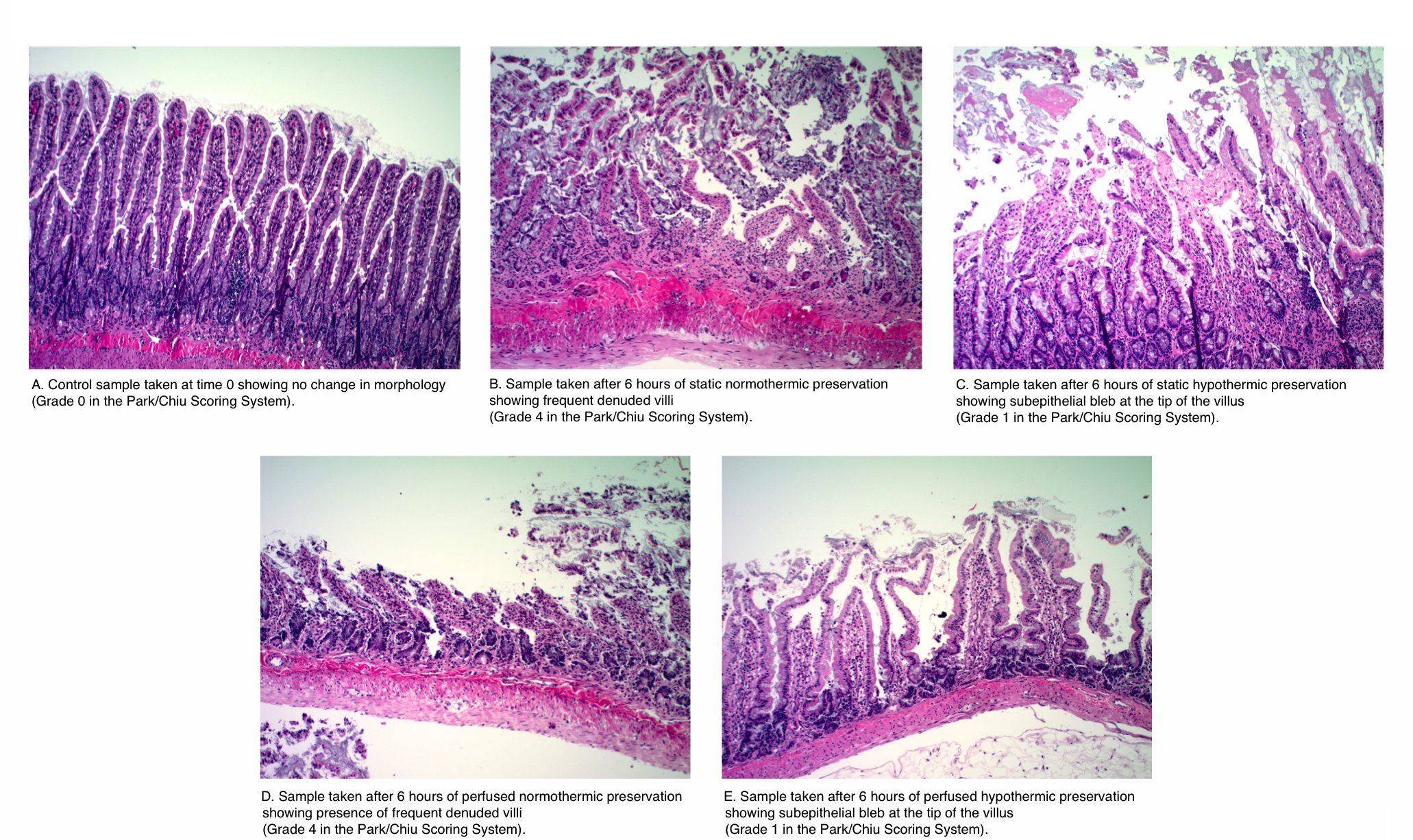
Methods:  Small intestine segments were procured from rats and connected to customized chambers and a perfusion system. Paired intestinal segments were perfused in the luminal and vascular side with University of Wisconsin Solution at two different temperatures (37 and 4 Celsius) over 6 hours. Intestinal tissue samples were processed and evaluated by a blinded pathologist using the Park/Chiu Scoring System for Grading Intestinal Ischemia.

Results: The histopathological analysis showed that continuous perfusion of the intestinal segments at 4 Celsius better preserves the small intestinal segment in comparison to perfusion at 37 Celsius at 6 hours. The differences among the means (hypothermic: 1.67 vs. normothermic: 3.5) in the scoring was significant at an alpha level of 0.05 (p-value=0.0048). The attached figure displays the pathological analysis of the samples. 

Conclusion: In conclusion, continuous hypothermic perfusion of small intestinal grafts with University of Wisconsin solution resulted in improved preservation of the intestinal tissue in comparison to continuous normothermic preservation. These results suggest that continuous perfusion at normothermic conditions shows no advantage to continuous hypothermic perfusion as judged by pathological evaluation. 


01.16 Alterations in Mitochondrial DNA Density in Right Ventricular Failure and Recovery

J. Winward1, M. E. Bowen1, H. Li1, S. H. McKellar1  1University Of Utah,Cardiothoracic Surgery,Salt Lake City, UT, USA

Introduction:
Increasing evidence indicates that abnormal energy metabolism plays a leading role in right ventricular failure (RVF), but the specific role in RVF and RV recovery (RVR) is unknown. Our analyses showed failed fatty acid oxidation (FAO) and changes in hypoxia markers in RVF and RVR. These observations led us to hypothesize that the buildup of FAO precursors was the result of insufficient mitochondrial (MT) biogenesis. Our objective was to discover how MT biogenesis is altered in RVF and RVR cardiomyoctes compared to healthy controls in a rabbit model.

Methods:
Fifteen rabbits were assigned to one of three groups—control, RVF, and RVR—and RV tissue samples were taken from each rabbit’s RV. MT gene expression and transcriptional variance were measured via RT-PCR and RNA sequencing. Immunoblotting of MT biogenesis activators was also performed.

Results:
MT DNA density decreased during RVF with fold changes of RVF=0.75 (p=0.22) returning to RVR=1.06 (p=0.84). Further division of the RVF group into compensated RVF (cRVF) and decompensated RVF (dRVF) revealed that MT density was qualitatively greater in cRVF (FC=0.88, p=0.58) than in dRVF (FC=0.62, p=0.26). RNA sequencing revealed significantly increased transcription of HIF in RVF when compared to the control (FC=0.93; p<0.04) and RVR (FC=0.90; p<0.03). However, the rate of transcription of downstream activators of MT biogenesis (i.e. PGC-1-α, NRF-1, NRF-2, Akt3, Perm-1, TFAM, VEGF, and AMPK) was not significantly altered between the groups. Despite its unaltered transcription, immunoblotting of activated AMPK revealed a significant fold increase in RVF (1.50) and RVR (1.61) compared to the control (1.06) (p=0.01). 

Conclusion:
Our data qualitatively suggest that MT DNA density decreases in a manner directly proportion to the severity of RVF, and that it increases in RVR.  Other studies show similar findings in human subjects. Additionally, the increased transcription of HIF in the RVF samples leads us to hypothesize that hypoxia plays a significant role in the MT pathogenesis of RVF. The discrepancy between the rate of transcription and the enzyme activation of AMPK, in conjunction with the altered MT DNA density in RVF and RVR despite the unchanged rate of transcription of upstream MT biogenesis regulatory enzymes, leads us to hypothesize that the functionality rather than the quantity of MT biogenesis regulators is leading to the variance in MT DNA density, as seen with the increased levels of the activated AMPK protein in RVF and RVR, despite its unaltered rates of transcription. Further research investigating the alterations of MT biogenesis markers at the transcriptional and translational level, and hypoxia’s role therein, is warranted.
 

01.15 Proof of Concept: Magnetic Chest Tube Positioning System

D. Laan1, D. Vu1, M. Hernandez1, J. Aho1, H. Schiller1  1Mayo Clinic,General Surgery,Rochester, MN, USA

Introduction: Chest tubes can serve as life-saving adjuncts in trauma. Unfortunately, suboptimal positioning frequently complicates tube placement and can lead to impaired function. Magnets have been shown to successfully guide and direct small catheters, but this technology has never been shown to be efficacious in chest tube positioning. We sought to demonstrate, in a deceased porcine model, the utility of a 2-magnet system in directing the intra-thoracic position of a chest tube (Figure 1).

Methods: In recently deceased cross-bred domestic swine we tested magnetic positioning of  a chest tube  The operator held one magnet on the outside of the chest.  The second magnet was introduced through a catheter to the distal tip of the chest tube.  The operator was then tasked with moving the tube to distinct pre-marked intrathoracic locations under blinded conditions. This was achieved by taking advantage of the magnetic force between the two magnets. The experiment was video-recorded through an open sternotomy incision to determine success of tube positioning. Five chest tube positioning maneuvers were attempted with this system. An attempt at chest tube positioning with no magnet (standard of care) to premarked intrathoracic locations was attempted as a control.

Results:The chest tube positioning system was successful in directing a chest tube from one pre-marked location to another on 4 of 5 attempts. The system demonstrated an ability to move the tube in the cephalad-caudad axis and the anterior-posterior axis. Magnetic coupling between the 2 magnet ends was confirmed at a distance of 10cm in this model. The control chest tube with no magnet failed to navigate intrathoracically from one pre-marked location to the next with 0 of 5 attempts successful.

Conclusion:Positional flaws in chest tube placement are common. We demonstrate the 2-magnet system’s efficacy as an alternative to the traditional hand-guided method under simulated placement conditions.  The pull between two magnets can be effective with up to 10cm separating magnet ends. Furthermore, we have shown with some reproducibility that a magnetic chest tube positioning system may be superior to the current standard of care technique of chest tube placement. Further study is needed to develop this emerging technology.


01.14 Plasma Heparan Sulfate is a Biomarker of Acute Kidney Allograft Rejection

L. Lin1, M. McRae1, A. MacDonald1, T. V. Brennan1  1Duke University Medical Center,Surgery,Durham, NC, USA

Introduction:
Non­invasive biomarkers of acute allograft rejection (AR) are needed to allow for early diagnosis. Serum creatinine (Cr) is an imperfect monitor of allograft dysfunction because it rises late in the process of immune injury, can be elevated for non-immunological reasons, and the magnitude of change above baseline in the setting of AR can be subtle. During AR lymphocytic invasion of allograft tissues requires the degradation of extracellular matrix. Activated CD4+ T cells express heparanase that degrade ECM and releases cleaved heparan sulfate (HS). We propose the use of plasma HS measurements to overcome the limitations of the serum Cr assay for the detection of AR.

Methods:
Serum and plasma samples from Duke University from consented kidney transplant recipients were collected and stored with approval of the Duke University IRB committee. HS concentrations in the plasma were determined by enzyme-linked immunosorbent assay. Patient groups were compared by two-tailed Student's t-test.

Results:
Patients with biopsy demonstrated AR had significantly higher plasma HS levels within 2 weeks of biopsy than patients with stable function (20.86+/-18.12 ug/mL, 95% CI=12.38-28.34 vs. 2.71+/-1.90 ug/mL, 95% CI=2.35-3.07; p<0.0001). Similarly, patients with biopsy demonstrated AR had significantly higher plasma HS levels than patients undergoing biopsy for cause, but without AR (20.86+/-18.12 ug/mL, 95% CI=12.38-28.34 vs. 3.23+/-1.61 ug/mL, 95% CI=2.26-4.20; p=0.0015) within 2 weeks of biopsy (Fig. 1A). No elevations of HS levels were found in patients with BK viremia (n=13, viral titer range 374 – 1.26×10^7 copies/mL). In comparison, elevations in serum Cr levels in patients with biopsy demonstrated AR versus patients undergoing biopsy for cause, but without AR were lesser in magnitude (5.35+/-3.18 mg/dL, 95% CI=4.09-6.60 vs. 2.64+/-1.15 mg/dL, 95% CI=1.95-3.33; p=0.0052) (Fig. 1B).

Conclusion:
Plasma HS is a sensitive biomarker of AR of kidney allografts because it is detectable prior to AR and has a greater magnitude of elevation compared with serum Cr.
 

01.13 Stem Cell and Granulocyte Colony Stimulating Factor to Promote Recovery in a Limb Transplant Model

K. Kniery1, M. DeHart1, S. Salgar1  1Madigan Army Medical Center,Clinical Investigation,Tacoma, WA, USA

Introduction:
During the global war on terrorism (2001-2008) in Iraq and Afghanistan, 737 US military service members sustained major limb amputations. Limb transplantation offers hope to improve the quality of life. We investigated whether mesenchymal stem cell (MSC) and G-CSF therapy can improve functional recovery in nerve transection-repair and limb transplant models.

Methods:
Under general anesthesia the sciatic nerve branches (tibial, peroneal and sural) were transected and repaired.  In another group, syngeneic right hind limb transplantation was performed. MSCs (5×10⁶; passage ≤6), G-CSF (50µg/kg), or Vehicle were administered topically and i.v./i.p. 

Results:
At two weeks post-nerve repair, sensory function (SF) in all groups was ~1.5 on a scale of Grade 0-3 (0=No function; 3=Normal function).  By 4 weeks it was 2.2±1.0, 2.0±1.2, and 1.8±1.3 in MSC, G-CSF and Vehicle treated groups, respectively. By 10 weeks, normal SF (~3) was restored in all groups (n=8/group). The sciatic nerve function index (SFI) a measure of motor function (0=normal; -100 =nonfunctional) during 5-16 weeks was markedly improved in G-CSF (-40 to -26) compared to MSC (-93 to -66) or Vehicle (-110 to -45) group (Figure1). In limb transplants, SF recovery ranged 0.8-1.3 by 8 weeks, and 1.5-2.0 by 16 weeks post-surgery.  Limb transplants (~60%) developed flexion-contractures and we were unable to calculate SFI. Fewer animals in G-CSF (37%) compared to Vehicle (66%) group developed contractures. Gastrocnemius muscle atrophy was evident in limb transplants. 

Conclusion:
G-CSF and MSC therapies appear to promote sensory and motor function recovery in nerve transection-repair and limb transplant models.