42.04 Characteristics Affecting Virtual Reality Distraction for Pain

J. Harder1, M. De Vries1, H. Van Goor1  1RadboudUMC,Nijmegen, GELDERLAND, Netherlands

Introduction:
Post-surgical pain (PSP) is a difficult to treat condition that requires alternative means to improve patient comfort, physical functioning and quality of life. Virtual reality (VR) has shown to be an distraction tool that can reduce pain perception and expectation and anxiety to suffer from pain. Most studies investigating VR distraction included mostly young patients and did not explore patient or VR characteristics. Our goal was to investigate the effects of two applications (passive or interactive) of VR distraction and their association with personal characteristics such as age, gender, visualisation, imagination, immersion in the virtual world and previous gaming or VR experience.

Methods:
Fifty healthy volunteers (25 M, 25 F, 19 – 66 years of age, mean age 40.9 years) underwent electrical and monofilament tactile perception tests while undergoing three study conditions (control (black screen, no audio), passive VR, interactive VR) in a randomized order using a placebo-controlled, three-way, crossover design. The in-house developed passive and interactive VR applications consisted of the same journey through a river-like landscape, but differed only by a cognitive task (shooting at various targets using head movements) in the interactive VR. Personal characteristics and immersion in the virtual world were gathered using a questionnaire with a 5-point Likert scale design.

Results:
A difference in overall mean of electrical detection threshold and monofilament threshold was observed between each study condition (F(2, 76) = 6.340, p = 0.003 | F(2, 76) = 20.174, p < 0.0005). Interactive VR showed a significant greater beneficial effect as a distraction tool compared to passive VR (p = 0.012). No gender effect was found. There was a positive correlation between age and interactive VR distraction (r = 0.333, p = 0.018). The amount of self-reported immersion in the virtual world showed a positive correlation with an increased effect of distraction by VR (r = 0.352, p = 0.012). Other personal characteristics and previous gaming or VR experience did not affect VR distraction (p > 0.1).

Conclusion:
Passive and interactive VR both distract from pain and tactile sensation, with interactive VR having the largest effect, independent of age and gender. Self reported presence in the VR world enlarged the distraction effect. Previous gaming/VR experience did not affect VR distraction. Future research focuses on personalizing VR applications for a maximum distraction effect to be used as an adjunct to painkillers for post-surgical pain treatment.
 

42.03 Wireless electrical power delivery for artificial human organs

P. Bonde1,2, J. Smith1  1University Of Washington,Department Of Electrical Engineering,Washington, WA, USA 2Yale University,Cardiac Surgery,New Haven, CT, USA

Introduction:
Models of power delivery within an intact organism have been limited to destructive ionizing radiation and to some extent sound & magnetic waves for diagnostic purposes. Traditional electrical power delivery within intact human body relies on implanted batteries that limit the amount and duration of delivered power. The efficiency of current battery technology limits substantial demand to be met such as continuous operation of an implantable artificial heart pump, artificial kidney or artificial liver. We have developed a Free Range Electrical Energy Delivery (FREE-D) for remote electrical power delivery to an intact biological system using magnetic resonant coupling technology up to a meter distance wirelessly. FREE-D system can allow continuous operation of artificial organs, which consume large amount of energy without being dependent on implanted batteries alone.


Methods:
The FREE-D system consists of two resonators efficiently exchanging energy by sharing magnetic fields that oscillate at a specific resonant frequency separated by an intervening distance. A power amplifier delivers an AC signal to the transmit resonator consisting of a single-turn drive loop and a multi-turn coil. Current flowing through the transmit coil induces a magnetic field that is emitted in all directions. When a second resonator consisting of a multi-turn coil and a single-turn receive loop is brought within range of the first, the magnetic field induces electrical current in the receive coil. Additional rectification circuitry connected to the receive loop converts the AC signal into a DC voltage compatible with the artificial heart pump system controller. Magnetic fields emitted by the FREE-D resonators can permeate through solid objects such as walls, household furniture, and human tissues, and can pass around highly conductive objects such as metallic furnishings and other electronic devices. Auto-tuning makes it possible for efficient wireless power transfer to occur through all of these interfering mediums. 


Results:
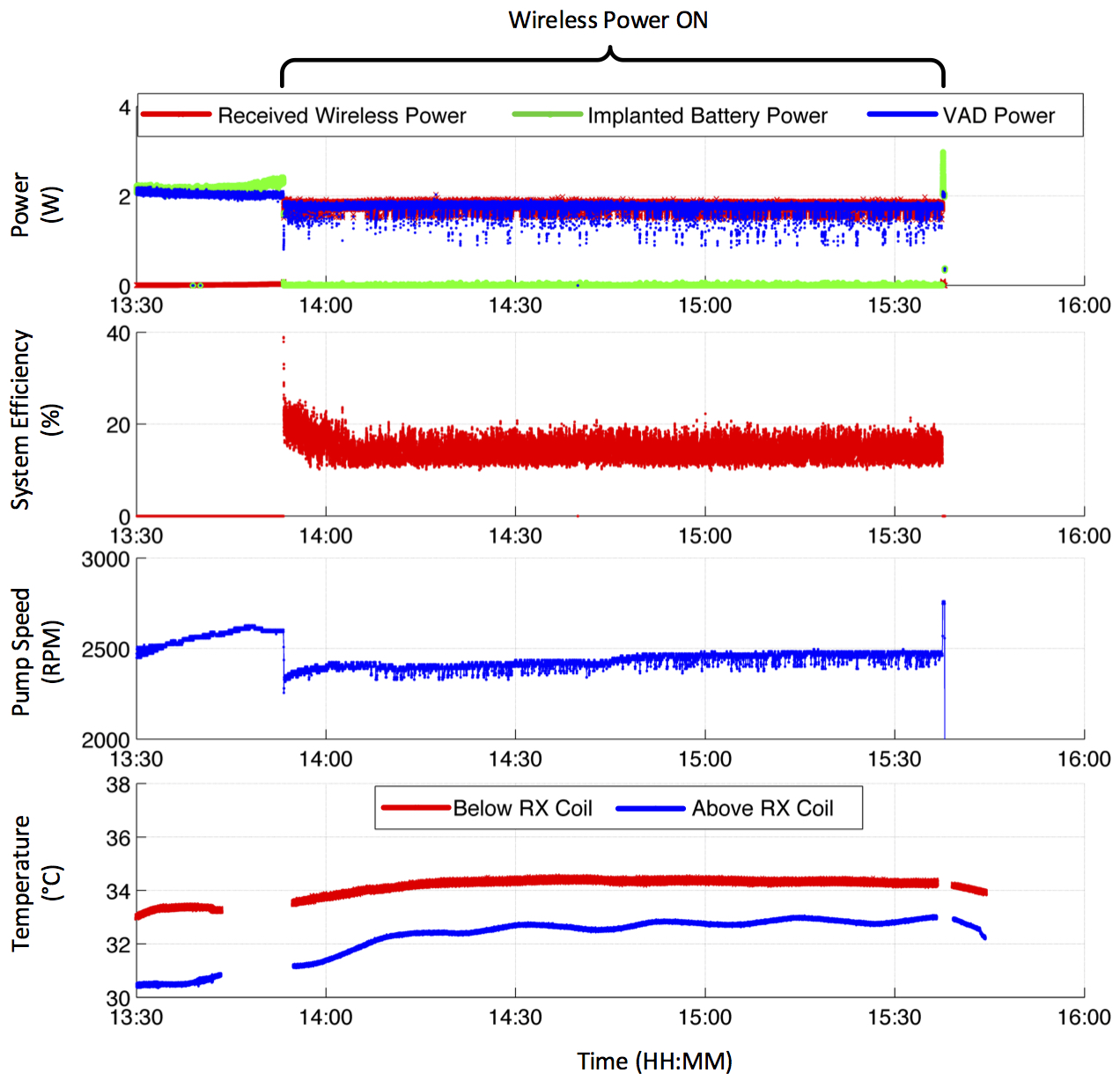
Artifical heart pumps were operated continuously for over 90 days in vitro without interruptions at constant speed. The system didnot have a single interruption or electrical delay or fault detected. The system was then tested in vivo in swine (n=4) to test for efficiency and any temperature changes (Fig 1); the operating temperature invivo of the receive coil remained below 35 degrees celsius for the duration of the experiemnts. Histological analysis near the implanted coil didnot show any thermal induced cell damage.

Conclusion:

Remote electrical power delivery is achieved with high reliability without any tissue damage. This technology has the potential to revolutionize and achieve total implantability for artifical human organs.

42.02 Enriched Probiotic Biofilms: Preservation of Gut Barrier Function in Necrotizing Enterocolitis

J. K. Olson1, C. J. McCulloh1, J. B. Navarro1, L. Mashburn-Warren1, S. M. Gartner1, S. D. Goodman1, G. E. Besner1  1Nationwide Children’s Hospital,Columbus, OH, USA

Introduction:  Probiotic administration for the prevention of necrotizing enterocolitis (NEC) traditionally utilizes planktonic, free-living bacteria and requires repetitive administration for efficacy. We have previously shown that a single-dose probiotic treatment in which Lactobacillus reuteri (Lr) is grown as a biofilm on the surface of biocompatible semi-permeable dextranomer microspheres (DM) prior to administration demonstrates efficacy against NEC, and that loading the lumen of DM with prebiotics such as sucrose (DM-Sucrose) further enhances intestinal protection. In vitro work has shown that sucrose is an inducer of GtfW, an enzyme key to biofilm production, and that maltose is the sole substrate of GtfW. Given that breakdown of the intestinal mucosal barrier is a central component of NEC, we sought to evaluate intestinal mucosal permeability in an experimental model of NEC following administration of this novel probiotic delivery system.

Methods:  A neonatal rat model of experimental NEC was employed. Rat pups were delivered via C-section then given a single enteral probiotic/prebiotic treatment. Treatment groups were as follows: (1) sterile water (vehicle control); (2) planktonic Lr; (3) Lr + DM-Sucrose; (4) Lr + DM-Maltose; or (5) unstressed breast fed pups. Pups were then subjected to repeated episodes of stress (hypercaloric feeds and periods of hypoxia/hypothermia). An enteral dose of fluorescein isothiocyanate (FITC)-labeled dextran was administered 48h after birth, pups were sacrificed 4h later, and serum FITC-dextran levels were determined.

Results: Serum FITC-dextran levels in pups exposed to NEC that received sterile water alone were significantly elevated compared to breast fed unstressed pups (32.0 ± 6.5 vs. 2.22 ± 0.3 µg/mL; p=0.003) (Figure 1), indicative of gut barrier breakdown. Serum FITC-dextran levels were statistically unchanged in pups that received a single dose of planktonic Lr (17.3 ± 5.0 µg/mL, p=0.08). However, levels were significantly reduced in pups that received a single dose of either Lr + DM-Sucrose (10.8 ± 1.2 µg/mL, p=0.004) or Lr + DM-Maltose (9.0 ± 3.2 µg/mL, p=0.007). There was no difference in efficacy between Lr + DM-Sucrose and Lr + DM-Maltose (p=0.569).

Conclusion: Intestinal mucosal barrier function is preserved during experimental NEC by the administration of a single dose of Lr coupled with sucrose- or maltose-loaded microspheres to optimize biofilm production. Improved gut barrier function is likely a key element in the enhanced protection against NEC seen with this novel treatment. These results further support the benefits of this novel probiotic delivery system for the clinical prevention of NEC in the future.

41.20 Smooth Muscle Cell LRP1 Deficiency Affects Genes Regulating Extracellular Matrix Composition

S. Tsai1,2, D. Ramnarain1, M. Kanchwala4, C. Xing4, J. Herz3  1University Of Texas Southwestern Medical Center,Vascular Surgery,Dallas, TX, USA 2Dallas Veterans Affairs Medical Center,Vascular Surgery,Dallas, TX, USA 3University Of Texas Southwestern Medical Center,Molecular Genetics,Dallas, TX, USA 4University Of Texas Southwestern Medical Center,Clinical Sciences,Dallas, TX, USA

Introduction: LDL receptor related protein 1 (LRP1) is an integral regulator of vascular wall structural stability.  A mouse model generated by targeted deletion of LRP1 in smooth muscle cells (smLRP1-/-) in the context of hyperlipidemia is characterized by aggressive aortic atherosclerosis and aneurysmal degeneration.  Even under normolipidemic conditions, smLRP1-/- mice have evidence of structural abnormalities in the aortic wall.  The objective of this study was to understand the enhanced gene expression patterns and signaling pathways driving the changes in aortic wall structure using whole transcriptome sequencing.

Methods:  The aortas of 6 smLRP1-/- mice and their wild type littermate controls were harvested into RNAlater.   The adventitia and peri-adventitial tissues were stripped using micro-dissection techniques.  RNA was extracted and pooled RNA from each genotype was submitted for whole transcriptome analysis, which was performed in duplicate.  Whole transcriptome data was mapped with TopHat and differential expression analysis was carried out using EdgeR.  Genes with false detection rate (FDR) <0.05 (999 genes) were used for pathway/network analysis using Ingenuity Pathway Analysis (IPA).   

Results: LRP1 deficiency in aortic smooth muscle cells (SMCs) was confirmed by immunohistochemistry of descending thoracic aortic cross sections from smLRP1-/- and wild type mice.  Elastin staining revealed diffuse elastin breaks in the smLRP1-/- aortas, accompanied by thickened medial layers of the aortic wall (Figure).  Among the genes that were significantly upregulated in the smLRP1-/-  aorta were 15 collagen genes, including the fibrillar collagens Col1a1 (14.92 fold change, FDR<0.01), Col1a2 (6.39 fold change, FDR<0.01), and Col3a1 (5.82 fold change, FDR<0.01), as well as collagens associated with the basement membrane and regulation of cell migration, including Col4a1, Col4a2, Col4a4, and Col15a1. Elastin gene expression was also upregulated (4.01 fold change, FDR<0.01), in addition to expression of MMP14 (34.19 fold change, FDR=0.01), an activator for MMP2, consistent with observed elastin degradation and partial compensatory upregulation of elastin.  Pathway analysis using IPA software demonstrated upregulation of genes involved in cell migration and cell proliferation pathways, converging on erk1/2, which is known to undergo enhanced phosphorylation/activation in the absence of LRP1.  

Conclusion: Our findings demonstrate that the major aortic wall structural changes associated with LRP1 deficiency in SMCs are driven by enhanced expression of pro-fibrotic genes, in addition to genes involved in cell migration and cell proliferation, as well as a shift towards elastin degradation.


41.19 Endogenously Expressed IL-10 Contributes to Wound Healing and Regulates Tissue Repair Response

M. M. Rae1, T. Lu1, C. M. Moles1, X. Wang1, M. Fahrenholtz1, H. Li1, P. Duann1, P. Bollylky2, S. Balaji1, S. G. Keswani1  1Texas Children’s Hospital And Baylor College Of Medicine,Division Of Pediatric Surgery,Houston, TX, USA 2Stanford University School Of Medicine,Infectious Diseases And Microbiology And Immunology,Stanford, CA, USA

Introduction:
Our lab has shown a significant role for the anti-inflammatory cytokine IL-10 in regulating inflammation and ECM production, thereby attenuating fibrosis in skin wounds. Previous reports have shown, paradoxically, that wounds in IL-10-/- mice heal faster, with increased rates of re-epithelialization as compared to wildtype mice. However, these wounds were not controlled for contraction and wound environment. Therefore, we sought to determine the role of contraction on IL-10’s wound healing and anti-fibrotic effects in a controlled moist wound environment.

Methods:
Full thickness excisional 6mm wounds were made in IL-10+/+ and IL-10-/- mice that were controlled for contraction using a silicone stent. A consistent and moist wound environment was provided by semi-occlusive dressing Tegaderm. Wounds were serially photographed at 3, 5 and 7d and harvested at 7d and 14d post wounding, then examined for epithelial gap, granulation tissue (H&E), myofibroblasts (a-SMA staining) and leucocyte infiltration (CD45). Data is presented as mean+/-SD, n=5 wounds/group/time point; p-value by ANOVA.

Results:
Macroscopic appearance of unstented wounds with no dressing showed accelerated wound closure in IL-10-/- mice by day 7 compared to controls(IL-10+/+). This effect was lost when a semi-occlusive wound dressing was applied to unstented wounds to create a consistent moist wound environment, with no significant differences observed in re-epithelialization(IL-10-/- 2380±508.4 vs IL-10+/+ 2480.4.7±824.6, p=ns), epithelial gap (IL-10-/- 1433.7±558.1 vs IL-10+/+ 1436.2±527.7, p=ns), granulation tissue (IL-10-/- 1.65±0.5 vs IL-10+/+ 1.21±0.4, p=ns), or CD45 positive cells (IL-10-/- 10.1%±6.2 vs IL-10+/+ 8.9%±4.2, p=ns). In unstented wounds, a-SMA was abundantly expressed at the wound margins, but in IL-10-/- wounds, a-SMA was present throughout the granulation tissue and extended into deep dermal layers, whereas a-SMA expression was less pronounced in the IL-10+/+ wound bed. Stenting of wounds, which controlled for the contractility of mouse skin, significantly delayed wound healing. However, there was no statistical difference in either epithelial gap (IL-10-/- 4883.5±610.8 vs. IL-10+/+  4152.2.7±480.6, p=ns) or granulation tissue (IL-10-/- 0.33±0.1 vs. IL-10+/+  0.55±0.2, p=ns) at day 7, although stented IL-10-/- wounds exhibited increased a-SMA density. Interestingly, the CD45+ cellular infiltrate significantly increased in stented IL-10-/- mice as compared to unstented (IL-10-/- 10.1%±6.2 vs. IL-10-/- with stent 32.8%±15.6, p<0.01), but did not change in IL-10+/+ mice. Finally, wounds in IL-10-/- mice also developed significantly more scar tissue and much thicker epidermis on day 14 compared to IL-10+/+ mice. 

Conclusion:
IL-10 expression does not delay normal wound healing of skin wounds when wounds are controlled for contraction and moist environment. However, the loss of IL-10 leads to increased fibrosis. This data signifies a previously unrecognized role for endogenously expressed IL-10 contributing to the tissue repair response.  
 

41.18 Comparison of a Modern Cohort to Lund & Browder suggests that Burn Estimate Errors are Operator Error

W. C. Ray1, R. W. Rumpf1, A. Rajab1, S. K. Martinez2, C. G. Gerrard2, N. L. Adolphi2, R. Fabia1  1Nationwide Children’s Hospital,Columbus, OH, USA 2University Of New Mexico HSC,Radiology-Pathology Center For Forensic Imaging,Albuquerque, NM, USA

Introduction:

Effectively treating burns relies on rapidly and accurately assessing the total body surface area (TBSA) and the proportion of skin that was burned (TBSA-B).  Without accurate measurements of TBSA and TBSA-B, the efficacy of treatment is compromised, increasing the potential for medical complications.  As such the burn treatment community requires a rapid and accurate means of assessing the extent and severity of burns.  Unfortunately, even burn experts admit that most initial burn assessments, even those performed by experts themselves, frequently have significant errors.

Current methods for determining TBSA-B rely on tables of average body statistics such as the Lund and Browder tables.  Today’s pediatric population differs in both size and shape from the population used to derive these charts, and estimates based on historical models are assumed to deviate from today’s reality.  In addition to potential errors in the tables themselves, Lund and Browder and similar tables can be procedurally difficult to apply, relying on subjective and relative comparisons that result in significant deviations from the correct value even when the charts themselves are accurate.

Methods:

We initially hypothesized that the preponderance of errors seen in TBSA-B estimates using Lund and Browder were because the chart could not adequately represent a cohort taken from today’s pediatric population.  In order to test this hypothesis, we compared the zone percentages as described in the Lund and Browder chart to actual TBSA percentages as measured using CT scans of human cadavers.  Previous attempts at evaluating the accuracy of the Lund and Browder chart have been self-referential (e.g., comparing the results that different individuals achieve when applying the chart to the same test subject); to our knowledge this is the first study using an empirical approach to evaluating the chart by comparison to an external “gold standard” measurement of surface areas.

Results:

Unexpectedly, the actually-measured zone percentages were in reasonable agreement with Lund and Browder, even with subjects in the 1% tails of the BMI range. The largest deviation was 5.4%, significantly less than the error seen in real-world TBSA-B estimations.  It would appear that while today’s population is morphometrically different from that studied by Lund and Browder, their body-region proportions scale surprisingly well. 

Conclusion:

Our data indicates that, contrary to common assumption, the primary source of error in TBSA-B estimation is in the application of the Lund and Browder table rather than in the contents of the table.


41.17 Subcutaneous Cefazolin to Reduce Surgical Site Infection in a Porcine Model

G. Dubrovsky1, N. Huynh1, J. Rouch1, J. P. Koulakis2, D. P. Nicolau3, C. A. Sutherland3, S. Putterman2, J. Dunn1  2University Of California – Los Angeles,Department Of Physics And Astronomy,Los Angeles, CA, USA 3Hartford Hospital,Center For Anti-Infective Research & Development,Hartford, CT, USA 1University Of California – Los Angeles,Division Of Pediatric Surgery, Department Of Surgery, David Geffen School Of Medicine,Los Angeles, CA, USA

Introduction:
In the US, FDA-approved administration routes for cefazolin include intravenous and intramuscular. Subcutaneous administration of antibiotics is not well described in the literature, but has been used in other countries and appears to be a convenient method of drug delivery in certain cases. During surgery, we hypothesize that subcutaneous injection may provide higher local concentrations of antibiotic at the incision site and thus will lower the rate of surgical site infections (SSIs). To better understand the kinetics following subcutaneous injection, we describe the serum concentration levels of cefazolin in a porcine model as an estimate of the subcutaneous concentrations.

Methods:
Inhaled gas anesthesia was administered to Yucatan pigs. A femoral arterial catheter was placed. The pigs were administered 20mL of 25 mg/kg cefazolin subcutaneously, and serial blood samples were taken for 2 hours. Blood samples were analyzed for cefazolin concentration using chromatography. The elimination rate constant was calculated by linear regression to the semi-log plot of the terminal phase of the serum concentrations. Area under the curve was calculated using the linear trapezoidal rule until the final measured concentration. The final concentration divided by the elimination rate constant was added to this.

Results:
Maximum serum concentrations of cefazolin are achieved 44.3 minutes after the time of subcutaneous injection, and are 23.7 µg/mL (Figure 1). The elimination rate constant is .004 minutes -1 and the half-life is 173 minutes. The area under the curve is 4600 µg*min/mL and the clearance is 5.4 mL/(kg*min). The relative bioavailability of subcutaneous injection is 88%.

Conclusion:
We describe the pharmacokinetics of subcutaneous administration of cefazolin. This route shows good bioavailability, although the maximum serum concentrations achieved with subcutaneous injection are not as high as those from IV injection. The serum levels have a lower peak, but the medication remains in the blood for a longer period of time. As a result, higher doses of antibiotic can be injected locally without incurring systemic toxicity. Subcutaneous administration will therefore result in higher concentrations of antibiotic at the incision site than standard IV administration and thus may be more effective in preventing SSIs. Further studies are needed to detail the exact effect of subcutaneous antibiotic injection on SSI rates.
 

41.16 Three Dimensional Confocal Imaging Analysis of Adipose Tissue

N. N. Chung1, C. P. Blackshear1, C. Ransom1, D. Irizarry1, D. Nguyen1, M. Longaker1,2, D. C. Wan1  1Hagey Laboratory For Pediatric Regenerative Medicine,Stanford University Medical Center,Stanford, CA, USA 2Institute For Stem Cell Research And Regenerative Medicine,Stanford University Medical Center,Stanford, CA, USA

Introduction:  Autologous fat grafting is frequently used to correct soft tissue defects, but outcomes remain unpredictable. Studies have shown stromal cell enrichment of fat grafts to improve volume retention, but the mechanism of this effect has been primarily explained through inferential gene expression analysis and two-dimensional histologic staining. In this study, we employed novel three-dimensional confocal imaging to determine how graft architecture changes with stromal cell enrichment.

Methods:  Confocal microscopy with optical sectioning was employed to reconstruct three-dimensional data of whole mounted tissue using Imaris. Unprocessed human abdominoplasty fat, as well as unenriched human fat grafts and stromal cell enriched fat grafts after two, four, six, and eight weeks of implantation in immunocompromised mice were evaluated. Supplemental stromal cells were fluorescently tagged to determine three-dimensional distribution throughout the fat graft. Isosurface rendering was employed to determine individual adipocyte size and variability, vascular density, and proximity of stromal cells to new vasculature.

Results: Preliminary data has shown that confocal imaging of adipose tissue results in superior images that elucidate tissue architecture and more closely approximates the graft in situ. Images allowed for clear visualization of supplemented ASCs within the graft environment, showing an intimate relationship with revascularization patterns in the fat graft. Heterogeneity in adipocyte size was also appreciated with fat grafts supplemented with stromal cells appearing more similar to unprocessed abdominoplasty fat.

Conclusion: Confocal imaging allows for direct three-dimensional visualization of the fat graft environment that approximates in situ, which holds promise for future study of CAL grafts and retention.


41.15 AMD3100: Single-Dose Stem Cell Mobilizing Agent In Vascularized Composite Tissue Allografts In Canine

B. J. Swearingen1,2, S. S. Graves2,3, R. F. Storb2,3, D. W. Mathes1,2  1University Of Colorado Denver,Plastic And Reconstructive Surgery,Aurora, CO, USA 2Fred Hutchinson Cancer Research Center,Clinical Research Division,Seattle, WA, USA 3University Of Washington,School Of Medicine,Seattle, WA, USA

Introduction:
Vascularized Composite Allograft (VCA) transplantation is a clinical reality but limited by toxicities of chronic immunosuppression and rejection. Current clinical tolerance protocols rely on recipient conditioning and donor cell mobilization that limits use to living donor transplants. We sought to design a clinically relevant protocol applicable to cadaveric organs. We modified our existing non-myeloablative stem cell canine VCA transplant model to use AMD3100 (Plerixafor) for stem cell mobilization.

Methods:
5 DLA-haploidentical, related canine recipients [Group I] received conditioning with 350cGy TBI, AMD3100-mobilized donor stem cells and VCA transplantation with a short course of immunosuppression (MMF:56 days/CSP 70 days +/- taper). 4 DLA-haploidentical, related canine recipients [Group II] underwent identical conditioning plus an infusion of Bone Marrow (BM) Aspirate in addition to AMD3100. CD34+ hematopoietic progenitor cells were quantified via flow cytometry. Peripheral blood chimerism was evaluated by PCR techniques weekly. VCA graft survival was followed clinically and histologically.

Results:
All 9 canines tolerated the conditioning regimen. 4 [Group I] and 3 [Group II] were followed long-term. Stem cell engraftment and donor chimerism were seen in all dogs. Median COBE apheresis counts were 6.21×10^8 (Group I) and 3.08×10^8 (Group II) cells/kg, respectively. Bone Marrow aspirate counts were 1.47×10^8 cells/kg (Group II). No acute rejection nor evidence of GVHD was seen.  An unexpected finding of persistent thrombocytopenia that resolved on loss of donor cell chimerism seen in Group I led to addition of BM aspirate for Group II in an attempt to address this.

Conclusion:
This study demonstrates proof of principle for AMD3100 as a single-dose stem cell mobilizing agent for a clinically relevant tolerance protocol. Use of AMD3100 led to stem cell engraftment in all animals transplanted with no evidence of acute rejection in the VCA. Current application of AMD3100 is limited by thrombocytopenia that may be resolved with the addition of BM Aspirate.
 

41.13 Smartphone Camera Viability For Skin Tone Measurement

J. Butts1, R. Seelaus1, D. Morris1  1University Of Illinois At Chicago,Plastic Surgery,Chicago, IL, USA

Introduction:

Accurate color measurement is essential to the plastic surgeon desiring optimal aesthetic outcomes. Variability of human perception and environmental lighting conditions limit observation or swatch-based methods, leading to use of spectrophotometers as the gold-standard for accurate color matching.

Advances in smartphones camera technology and the utilization of device-independent color-spaces may provide an opportunity for replacement of spectrophotometers in resource-limited settings, without significant calibration or processing.

We investigated the utility of a consumer smartphone camera against a spectrophotometer for the evaluation of skin tones in a variety of illuminant environments.

Methods:

Silicone disks pigmented to match a representative skin sample from a fair-skinned subject were created using spectrophotometer measurements in standard fashion. Skin and disk samples were measured with the spectrophotometer and imaged with a smartphone under three illuminant conditions: D65 (simulated daylight), incandescent, and white fluorescent TL84.

Average pixel RGB values of each silicone and skin sample were calculated from each smartphone image. Samples were converted from sRGB to Lab color-spaces, and Euclidian distances (dE) were calculated between matched skin and silicone Lab samples.

Results:

A two-way one sided t-test was performed to assess equivalence between mean spectrophotometer and smartphone values. A magnitude threshold of 1.0 was chosen as a maximally-stringent, just noticeable difference in fair-skinned individuals.

Images taken under D65 were statistically equivalent to the spectrophotometer. Those under incandescent and fluorescent were not, with incandescent providing lower, and fluorescent higher, dE values.

Conclusion:

Equivalence between smartphone and spectrophotometer under simulated sunlight agrees with recommendations for indirect sunlight as an ideal condition for color matching. Although incandescent and fluorescent narrowly-missed equivalence, their precision suggests utility if measurements are taken in consistent lighting conditions or for applications with greater tolerances.

41.12 Identification and Characterization of a Long Noncoding RNA Involved in Osteogenic Differentiation

C. P. Blackshear1, A. Luan1, E. A. Brett1, J. S. Flacco1, D. Nguyen1, M. T. Longaker1,2, D. C. Wan1  1Stanford University School Of Medicine,Hagey Laboratory For Pediatric Regenerative Medicine, Department Of Surgery, Plastic And Reconstructive Surgery Division,Stanford, CA, USA 2Stanford University School Of Medicine,Institute For Stem Cell Biology And Regenerative Medicine,Stanford, CA, USA

Introduction:  Adipose-derived stromal cells (ASCs) hold great promise for the treatment of tissue deficits. Increasingly, long noncoding RNAs (lncRNAs) have been shown to be capable of altering gene transcription. This study sought to identify and characterize specific lncRNAs involved in osteogenic differentiation.

Methods:  Directional RNA sequencing libraries from ASCs and human induced pluripotent stem cells (iPSCs) undergoing osteogenic differentiation identified a lncRNA, LOC100505806, with a role in bone formation through guilt-by-association analysis. A lentiviral vector for lncRNA overexpression was generated and ASCs and iPSCs were transduced. Osteogenic capacity and pluripotency were then evaluated, respectively. Finally, knockdown of our lncRNA also was performed to evaluate for effects in iPSCs.

Results: Overexpression of our lncRNA resulted in enhanced osteogenic differentiation by ASCs, as demonstrated by gene analysis and histologic staining. Lentiviral transduction and puromycin selection of induced pluripotent stem cells also revealed changes in pluripotent state following LOC100505806 upregulation. Importantly, siRNA knockdown of LOC100505806 did not result in maintenance of pluripotent gene expression when iPSCs were subsequently cultured in osteogenic differentiation medium.

Conclusion: lncRNAs may serve as potential targets to manipulate osteogenic differentiation in multipotent stem cells. Upregulation of LOC100505806 enhanced bone differentiation with ASCs and altered pluripotent state with iPSCs. Knockdown of LOC100505806, however, was not sufficient to maintain pluripotency when an osteoinductive environment was introduced.
 

41.11 The Effects of Smooth Muscle Cells and Subpopulations of Fibroblasts on Breast Capsular Contracture

D. M. Irizarry1, C. D. Marshall1, D. Nguyen1, M. T. Longaker1,2, D. C. Wan1  1Stanford University Medical Center,Hagey Laboratory For Pediatric Regenerative Medicine,Palo Alto, CA, USA 2Stanford University Medical Center,Institute For Stem Cell Research And Regenerative Medicine,Palo Alto, CA, USA

Introduction:
Despite continued improvements in breast implant design, capsular contracture remains a significant problem. This is further worsened by adjuvant radiation therapy for breast cancer. Several studies have correlated fibroblast density with capsular contracture, and more recent reports have also shown a functional difference in fibroblasts from non-contracted and contracted breast implant capsules.  Emerging data has now shown specific fibroblast subpopulations to exist, and in particular, CD26+ fibroblasts have been implicated in fibrosis/scarring following radiotherapy. The role of this fibroblast subpopulation in radiation-induced contracture was thus evaluated. 

Methods:
Human breast capsule specimens were taken from patients following mastectomy with tissue expander placement and post-operative radiation therapy. The contralateral non-irradiated side served as a control. Capsules were mechanically and then enzymatically digested to obtain fibroblasts. Fluorescence-activated cell sorting and immunohistochemistry staining were performed. Cell surface marker expression and gene expression were compared between irradiated and non-irradiated specimens.  Proportions of CD26+ fibroblasts were compared between the irradiated side and non-irradiated side. 

Results:
Histology of the specimens revealed that irradiated, contracted capsule contained more dense eosinophilic material suggestive of smooth muscle tissue. Furthermore, irradiated, more severely contracted capsules were found to have more vimentin+/CD26+ cells by histology and flow cytometry. Gene analysis demonstrated differences in fibrotic gene expression between fibroblasts harvested from each side. 

Conclusion:
These results suggest a role for CD26-expressing fibroblasts and smooth muscle cells in breast capsule contracture following radiation. These are both potential future targets for intervention aimed at reducing capsular contracture severity.
 

41.10 Cellular Evidence of Polyethylene Glycol Fusion in Cultured Neuroblastoma Cells

A. N. Hoffman2, R. Bamba1, M. Stephanides1, A. C. Pollins1, W. P. Thayer1  1Vanderbilt University Medical Center,Plastic Surgery,Nashville, TN, USA 2Vanderbilt University School Of Medicine,Nashville, TN, USA

Introduction:
The degree of functional recovery after a peripheral nerve injury is rarely satisfactory.  Peripheral nerves have the capacity to regenerage, but unfortunately this regeneration is very slow, and there is usually significant loss of function. Current methods in nerve repair depend on the type of nerve injury and include direct repair, nerve conduits, nerve autografts and allografts. These techniques focus on optimizing the process of axonal regeneration. Polyethylene glycol (PEG) fusion is a new technique in peripheral nerve repair that aims to fuse damaged axons in order to provide some immediate restoration of axonal function. The PEG fusion technique involves reconnecting the cut ends of severed axons with microsutures and applying a sequence of solutions including PEG. It is thought to delay Wallerian degeneration, and animal studies have shown immediate restoration of compound action potentials and improved behavioral outcomes.

Methods:
Rat B35 neuroblastoma cells were grown using standard cell culture techniques. The cells were labeled separately with CellTracker Green CMFDA and CellTrace Violet fluorescent dyes. These dyes were chosen because they freely diffuse across cell membranes, and are not transferable between cells except to daughter cells. Equal concentrations and volumes of the green- and violet-labeled cells were then combined and PEG solutions were added to the cell pellet at concentrations of 50%, 75% or 100% PEG by weight with 5% DMSO. A control sample consisted of the two populations of fluorescently labeled cells combined without addition of PEG. Other controls included unstained and single stain groups. The samples were analyzed using the BD LSRFortessa flow cytometer. The amount of fusion was assessed by the percent of double positive cells.

Results:

Results show that there is increasing cell fusion with increasing concentrations of PEG. Viability tests were also performed with the viability factor PI, and they showed decreasing viability with increasing concentrations of PEG (Table 1).

Conclusion:
The results of this experiment provide evidence of PEG fusion at the cellular level. Using the double-positive population as an indicator of cell fusion is not a perfect measure, as cells are equally likely to fuse with other cells of the same color. Therefore, it is most likely an underestimate of the amount of fusion. Assessment of cell viability indicated that there is increasing cell death with increasing concentration of PEG. This trade-off between increased fusion and increased cell death represents a significant balance that must be assessed for applicability in patients with nerve injuries. Future directions of this experiment include studying the long term viability of fused cells.

41.09 Tracking CD Marker Drift of Fresh Adipose Derived Stem Cells Induced by 2-Dimensional Cell Culture

E. Brett1, D. Wan1  1Stanford University,Palo Alto, CA, USA

Introduction:  The influence of static, 2D culture on cell types has been investigated on cell lines and primary cultures. Long term subcultures of cell lines have shown susceptibility to ‘drift’, both phenotypically and genotypically. CD marker drift is pertinent to primary cell types which require fluorescent assisted cell sorting for isolation. Adipose derived stem cells (ASCs) are an example of such cells, relying on FACS selection of CD45-/CD31-/CD34+ cells from processed lipoaspirate. 

Methods:  Choosing CD90, CD105, and BMPR1b, we plated FACS-sorted ASCs positive and negative for these markers, and analysed the extent of CD marker drift over 0hr, 12hr, 24hr, 36hr, 48hr, D3, D5, and D7 after standard cell culture. It was consistently observed that ASCs which were sorted for a presence or absence of specific marker at 0 hour have a significantly different CD expression profile after 36 hours of cell culture (*p<0.05). We also analysed the effect of CD positive and negative populations on one another in a co-culture setting. A decrease of CD marker expression was largely observed across the cell populations, caused by originally CD-positive cells drifting towards a negative phenotype. The osteogenic potential of purified positive and negative populations was assessed using a standardized in vitro model of osteogenic differentiation. Significant differences were observed between the osteogenic capacities of fresh ASCs versus ASCs whose CD marker profiles had drifted over 36 hours of culture (*p<0.05). This drift was characterized further using qRT-PCR, which detected concomitant increases in expression of osteogenic and mechanotransductive genes in plated cells. 

Results: These observations allude to the volatile nature of CD expression by FACS-sorted ASCs once in culture, and suggest an influence of cell culture on capabilities of ASCs to commit to lineage. 

Conclusion: In vitro culture has governance on phenotypic and genotypic profile of ASCs. This has a significant downstream impact on the osteogenic capacity of the cells in vitro; an important consideration for all in vitro assays. 


41.07 Peptide Amphiphile Nanofibers Amplify Effects of BMP-2 on Craniofacial Bone Regeneration

R. Bakshi1, A. Hokugo1, S. Zhou1, K. Rezzadeh1, L. A. Segovia1, S. Stupp2, R. Jarrahy1  1University Of California – Los Angeles,Regenerative Bioengineering And Repair (REBAR) Laboratory, Division Of Plastic And Reconstructive Surgery, Department Of Surgery,Los Angeles, CA, USA 2Northwestern University,Simpson Querrey Institute For BioNanotechnology,Chicago, IL, USA

Introduction:
Bone morphongenic proteins (BMPs) have numerous applications in craniofacial surgery and play a central role in the development of regenerative therapies for bone reconstruction. Unfortunately, the broad applications of BMPs are limited by its side effect profile and high cost. Nanoscale peptide amphiphiles (PAs) are a novel biomaterial that can self-assemble into nanofibers. These PA gels have demonstrated the ability to function as a controlled release carrier of BMP-2 and support bone regeneration. In this study, we examined the effect of PA gels on in vitro osteogenic differentiation of bone marrow stromal cells (BMSCs) and in vivo bone regeneration in cranial defects.

Methods:
BMSCs were cultured and treated either with control media or media with BMP-2 and PAs. Quantitative RT-PCR of genes specifically related to osteogenesis was used to assess for osteogenic differentiation in vitro. To study the in vivo effects, cleft palates were created in rat maxilla. The defect was treated with one of the following: 1) collagen with BMP-2 (high dose), 2) collagen with BMP-2 (low dose) and PAs 3) collagen with inert vehicle (control) or 4) no treatment. After 8 weeks, the maxilla was harvested for radiographic analysis with micro-computed tomography and for histology.

Results:
BMSCs treated with BMP-2 with PAs had increased up-regulation of markers of osteogenesis demonstrating osteogenic differentiation. The cleft palate implanted with collagen sponge and low dose BMP-2 showed bone regeneration comparable to the collagen with high dose BMP-2.

Conclusion:
PAs with BMP-2 have the ability to induce osteogenic differentiation in BMSCs as demonstrated by these results. The in vivo and in vitro studies demonstrate the osteogenic capacity of PAs with BMP-2. This data may represent the use of PAs with low dose BMP-2 as a viable alternative to current uses of clinical BMP-2 in tissue engineering.

41.06 Topical Application of Sphingosine-1-Phosphate Promotes Deep Dermal Burn Healing.

T. Tsuge1, M. Aoki1, K. Kubomura1, S. Akaishi1, K. Takabe2, R. Ogawa1  2Roswell Park Cancer Institute,Breast Surgery,Buffalo, NY, USA 1Nippon Medical School,Plastic, Reconstructive And Aesthetic Surgery,Tokyo, , Japan

Introduction:
Deep dermal burn (DDB) is an injury with epidermal, superficial and deep dermal damages. Its healing often requires longer than a month despite appropriate treatment, and sometimes results in hypertrophic scar. During such a long period of healing process, DDBs can easily proceed to deeper burn wound due to infection through necrotic tissues. Therefore, a novel treatment that can activate macrophage functions to promoting epithelialization is anticipated as a breakthrough in burn treatment. Sphingosine-1-phosphate (S1P) is a lipid mediator that is involved in immune cell migration, angiogenesis, and cell proliferation. In particular, S1P plays an important role in inflammatory cell recruitment into local tissues. Further, S1P receptor 1 signaling is essential for angiogenesis. We focused on S1P as a new approach for wound treatment. We investigated the therapeutic effects in rat DDB model.

Methods:
Four of 10 mm-diameter hair-removed skin areas were contacted with 78 ºC water for 10 seconds to create DDBs in F344 rats (male, 8-12 week). Topical application with control or S1P ointment was performed every other day until day 14 after injury. We performed burn area analysis, and analyzed macrophage recruitment and microvessels by immunohistochemistry. 3D photo acoustic imaging system was used to estimate angiogenesis in burn area. Microvascular integrated densities were calculated using ImageJ.

Results:
Topical application of S1P significantly promoted deep burn wound healing of DDBs comparing with control. Average of burn wound areas treated with vehicle at day 12 was 32.2 % of day 0, whereas average of those treated with S1P was 17.1 % (p=0.039). Surprisingly, we found that autolysis of necrotic tissues was promoted in DDBs treated with S1P. This results suggests that macrophage recruitment is increased by S1P treatment. Further, by analysis with 3D photo acoustic imaging, we found that microvascular integrated density in burn areas treated with S1P was significantly higher than that treated with control at day 14 after injury. This result demonstrates that topical S1P promote angiogenesis.

Conclusion:

Our results show that topical application of S1P promote deep dermal burn wound healing by recruitment of macrophages, promoted autolysis of necrotic tissues, and enhanced angiogenesis. It is suggested that increased macrophages by S1P treatment may remove necrotic tissues, prevent infection, and promote angiogenesis. Thus, S1P may have a role as novel approach for wounds with necrotic tissues such as DDBs.

41.05 Transplantation of IL-10 Polarized M2 Macrophages Improves Wound Repair and Skin Regeneration In Vivo

L. Barnes1,2, M. S. Hu1, A. T. Cheung1, A. L. Moore1,3, C. D. Marshall1, T. Leavitt1, A. Dubnika1, M. Ahmed1, G. C. Gurtner1, H. Lorenz1, J. Rajadas1, M. T. Longaker1  1Stanford University,Palo Alto, CA, USA 2Howard Hughes Medical Institute,Chevy Chase, MD, USA 3Brigham And Women’s Hospital,Boston, MA, USA

Introduction:
Skin defects resulting from trauma, tumor resections, and congenital anomalies pose a significant reconstructive challenge. Thus, it is necessary to develop novel approaches in regenerative medicine to improve tissue repair. Macrophages can be polarized to two phenotypes (M1 and M2) that have been shown to have different functions: M1 phenotype macrophages are pro-inflammatory, while M2 phenotype macrophages are pro-regenerative. Using an established humanized splinted excisional wound model in mice, we now demonstrate the ability of supraphysiologic levels of murine M2-polarized macrophage subpopulations to further promote skin regeneration in vivo.

Methods:
In this study, monocytes were isolated from the bone marrow of mice expressing green fluorescent protein (GFP) constitutively in the cytoplasm of all cells (FVB-Tg(CAG-luc,-GFP)L2G85Chco/J mice (JAX, Bar Harbor, Maine)). They were subsequently differentiated into macrophages using macrophage colony-stimulating factor (M-CSF), seeded onto biomimetic scaffolds containing recombinant interleukin-10 (IL-10) to polarize them, and transplanted onto splinted excisional wounds on the dorsa of wild-type (FVB/NJ) mice. Gross wound progression and size was assessed from digital photographs. Wound tissue was harvested for histologic, immunohistochemical, gene expression, and cellular analysis.   

Results:
Gene expression analysis confirmed that macrophages seeded onto the biomimetic scaffold containing IL-10 were polarized to the M2-phenotype in vivo within 48 hours. Digital image and histologic analysis demonstrated that supraphysiologic levels of M2-polarized macrophages significantly accelerated wound healing without adverse effects on scar size and quality when compared to controls (*p<0.05). 

Conclusion:
We have shown that polarizing M2-macrophages on a biomimetic scaffold in vivo accelerates wound healing. This method minimizes the time cells spend in culture and has been shown to be optimal for cell delivery. Given that monocytes can be readily isolated from peripheral blood, this technique could allow for the rapid delivery of a patient’s own cells to enhance cutaneous wound healing.
 

41.04 A mouse model of mandibular distraction osteogenesis

R. C. Ransom1,2, T. Leavitt1, C. D. Marshall1, L. Barnes1, D. C. Wan1, M. T. Longaker1,2  1Hagey Laboratory For Pediatric Regenerative Medicine,Department Of Surgery, Division Of Plastic Surgery, Stanford University School Of Medicine,Palo Alto, CA, USA 2Institute For Stem Cell Biology And Regenerative Medicine,Stanford University,Stanford, CA, USA

Introduction: Distraction osteogenesis (DO) refers to the gradual mechanical lengthening of a bone segment, resulting in the growth of new bone. DO is a powerful method of endogenous bone tissue engineering that has been applied to the craniofacial skeleton in patients with critical-sized bone defects. However, the cellular and molecular signaling that regulates this process of de novo bone formation is not well understood. We aimed to develop a rigorous and genetically dissectable mouse model of mandibular DO.

Methods: Mandibular DO devices were manufactured using computer-aided design (CAD) software and 3D printing. Animals were divided into four groups: sham-operated (n = 6 per time point), fracture (n = 6 per time point), acutely lengthened (n = 8 per time point) and gradually distracted (n = 8 per time point) right hemimandibles. Gradual DO was performed at a rate of 0.15 mm every 12 h over the course of 10 days to a total length of 3 mm.  Acute lengthening was performed at a single time point to a total length of 3 mm. Therefore, the total amount of distraction lengthening (3 mm) was maintained across both groups. Specimens were harvested at mid-consolidation (POD 29) and end consolidation (POD 43) time points for micro-computed tomography (CT) and histological analysis.

Results: Histologic and CT analyses confirmed that fractured mandibles healed normally through a cartilaginous callous with complete bone bridging by POD 43. However, gradually distracted mandibles revealed a remarkable pattern of new bone formation. Two weeks into consolidation (POD 29), greater than half of the distraction gap was filled with trabecular bone. No cartilage was seen within the distraction gap, indicating that the regenerate was produced through direct intramembranous bone formation. At the end of consolidation (POD 43), trabecular bone completely bridged the osteotomy site of all gradually distracted specimens. Trabecular bone along the edges of the osteotomies was remodelled to lamellar, cortical bone. Three-dimensional reconstruction of the CT images of gradually distracted specimens clearly showed complete osseous bridging of the osteotomy gap. In marked contrast to the above data, the distraction gap of all mandibles undergoing acute lengthening was filled with fibrous tissue at end-consolidation and there was minimal histological or radiographic evidence of new bone formation at the osteotomized bone.

Conclusions: We have developed and a model of mandibular DO in mice, and begun to characterize the processes that lead to the formation of new bone. This model will afford tremendous opportunities to utilize transgenic mouse systems that are not available in larger animals. Experiments utilizing this unique model will offer valuable insight into the biology of de novo bone formation and mechanical forces guiding distraction osteogenesis, speeding the development of clinical therapies for patients suffering from critical-sized bone defects.

41.03 Characterization of Transcriptional Alterations within Adipose-Derived Stromal Cells in Culture

P. A. Mittermiller1, M. Rodrigues1, R. E. Shor1, A. Y. Li1, B. A. Kuehlmann1, G. C. Gurtner1  1Stanford University,Plastic And Reconstructive Surgery,Palo Alto, CA, USA

Introduction:

Adipose-derived stromal cells (ASCs) are a valuable source of cells for use in regenerative medicine. Our laboratory has been determining the ideal ASC subpopulation to be used in clinical trials. We have previously identified a subset of ASCs with a favorable gene expression profile that demonstrates improved wound healing in diabetic mice. This subset of cells can be isolated using a unique surface marker combination (CD26+/CD55+). In order to obtain sufficient numbers of these cells for therapeutic purposes, expansion of the cell line is necessary. Therefore, we tested whether expansion would result in changes to their transcriptional profiles.


Methods:

Adipose tissue was surgically extracted from three wild-type mice. The tissue was digested and single cells were subjected to fluorescence-activated cell sorting (FACS). Using this technique, cells were sorted for non-hematopoietic (CD45-), non-endothelial (CD31-) single cells with the progenitor cell marker (CD34+). This marker profile isolates all ASCs. Cells were also sorted for the ASC subpopulation of interest with the additional markers CD26 and CD55. Two sets of cultures were performed, one with parental ASCs and the other with CD26+/CD55+ ASCs. The cells were passaged every five days for a total of three passages (P0 to P3). Cells were trypsinized after each passage and analyzed by FACS to measure absolute cell numbers. Freshly isolated CD26+/CD55+ cells were subjected to single-cell transcriptional analysis and compared to the same group in culture after passages P0 and P1.


Results:

Using the methods described above, three thousand parental ASCs and three thousand CD26+/CD55+ ASCs were isolated. The number of cells from the parental group increased by two-fold in P0, nine-fold in P1, and over one hundred-fold in P2. The cells from the CD26+/CD55+ ASC group proliferated more than those in the parental group after each passage. Single-cell transcriptional analysis revealed a drastic drift in gene expression profiles between freshly isolated CD26+/CD55+ cells and those cells in culture. There were notable, statistically significant increases in genes VEGF, FGF2, FGF7, TGFB1, FGFR1, FGFR2, EGFR, CCND1, PCNA, ADAM10, and HB-EGF in the sorted ASCs in culture.


Conclusion:

Through single-cell transcriptional analysis, we have identified an ASC subpopulation that improves wound healing in diabetic mice. This study has offered insight into the changes that occur in the cell line with expansion. CD26+/CD55+ ASCs demonstrate an increased rate of proliferation in culture when compared to parental ASCs. Furthermore, during in vitro proliferation these cells demonstrate an increase in expression of genes responsible for growth factors, growth factor receptors, proliferation and production of extracellular matrix. These findings continue to suggest that the CD26+/CD55+ ASC cell line is favorable for regenerative purposes and can be expanded in vitro for cell-based therapies.

41.02 Analyzing the Effects of Desferal Treatment on Irradiated Tissue and Fat Graft Retention

D. M. Irizarry1, J. Flacco1, C. P. Blackshear1, G. C. Gurtner1, M. T. Longaker1,2, D. C. Wan1  1Stanford University Medical Center,Hagey Laboratory For Pediatric Regenerative Medicine,Palo Alto, CA, USA 2Stanford University Medical Center,Institute For Stem Cell Research And Regenerative Medicine,Palo Alto, CA, USA

Introduction:
Radiation therapy induces hypovascularity causing the skin to become fibrotic and soft tissue to atrophy. Fat grafting has been shown to improve the quality of irradiated skin, but volume retention of the graft is significantly decreased. Desferal, an FDA-approved iron-chelating medication, has been shown to increase angiogenesis, and treatment of irradiated sites with this compound may reduce radiation induced ischemia and enhance fat graft survival. 

Methods:
Immunocompromised mice underwent 30-Gy external beam irradiation of the scalp. Four weeks later, mice either received seven desferal treatments (1mg in 100ul) or saline subcutaneously to the irradiated area. Laser Doppler analysis (LDA) was recorded prior to irradiation, following irradiation, and following each treatment. Human fat grafts were then injected in the subcutaneous plane of the scalp and volume retention measured by CT scan over 8 weeks. Finally, skin and fat samples were evaluated histologically. 

Results:
After 4 treatments with desferal, a significant increase in microvasculature was observed using LDA (p<0.05).There was also a positive trend observed with the development of microvasculature in the fat graft with LDA. Using microCT, we observed a significant increase in fat graft volume retention with the desferal treated group compared to the saline treated group, and this was paralleled by improved histologic staining of skin and fat grafts (p<0.05). 

Conclusion:
Our results show increased microvasculature and increased fat graft volume retention with desferal treatment. Desferal treatment may also promote beneficial effects in dermal thickness and in quality scoring of the fat grafts, thus leading to a potential clinical application in radiation damaged soft tissue.