88.20 Hepatic ischemia reperfusion induction of acute renal insufficiency:

T. L. Munsch1, N. J. Skill1, M. A. Maluccio1, S. Mangus1, C. A. Kubal1  1Indiana University School Of Medicine,Transplant,Indianapolis, IN, USA

Introduction:
Hepatic ischemia reperfusion injury (IRI), associated with liver transplant, is linked to acute kidney injury and an increase in morbidity and mortality. The purpose of this study was to evaluate renal impairment in response to the previously established murine 70% hepatic ischemia reperfusion model.  

Methods:
In FVB mice the left hepatic artery and left portal vein was clamped to induce ischemia in the medial (ML) and left lateral lobe (LLL). Flow to the caudate (CL) and right lateral lobe RLL) was not impeded. After 30min the clamp was released. Perfusion of the LLL, RRL, spleen, and kidneys was measured using a laser Doppler flow meter before and after clamping, and before and 5 min after clamp release. 24hr post ischemia/reperfusion perfusion rates were recorded and serum and kidneys were collected. Serum ALT and creatine were quantitated using a coupled enzyme assay. Renal kidney injury molecule-1 (KIM1), a marker of renal injury, was quantitated by RT-PCR. 

Results:
Perfusion rates to the LLL, spleen and kidneys were markedly reduced following clamping of left hepatic artery and left portal vein. In contrast, perfusion to the RLL was increased. Except for the spleen, perfusion rates did not normalize within 24hr of reperfusion and remained below pre ischemia levels. Serum ALT and creatinine and renal KIM-1 expression were significantly increased in IRI mice when compared to sham operated controls.

Conclusion:
The mouse 70% hepatic IRI model is appropriate for the study of transplant mediated acute kidney injury.  Additional studies are required to evaluate the mechanisms connecting hepatic ischemia to renal perfusion and injury. 
 

88.19 The Association Between Abdominal Aortic Aneurysm Expansion Rates and Inflammation

E. S. Lee1, K. C. Chun2, Z. T. Irwin2, A. T. Nguyen2, K. J. Dolan2, R. E. Noll1, P. A. Ghosh2  1Sacramento Veterans Affairs Medical Center,Surgery,Mather, CA, USA 2Sacramento Veterans Affairs Medical Center,Research,Mather, CA, USA

Introduction: Predicting abdominal aortic aneurysm (AAA) expansion rates upon first clinical presentation is challenging to clinicians. AAA is an inflammatory disease and monocyte/ macrophage infiltration is important for AAA development. However, a link between inflammation and AAA expansion has not been well defined. We hypothesize that expanding AAA will have increased inflammation versus stable AAA.  The purpose of this study is to explore the relationship of inflammation and AAA expansion rates.

Methods: Patients screened for AAA with 1-yr minimum follow up were recruited for this study. Subjects received a one-time study abdominal ultrasound and 30mL blood draw after ultrasound. The expansion rate (cm/yr) was calculated from the AAA screening results and the study ultrasound. Patients were then divided into stable (<0.1 cm/yr) or expanding (≥0.1 cm/yr) AAA groups. Inflammation status was determined by monocyte activity via activated RhoA protein levels from collected patient blood using Western-blot assay. RhoA was then compared to current cardiovascular risk factors such as age, race, blood pressure, total cholesterol, body mass index, hypertension, diabetes, current smoking status, statin use, coronary artery disease, peripheral vascular disease, chronic obstructive pulmonary disease, stroke, and estimated glomerular filtration rate for analysis between stable and expanding groups. A univariate analysis of risk factors and backwards selection logistic regression of significant univariate variables were then used to determine statistical significance.

Results: A total of 83 patients (mean±stdev: 73.4 ± 7.2 years old) were recruited for the study. There were 33 patients with stable AAAs and 50 patients with expanding AAAs. Average maximum aortic diameter was smaller in the stable AAA group versus the expanding AAA group (3.3 ± 0.8 cm vs 4.9 ± 0.9 cm, p<.01). Only increased RhoA (p=.01; OR=2.75, 1.26-5.98) and eGFR<60 ml/min (p=.01; OR=.27, 0.10-0.77) were significantly associated with AAA expansion.

Conclusion: Patients with stable AAA have smaller aortic diameters, less monocyte RhoA activity, and greater renal function when compared to patients with expanding AAA. Patients with larger AAA may have higher inflammation levels and expansion rates versus patients with smaller AAA. There is insufficient data to suggest that RhoA would serve as a viable biomarker for AAA expansion.

88.18 Developing Ex-Vivo Model of Left Ventricular Outflow Tract to Understand Disease Etiology?

K. Brown1, Y. Ning2, M. Kang1, M. Farenholtz2, J. Grande-Allen1, S. Keswani1,2  1Rice University,Bioengineering,Houston, TX, USA 2Texas Children’s Hospital,Department Of Surgery,Houston, TX, USA

Introduction: Turbulent flow in the Left Ventricular Outflow Tract (LVOT) is considered the main cause of Discrete Subaortic Stenosis (DSS). DSS is a heart disease characterized by the formation of a fibrotic membrane encircling the LVOT. Our lab is using DSS patient echocardiographic data to mimic pathologic conditions ex-vivo in a parallel plate flow loop bioreactor. We aim to understand the mechanism of DSS by studying cellular behavior under varying flow conditions. To understand cellular behavior, we must have an appropriate system for cells to adhere to. In this study, we compare the efficiency of cell adherence to different matrices when exposed to flow as modeled on patient echo data.

Methods:  To compare the efficiency of cell adherence with different matrices, we created gelatin, gelatin methacryloyl (gelMA), and fibronectin coatings on functionalized glass slides. Organic residues were removed from glass slides with sulfuric acid. The slides were then treated with 3-(Trimethoxysilyl)propyl methacrylate to functionalize the surface. Endocardial endothelial cells (EECs) were isolated from porcine left ventricle tissue. We then plated 1×10^6 of EECs on each slide and subjected them to flow rates observed in the LVOT.  We analyzed cell adherence using a computational algorithm to assess the efficiency of each matrix with an image analysis pipeline to assess cell morphology and cell counting. CD31 IHC is used to validate endothelial phenotype. 

Results: We created a bioreactor that could mimic flows at high, low, and static conditions. EECs were confirmed to be near 100% endothelial lineage by CD31 and DAPI staining. Cells were seeded onto gelatin, gelMA, and fibronectin matrices. Gelatin was observed to have 100% adherence at static conditions, 80% adherence at low shear, and 75% adherence at high shear. Cells under high shear flow on gelMA matrix showed a dissociation of the CD31 from the cell membrane in response to high shear as compared to low shear.

Conclusion: Cells adhered to gelatin with optimal adherence under high flow. This gel composition in the bioreactor allows us to the investigate flow disturbances in the LVOT. Interestingly, CD31 is suggested to be affected by differential shear forces and may have a role in mechanotransduction in LVOT pathology. We hope this project will open new avenues for studying DSS and other heart diseases influenced by turbulent flow.

88.17 Hypothermic Machine Perfusion with Oxygen Pre-Charge of Perfusate in a Porcine DCD Model

J. Rosado1, A. Fano1, N. Dhaduk1, G. Dikdan1, J. V. Guarrera1  1Rutgers New Jersey Medical School,Surgery,Newark, NJ, USA

Introduction:  While the “gold standard” for preservation of donor livers is static cold storage (SCS), recently hypothermic machine perfusion (HMP) has entered the clinical arena. Furthermore, the role of oxygenation in machine perfusion has been a subject of debate. Current devices for HMP require the use of continuous O2 via an oxygenator which is cumbersome and limit ability to travel. Our group’s prior work has shown improved outcomes and utilization of “marginal” livers with HMP without active oxygenation. We developed a novel approach to oxygenation using a  perfusate “pre-charge” in conjunction with our closed portable perfusion device. The aims of the study were to: 1) Evaluate HMP in a porcine model of DCD recovery and preservation by evaluating perfusate markers. 2) Evaluate the efficiency and effects of O2 delivered with a novel technique of oxygen “pre-charge”.

Methods:  23 Porcine livers where obtained from a local USDA slaughterhouse. In this protocol livers mimic a model of DCD procurement due to the warm ischemia time. Livers were flushed with UW on site roughly 20 min after slaughter via the Portal vein and Hepatic artery and underwent (SCS) during transport to our laboratory. Livers were placed into one of 4 treatment arms: SCS (8 livers), HMP with oxygen pre-charge (HMP-O2) (5 livers), HMP without oxygen with inner lid on (HMP-L) (5 livers), or HMP with inner lid off open to air (HMP-A) (5 livers). All HMP was carried out using the ORS LifePort liver transporter using Vasosol™ solution at 4ºC. Pre-charge of the solution was performed using O2 running at 10L/min over 15 mins. SCS and HMP were undertaken for 4hrs from the time of arrival at lab. Measurements for oxygen levels in the perfusate and within tissue were recorded. Perfusate samples were obtained at 0, 2hr, and 4hrs and analyzed for AST, ALT, and LDH levels as markers for liver injury. 

Results: ALT showed a decrease in all HMP groups compared to SCS in the final time point, and was statistically significant in the HMP-O2 and HMP-L groups (SCS = 20.24, HMP-L=9.26 p=0.011, HMP-A=14.0 p=0.22, HMP-O2=9.18 p=0.019). LDH was also noted to be significantly in all HMP groups in comparison to SCS in the final time point (SCS = 3018.1, HMPL=1274.7 p=0.03, HMP-A=970.6 p=0.011, HMP-O2=1106.36 p=0.0451). AST levels were similar in all groups.

Conclusion: HMP appears to confer a protective advantage over SCS as evidenced by perfusate AST, ALT, and LDH levels. The technique of Oxygen Precharge is effective and not deleterious to the graft.  This simplified approach may facilitate logistics and portability of HMP which would encourage further adoption in clinical liver transplantation.


88.16 Recovery for Post-Operative Coagulopathy in Liver Transplantation: Not A Platelet Mediated Event

P. J. Lawson1, H. B. Moore2, R. Choudhury2, C. Chang2, A. Kam2, E. Pomfret2, S. Mandell3, J. Pomposelli2, M. Chapman4, T. L. Nydam2  1University Of Colorado Denver,School Of Medicine,Aurora, CO, USA 2University Of Colorado Denver,Surgery-Transplant,Aurora, CO, USA 3University Of Colorado Denver,Anesthesiology,Aurora, CO, USA 4University Of Colorado Denver,Radiology,Aurora, CO, USA

Introduction:

Thrombocytopenia persists following liver transplantation (LT) and platelet recovery after LT remains poorly understood. Therefore, the aim of this study is to characterize the arachidonic acid (AA), thrombin (TRAP), and adenosine diphosphate (ADP) platelet functions during LT, in contrast to clot strength and fibrinogen contributions. We hypothesize that platelets regain functionality following transplantation, but not all agonists at the same time.

Methods:

Whole blood samples were drawn from baseline phase preoperatively through post-operative day 5 (POD5). Eleven liver transplant recipients consented for analysis with both thrombelastograpahy (TEG) and rotational thromboelastometry (ROTEM) platelet aggregometry. Assays included, TEG MA (clot strength), TEG angle (fibrinogen), and platelet counts in addition to platelet aggregometry with added AA, ADP, and TRAP agonists. Platelet function was estimated by the area under the curve (AUC). Measurements were contrasted to five healthy individuals. A paired Wilcoxon test was used to contrast transplant patients over time, and a Mann Whitney test was used to contrast transplant and control samples.

Results:

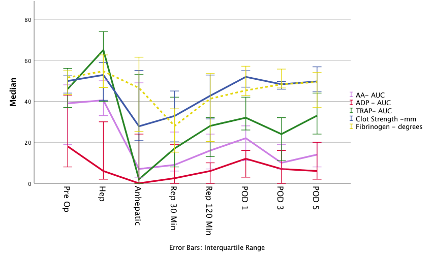
The median MELD score was 18. 64% of patients had baseline platelet counts less than 100,000. Clot strength was lowest during the anhepatic phase (P<0.05), returning to baseline by POD1 through POD5 (figure). Fibrinogen was lowest at 30 minutes after reperfusion (P<0.05), but regained baseline by POD1 (figure). Platelet counts remained low relative to baseline for POD1, 3, and 5 (85,000; 45,000; 51,000; 45,000. P<0.05). Platelet function of all three pathways reached their nadir during the anhepatic phase, and recovery began during reperfusion (figure). Platelet function improved by POD1 in all 3 pathways, but ADP was significantly low for POD3 (p=0.009) and 5 (p=0.036). AA and TRAP functions were depressed from POD1-5 but did not reach significance. Baseline platelet function did not differ from controls, but all three pathways were depressed by POD1 in transplant patients (p<0.05). TRAP regained function comparable to controls by POD5, but AA and ADP remained lower (P<0.001).

Conclusion:

Platelet agonist pathways were not upregulated following surgery in LT recipients. Patients regain baseline clot strength by POD1, despite a lower platelet count and suppressed function. Recovery in clot strength parallels an increase in fibrinogen while platelet function of the ADP and AA pathways remains suppressed through POD5. These measured coagulation derangements during and after LT warrant further investigation as current post-operative anti-thrombotic prophylaxis may be off target.

88.15 An Animal Model of Human Peripheral Arterial Bending and Deformation

R. El Khoury1, A. Nikanorov2, E. McCarroll3, G. LeClerc4, L. Guy4, M. Laflamme4, A. Mailloux4, L. B. Schwartz1,3  1Advocate Lutheran General Hospital,Department Of Surgery,Park Ridge, IL, USA 2National Coalition on Healthcare,Washington, DC, USA 3EFemoral Medical,Los Altos, CA, USA 4AccelLAB,Boisbriand, QUEBEC, Canada

Introduction:  Endovascular recanalization has become the treatment-of-choice for peripheral arterial disease. Although a wide variety of intravascular devices are approved for this purpose, anatomic and clinical outcomes following catheter-based intervention remain suboptimal as implanted stents will repetitively deform and fatigue during lower extremity movement. Pre-clinical testing of devices is often inadequate, given the lack of relevant animal models that genuinely mimic the complex motion of peripheral human arteries. The purpose of this study was to test the hypothesis that deformation of the human peripheral vasculature could be qualitatively and quantitatively modeled using an experimental animal. 

Methods:  Pelvic and lower extremity contrast angiography was performed in domestic Landrace-Yorkshire farm pigs weighing 25-35 kg. Anteroposterior images were obtained with the hind limbs naturally extended then repeated, (1) flexed approximately 90° at the hip and knee, (2) over-flexed in a non-physiological fashion. Two separate peripheral arterial segments were studied: an iliac segment between the circumflex iliac and deep femoral arteries and a femoral segment between the deep femoral and circumflex femoral arteries. Quantitative vascular angiographic analysis was utilized to measure arterial diameter, length and deformation. Percent axial arterial compression was calculated as the distance between constant arterial branches before and after hind limb flexion; bending was measured by planimetry and expressed in degrees.

Results: Eight iliofemoral arteries in four animals were imaged. Mean luminal diameters of the iliac and femoral segments in the neutral position were 5.4±0.5 mm and 4.6±0.5 mm. Hind limb flexion induced profound arterial compression, 17±8% and 29±6% within the iliac and femoral segments. Bending was also severe; with physiologic flexion to 90°, iliac and femoral arteries bent 36°±10° and 76°±13°. With extreme flexion of the porcine hind limb, the femoral artery could be reliably bent >90°. The observed deformation within the porcine iliac artery quantitatively exceeded the deformation observed historically within the human superficial femoral artery (~5% compression and 10° bending). Similarly, the degree of deformation within the porcine femoral artery quantitatively exceeded the deformation observed historically within the human popliteal artery (~10% compression and 70° bending).

Conclusion: Significant non-radial deformation of the porcine iliofemoral arteries was observed during manual hind limb flexion. The measured axial compression and bending deformation exceeded that observed in human peripheral arteries. The porcine iliofemoral model constitutes a “worst case” scenario for testing deformation and fatigue of intravascular devices indicated for the human peripheral vasculature.


88.14 Decreased Patency in Left-Sided Arteriovenous Grafts in a Porcine Model

S. Liu1, T. Wang1, J. Wang2, T. Isaji1, A. Feher4,5, N. Boutagy4,5, A. Sinusas4,5, L. Niklason2,3, A. Dardik1  1Yale University School Of Medicine,Surgery,New Haven, CT, USA 2Yale University School Of Medicine,Anesthesiology,New Haven, CT, USA 3Yale University School Of Medicine,Biomedical Engineering,New Haven, CT, USA 4Yale University School Of Medicine,Cardiology,New Haven, CT, USA 5Yale University School Of Medicine,Translational Research Imaging Center,New Haven, CT, USA

Introduction:
Complications of vascular access for hemodialysis remain a major source of morbidity and cost. Patients unable to have a fistula require a prosthetic graft, most commonly polytetrafluorethylene (PTFE), despite poor primary patency and increased thrombosis, stenosis, and need for reinterventions. Choice of laterality of arteriovenous graft placement is typically dependent on patient handedness, after consideration of adequate vessel diameters. Since temporary dialysis catheters may have reduced patency on the left side, we hypothesized that left-sided arteriovenous grafts may have reduced patency in a pre-clinical model.

Methods:
Ten Yorkshire male pigs (mean weight 48 kg, age 3.4 months) underwent ipsilateral or bilateral placement of arteriovenous grafts from the proximal common carotid artery (CCA) to the distal internal jugular vein (IJV) using PTFE (6 mm diameter, 6-7 cm length). Pigs were observed for 1, 2, or 3 weeks. Select pigs underwent ultrasound measurements of flow and ultrasound and caliper measurements of vessel diameters prior to graft placement, and some pigs underwent computed tomography angiography prior to the terminal procedure. Grafts and peri-anastomotic vessels were excised and analyzed with histology and immunostaining.

Results:
At baseline there was no significant difference in peak systolic or end diastolic velocities between the left and right CCA and IJV but the outer diameters of the CCA were smaller on the left side (4.2 versus 4.7 mm; p=0.0354). 10 left-sided and 8 right-sided PTFE grafts were placed; only 4/10 (40%) were patent on the left and 7/8 (88%) were patent on the right (p=0.03996, Chi-square). Post-operative histology showed thicker peri-anastomotic arterial walls on the left side (0.7 vs. 0.6 mm; p=0.0383) with greater intima-media surface areas (1.1 vs. 0.8 mm2; p=0.0286) compared to the right side. These differences were not seen between the left and right IJV. There was no significant difference in the number of smooth muscle cells, total proliferative cells, or extracellular matrix composition between the left and right sides; however, left-sided grafts had increased luminal macrophages at the arterial anastomosis compared to right-sided grafts (8.4 vs. 2.8 cells/hpf; p=0.0007). 

Conclusion:
Left-sided arteriovenous grafts are associated with significantly lower short-term patency compared to right-sided grafts; left-sided peri-anastomotic carotid arteries had increased wall thickness, medial area and increased numbers of macrophages near the arterial anastomosis despite similar blood flow.  These results suggest that left and right-sided arteries used in arteriovenous grafts may have different remodeling that translates to altered patency, and these differences should be considered when planning graft placement.
 

88.12 Human Platelet Lysate Improves In vitro Survival of Human Diabetic Mesenchymal Stem Cells

L. P. Brewster1,2, J. Raykin1,2  1Emory University School Of Medicine,Surgery,Atlanta, GA, USA 2Atlanta VA Medical Center,Surgery And Research Services,Decatur, GA, USA

Introduction:  Diabetic persons undergo premature aging of their vasculature leading to an earlier onset and more severe presentations of cardiovascular disease. Thus, they have great need for regenerative therapies. Mesenchymal stem cells (MSC) are one promising regenerative therapy that may help prevent vascular complications of diabetes. The objective of this work is to identify whether MSC survival pathways are improved by platelet lysate (PL), and if so, whether PL gel confers benefit to MSC survival and regenerative function on endothelial cells (EC).

Methods:
MSCs from Diabetic PAD (dMSC) and healthy patients were used. Luminescence studies were performed in MSCs transfected with luciferin lentivirus. Secretome analysis was performed with RayBiotech angiogenesis assays. Akt cell survival pathways were quantified by multiplex analyses.

Results:  dMSCs and healthy MSCs have increased retention and survival (2-3x) in PL gel than that seen in saline injection (most commonly used clinically). EGF expression in dMSCs is significantly increased (>5x; P<.0001) in PL gel over that in control groups. dMSCs had significant down-regulation of pAKT compared to healthy MSCs and MSCs from PAD patients without diabetes. Further, PL drastically changes the secretome profile of dMSCs compared to FBS. (Figure

Conclusion:

Initial concerns with cells from cardiovascular patients have now come into question for PAD patients. Improving dMSC survival could be important to PAD patients. In this work, we show that PL gel improves MSC survival in vivo and dMSC EGF expression, and that dMSCs have Akt signaling defects that may be reversible with PL supplementation.

88.11 Vein Graft Failure And Single Cell Genomics

N. Momi2,6, P. Liang2,6, S. Bhasin2,5, F. W. LoGerfo2,6, C. Ferran2,4, L. Pradhan-Nabzdyk2,6, M. Bhasin2,5  2Harvard School Of Medicine,Brookline, MA, USA 4Beth Israel Deaconess Medical Center,Center For Vascular Biology Research And Division Of Nephrology, Department Of Medicine,Boston, MA, USA 5Beth Israel Deaconess Medical Center,Genomics And Proteomics Center, Division Of Interdisciplinary Medicine And Biotechnology,Boston, MA, USA 6Beth Israel Deaconess Medical Center,Division Of Vascular And Endovascular Surgery, Department Of Surgery,Boston, MA, USA

Introduction:  Bypass grafting using autologous vein conduits is the cornerstone therapy for arterial occlusive disease. However, 30–50% of lower extremity vein grafts (VG) fail within 5 years from surgery. We hypothesize that VG implantation injury causes spatial and temporal genetic changes in the VG, triggering a cascade of interrelated molecular events starting with inflammation and culminating in vessel wall remodeling eventually leading to Intimal Hyperplasia (IH). This study aims to investigate the genomic contribution of individual cells including smooth muscle cells (SMC), endothelial cells (EC), adipocytes (Adipo), fibroblasts (FB), immune cells (T-Cells, Macrophages, NK etc.) towards VG failure and IH. 

Methods:  Canines underwent cephalic vein to common carotid artery interposition surgery. Cephalic VG and contralateral vein (CV) were harvested 24hrs post-surgery. Upon collagenase-I digestion, samples were subjected to 10X Single Cell (sc) genomics-based droplet sequencing to quantify and compare cell enrichment by ImmuneQuant software-based annotation and t-SNE clustering. Further, individual cell pools were validated by IHC. Additionally, bulk RNA-seq and ingenuity pathway analysis (IPA) was performed to elucidate key mediators/pathways. 

Results: Our results show a dramatic difference in the cell types present in VG vs. CV 24hrs post-surgery. CV predominantly had non-immune cells (93% of total distributed in 9 clusters), including EC, SMC, FB and Adipo, with fewer immune (7%, T-Cells). In striking contrast, VG demonstrated partial loss of ECs, Adipo and FB with simultaneous infiltration of immune cells, accounting for 89% of total cells, with 4 T-cell clusters (52%) (CD3D+PTPRC+CD19+IL3RA+), primarily Th1 cells (67% of T-Cells), 4 monocyte/macrophages clusters (34%) (CD68+TGFB1_CANFA+IL1R1+) and 1 B-cell cluster (3%). Further, supervised analysis of individual clusters highlighted differential expression of several genes including, chemokines (IL-8, CCL4/2), TFs (FOS) and extracellular matrix/vascular components (collagen/COL1A1, endothelin/EDN). Intriguingly, these findings were corroborated by bulk-RNA-seq indicating significant activation of inflammatory pathway, Th1 pathway, CD28 signaling in Th1 cells, PI3K signaling in B-cells, IL-8 and chemokine signaling. Finally, IHC analysis also validated the higher number of CD3T-cells in the VG. 

Conclusion: Significant monocytes/macrophages and T-cell infiltrates in VG vs CV reveal an early contribution of both innate and adaptive immune response towards the pathophysiology of VG remodeling. Sc-genomics provide a better understanding of the complex ecosystem that governs implantation injury and unsuccessful adaptations of the VG leading to graft failure, thereby opening avenues for plausible effective preventive measures and early therapeutic targets. 


88.10 Molecular Mechanism of IL-1β Induced Endothelial Dysfunction After Vascular Injury

M. Polcz1, P. Komalavilas1, J. Cheung-Flynn1, C. Brophy1  1Vanderbilt University Medical Center,Department Of Surgery,Nashville, TN, USA

Introduction:

Exposure of human saphenous vein to normal saline (NS), a non-buffered, acidic solution (pH 5.0-5.6) results in endothelial injury and impaired endothelial function, potentially resulting in decreased patency of vein grafts used for bypass. Previous data have suggested that NS induced injury leads to membrane disruption, ATP release, activation of the P2X7 receptor and p38 MAPK pathway.  This leads to an amplifying response resulting in inflammation and endothelial dysfunction. We hypothesized that increases in proinflammatory cytokine IL-1β, after activation of the P2X7R/p38 MAPK pathway, directly participates in endothelial dysfunction after injury.

Methods:

To determine the effects of IL-1β on endothelial-dependent relaxation, rat aorta (RA, n=12-20) were treated with IL-1β at 10, 50, and 100ng/ml in a muscle bath for 3 hours. Additionally, co-treatment with SB203580 (SB; 20µM), a p38 MAPK inhibitor, was performed.

To determine whether endothelial dysfunction associated with IL-1β is related to decreased nitric oxide (NO) bioavailability, human saphenous vein endothelial cells (HSVEC; n=4-5) were treated with IL-1β (10ng/ml) for 3, 6, and 24 hours. Western Blot analysis of endothelial nitric oxide synthase (eNOS), phosphorylated eNOS, and arginase II levels were performed. Protein-antibody complexes were visualized and quantified using the Odyssey Infrared Imaging System (LiCor Biosciences, Lincoln, NE). Protein levels were normalized to GAPDH level and phosphorylation was calculated as a ratio of the phosphorylated protein to total protein.

Results:

Treatment of RA with IL-1β led to a dose-dependent impairment of endothelial relaxation of pre-contracted tissues. This effect of IL-1β on endothelial dysfunction was prevented by cotreatment with SB, suggesting that IL-1β signals via a p38 MAPK-associated pathway. Additionally, treatment of HSVEC with IL-1β resulted in increased relative levels of arginase II at 6 and 24 hours but had no effect on eNOS levels or phosphorylation in HSVEC (Figure 1).

Conclusion:

IL-1β promotes endothelial dysfunction through increased arginase II levels resulting in decreased NO bioavailability.  These data provide a plausible hypothesis by which NS-induced injury leads to inflammation and endothelial dysfunction which may be associated with eventual vein graft failure.
 

88.09 Lower Shear Stress Magnitude in Female Mice During Arteriovenous Fistula Maturation

S. Ono1, T. Kudze1, T. Isaji1, T. Hashimoto1, B. Yatsula1, H. Liu1, T. Nishibe2, J. Koizumi3, A. Dardik1,4  1Yale University School of Medicine,Vascular Biology And Therapeutics Program And Department Of Surgery,New Haven, CT, USA 2Tokyo Medical University,Department Of Cardiovascular Surgery,Shinjuku, Tokyo, Japan 3Tokai University School of Medicine,Department Of Diagnostic Radiology,Isehara, KANAGAWA, Japan 4VA Connecticut Healthcare System,Department Of Surgery,West Haven, CT, USA

Introduction: The arteriovenous fistula (AVF) is the preferred method of dialysis access due to its proven superior long term outcomes. However, women have lower rates of AVF maturation than men (38% vs. 60%), preventing optimal AVF use. We used a novel mouse AVF model that recapitulates human AVF maturation to test the hypothesis that there is a difference in male and female AVF maturation.

Methods:   Aortocaval fistulae were created in male and female C57BL/6 mice (9-10 wks). At days 0, 3, 7, 14 and 21, infrarenal aortic and IVC diameters and flow velocity were monitored by Doppler ultrasound and used to calculate the resistance index, blood flow and shear stress. AVF were harvested at day 21 and AVF wall thickness was measured by computer morphometry; proteins were examined using immunofluorescence and mRNA by qPCR.

Results:  Female mice weighed less throughout the whole period (p<0.0001). At baseline, female mice had lower infrarenal IVC velocity (p=0.0005) and smaller magnitudes of shear stress (p=0.0003); although female mice had smaller infrarenal aortic diameter (p=0.0198), there was no significant difference in infrarenal IVC diameter (p=0.5112).  After AVF creation, both the female and male aorta (p=0.5681) and IVC (p=0.5680) dilated similarly and the aortic resistance index decreased similarly (p=0.0743). However, female mice had lower aorta (p=0.0187) and IVC mean velocity (p<0.0001); female mice also showed less blood flow volume in the aorta (p=0.0069) and IVC (p=0.0087) and lower shear stress magnitude in the IVC (p<0.0001) without any significant differences in aortic shear stress magnitude (p=0.31).  There were no significant differences in infrarenal IVC wall thickness either at baseline (p=0.9617) or at day 21 (p=0.2931). Although KLF2 mRNA was decreased in the female AVF on day 21 (p=0.048), there were no differences in protein expression (p=0.5224).

Conclusion: AVF in female mice have lower velocity, blood flow volume and magnitudes of shear stress without any differences in wall thickness or protein expression.  These findings suggest that hemodynamic changes in the fistula may play an important role underlying the diminished rates of AVF maturation in women.

88.08 Topographic Vascular Grafts – A Biomimetic strategy for Anti-thrombotic Surfaces

L. Pocivavsek1, J. Pugar3, N. N. Nath2, K. Salem2, W. Wagner2, S. Ye2, E. Tzeng2, S. Velankar3  1The University Of Chicago,Vascular/Surgery,Chicago, IL, USA 2University Of Pittsburgh,Vascular/Surgery,Pittsburgh, PA, USA 3University Of Pittsburgh,Chemical Engineering,Pittsburgh, PA, USA

Introduction:  The inner surfaces of arteries and veins are naturally anti-thrombogenic, whereas synthetic materials placed in blood contact commonly experience thrombotic deposition that can lead to device failure or clinical complications. We present a bioinspired strategy for self-cleaning anti-thrombotic surfaces using actuating surface topography motivated by our biomechanical study of arterial topography. 

Methods:  Utilizing finite element simulations, we studied the evolution of arterial topography as a function of pressure. Arterial luminal and wall geometries were segmented from histology slides of un-fixed human and mouse muscular arteries. The reconstructed arteries were imported into the finite element software ABAQUS, and simulations at physiologic conditions were conducted. Experimental counterparts were constructed utilizing silicone composites cylinders, 3 mm in diameter, and with varying luminal wrinkle wavelengths between 50 and 1000 microns. The cylinders were actuated at 1 Hz between diastolic and systolic pressures while filled with whole blood. At the end of 3 hours of actuation, the adhered surface platelet density was measured. 

Results: The luminal surface at zero pressure is highly wrinkled with a large local curvature that is proportional to the inverse of the wrinkle wavelength. Computationally, we found that upon inflation, the arterial luminal topography changes from this highly wrinkled state to a nearly flat surface upon reaching systolic pressures (see figure 1I). Experimentally, we found that topographic surface actuation dramatically decreased surface platelet adhesion: 90%, 95%, and 98% decrease in platelet deposition relative to unactuated surfaces for 1000, 250, and 80 micron wavelength surfaces, respectively (see figure 1 II A-C). Furthermore, we found a strong correlation between the degree of surface self-cleaning and wavelength; shorter wavelengths proved to be far more efficient at preventing un-wanted platelet adhesion than longer wavelengths. 

Conclusion: We show that arterial topography in native vessels can actuate as a function of physiologic pulse pressure. Furthermore, such active surface topography is shown in an experimental system to prevent platelet adhesion, with smaller wavelengths being more effective than longer ones at surface renewal. This work presents a novel bio-mimetic strategy geared towards creating durable small caliber vascular grafts with long term patency by preventing unwanted platelet surface fouling. 


88.07 Differential expression of Cathepsin L in the coronary arteries of atherosclerotic swine.

P. Gunasekar1, J. Dabestani1, D. K. Agrawal1, J. A. Asensio1  1Creighton University Medical Center,Department Of Surgery, Div. Trauma Surgery, Department Of Clinical & Translational Science,Omaha, NE, USA

Introduction:
Neointimal hyperplasia and restenosis following interventional procedures, including percutaneous transluminal coronary angioplasty (PTCA) and intravascular stenting still remain a significant clinical problem. These interventional procedures cause endothelial denudation and damage to intimal and medial layer which stimulates intimal smooth muscle proliferation and extracellular matrix deposition resulting in intimal hyperplasia (IH) and restenosis. Vascular sterile inflammation has been attributed to the formation of IH. Cathepsin L (CTSL), a member of lysosome protease, is highly associated with diet-induced atherogenesis and IH in animal studies. Vitamin D regulates several proteases and protease inhibitors in different cell types, contributing to its regulatory effects of cell physiology. In our study, we examined the effect of vitamin D on CTSL activity in the coronary arteries of atherosclerotic swine.

Methods:
Yucatan microswine were fed with a high cholesterol atherosclerotic diet. The swine received approximately 500 IU of vitamin D3/per day on the vitamin D-deficient diet, 2,500-3,500 IU of vitamin D3/per day on vitamin D-sufficient diet, and 4,500-5,500 IU of vitamin D3/per day on the vitamin D-supplement diet. After 5-6 months of the experimental diet, PTCA (percutaneous transluminal balloon angioplasty) was performed in the left circumflex coronary artery (LCX) in each swine.  After a year of the diet, angiography and optical coherence tomography (OCT) imaging were performed and swine euthanized. Coronary arteries were embedded in methyl methacrylate or paraffin. Tissue sections were stained with H&E, trichrome, and Movat’s pentachrome.  The expression of Ki67 (proliferation marker), CCR7 (Macrophage marker), Cathepsin L (lysosomal proteases) was evaluated by Immunofluorescence and Immunohistochemistry.

Results:
There was significantly greater density of Ki-67+ cells in post angioplasty LCX in vitamin D-deficient swine compared to vitamin D-sufficient swine and vitamin D-supplemented swine. CCR-7 was found to be higher in vitamin D deficient than vitamin D-sufficient swine and vitamin D-supplemented swine. CTSL expression and its activity were significantly increased in post angioplasty LCX of vitamin D-deficient swine than the supplemented swine.

Conclusion:
Cathepsin L drives IH and macrophage infiltration in coronary arteries after angioplasty in atherosclerotic swine. These findings suggest that vitamin D inhibits CTSL and thus has direct effect on neointimal hyperplasia after coronary intervention.
 

88.06 Novel “TopoGraft”: A Potential Solution for Platelet Aggregation

N. Nath1, L. Pocivavsek2, Y. Gao3, K. Salem1, S. Velankar3, E. Tzeng1  1University of Pittsburgh,Vascular Surgery,Pittsburgh, PA, USA 2University Of Chicago,Vascular Surgery,Chicago, IL, USA 3University of Pittsburgh,Department Of Chemical Engineering,Pittsburgh, PA, USA

Introduction: The tissue-blood interface represents a dynamic interaction between the endothelial cells (EC) and circulating RBCs, platelets and inflammatory cells. While ECs and underlying smooth muscle cells are vital to vascular homeostasis, the contribution of an artery’s dynamic luminal surface to prevent vascular thrombosis has not been previously evaluated. It has been reported in literature that vasoconstricted or relaxed arteries exhibit a corrugated luminal surface that flattens uniformly under distention, a transition, however, with unclear significance. We have shown that the arterial lumen possesses a regular wrinkled pattern that undergoes phasic wrinkling and flattening, offering a unique surface renewal mechanism. We hypothesize that uniquely incorporating this dynamic topography into vascular grafts may reduce platelet aggregation.

Methods:  Pig carotid arteries were subjected to pressures ranging from 40-140 mmHg, fixed, and examined histologically for luminal surface wrinkle amplitudes. Synthetic grafts were created using strain mismatch between two layers of implantable silicone of different stiffness to produce grafts with wrinkled or smooth luminal surfaces. An ex vivo pulsatile pump circuit was designed to cyclically distend wrinkled and smooth grafts to create dynamic topography. Activated platelets were circulated through the pump system at systolic pressures of 100-120 mmHg. Optical coherence tomography was used to capture cyclic changes of luminal surface. Grafts that were restricted by an external constraint to prevent cyclical distention were also tested. After platelet exposure, grafts were stained with Wright stain to quantify platelet adherence.

Results: Arteries distended at 40 mmHg had an average wrinkle amplitude of 20.52 ± 4.13 µm which decreased to 5.86 ± 1.69 µm at 140 mmHg (p<0.05). Incorporating luminal wrinkles into vascular grafts (Fig. 1A) with pulsatile flow, undergoing dynamic surface wrinkling and flattening (Fig. 1C) , reduced platelet deposition by 83% compared to smooth grafts (547 ± 96 vs. 4704 ± 573 per HPF, respectively; p<0.05). Constrained wrinkled grafts with static topography exhibited a doubling of platelet accumulation similar to that of smooth surface grafts constrained or undergoing cyclical distention (p<0.05 vs. wrinkled grafts with dynamic topography, Fig. 1B).

Conclusion: Arteries possess luminal topography that consists of a wrinkled surface that flattens under systolic pressures. Incorporating this dynamic luminal topography onto a synthetic graft reduced platelet aggregation. Dynamic luminal topography offers a unique method of preventing thrombus formation and may improve prosthetic vascular graft patency.


88.05 Ex vivo Isolated Vessel Perfusion for Assessment of Vascular-Targeted Nanomedicines

T. Lysyy1, L. Bracaglia2, A. Vaish1, O. Abousaway1, J. S. Pober4, G. Telides1, M. W. Saltzman2,3, G. T. Tietjen1  1Yale University School Of Medicine,Department Of Surgery,New Haven, CT, USA 2Yale University,Department Of Biomedical Engineering,New Haven, CT, USA 3Yale University,Department Of Chemical Engineering,New Haven, CT, USA 4Yale University School Of Medicine,Department Of Immunobiology,New Haven, CT, USA

Introduction: Vascular endothelial cells (ECs) are primary targets of and active participants in transplant rejection. We hypothesize that ex vivo treatment of graft ECs with anti-inflammatory or immunosuppressive agents can improve anti-rejection efficacy and reduce off target effects. Nanoparticle (NP) carriers have the potential both to deliver and mediate sustained release of drugs. Retention of polymer NPs in ECs can be improved with antibody-mediated targeting, but clinical optimization of this approach requires relevant experimental models. Cell culture alters the properties of ECs.  We aim to establish a reliable experimental model both to investigate NP targeting in isolated human vessels and to quantitatively evaluate effects of endothelial targeted nanomedicines.

Methods: Twelve de-identified human umbilical cords were obtained after Caesarean sections under a protocol approved by Yale Human Investigations Committee. Umbilical arteries were isolated and 10 cm vascular segments were subsequently connected to the perfusion system (Fig. 1 A). Fluorescent polymeric NPs (150nm) were conjugated to either Ulex europaeus agglutinin I lectin (ULEX) or to mouse IgG1 isotype antibody using EDC-NHS chemistry, using approaches we have optimized (Sci Transl Med 2017). NPs were spiked into intravascular perfusate (M199 media+ serum) at a concentration of 0.5 mg/mL. After 90 min of perfusion, vascular samples were washed and whole-mount specimens were analyzed by quantitative microscopy (n=12 images each).

Results: The perfusion loop design can house six vessel segments with independently manipulated flow rates and intravascular perfusate composition. To demonstrate the utility of this platform for quantitative analysis of NP targeting, we evaluated relative accumulation of ULEX-conjugated NPs compared to a nontargeted formulation (control).  ULEX-NPs were retained to a higher degree compared to nontargeted NPs as detected by quantitative microscopy (Fig. 1 B and C; Area of NP (pixels) 13 454±6796 versus 129.4±61.66 respectively, P<0.0002). These preliminary results demonstrate the capacity of this platform to quantitatively evaluate NP targeting efficacy.

Conclusion: We have developed a medium throughput isolated vessel perfusion system that is inexpensive and easy to produce. It can be adapted to test a variety of relevant targeting parameters, such as vessel size, type, pressure, flow rate, and shear stress. This system has the potential to improve clinical translation of endothelial-targeted nanomedicines by providing a native vascular context that retains the capacity for robust quantification without sacrificing translational relevance. Moreover, this ex vivo approach can facilitate subsequent in vivo experiments by treating human vessels prior to implantation in humanized mouse models.


88.04 Improvement of Bile Metabolism in Hepatic Ischemia-Reperfusion Injury by NRF2 Activation in Rats

J. Kim1, A. Martin1, J. Yee1, L. Fojut1, M. A. Zimmerman1, J. C. Hong1  1Medical College Of Wisconsin,Transplant Surgery,Milwaukee, WI, USA

Introduction: Cholestasis is an inevitable consequence of severe hepatic ischemia-reperfusion injury (IRI), and no treatment is available to prevent it. The detrimental function of membrane bile transporters is presumed to be the underlying precedent condition of cholestasis that arises during ischemia. Nuclear factor erythroid 2-related factor 2 (NRF2) mediates antioxidant gene transcription following oxidative stress. Previous studies suggest that bile transporter genes are regulated by NRF2 in a drug-induced liver injury model. However, the role of NRF2 in bile metabolism during hepatic IRI and its therapeutic potential are unknown. Bile metabolism is mediated by membrane transporters, such as bile salt export pump (BSEP), multidrug resistance-associated protein 2 (MRP2), MRP3, and anion exchange protein 2 (AE2). We investigated the validity of NRF2 activation by using bardoxolone methyl (BARD) as a treatment to increase the transcriptional activities of bile transporters thereby to improve bile metabolism in hepatic IRI.

Methods: An empty vehicle (control) or bardoxolone methyl (2 mg/kg) was infused into the inferior vena cava of Sprague-Dawley rats 5 min before the ischemia. Hepatic warm ischemia was induced by clamping the median and left lobes. Sham surgery, 60 min or 90 min periods of hepatic warm ischemia were applied, and samples were obtained after 24 h of reperfusion (n=5 per each group). Serum levels of bile acids were tested, and hepatic tissue was sampled to determine the quantitative polymerase chain reactions (qPCR) of bile transporters. The results of qPCR were presented as mRNA levels relative to those of the glyceraldehyde 3-phosphate dehydrogenase gene (Gapdh).

Results: After 60 min of ischemia and 24 hours of reperfusion, the hepatic tissue mRNA levels of Mrp2 and Mrp3 in the BARD group were higher when compared to those of the control (P=0.032 and 0.008, respectively; Figure A). After 90 min of ischemia and 24 hours of reperfusion, the mRNA levels of Bsep and Ae2 in the BARD group were lower than those of the control (P=0.008 and 0.016, respectively; Figure B). The serum bile acid levels were significantly lower in the BARD group after 60 min of ischemia and 24 hr of reperfusion (P=0.016, Figure A).

Conclusion: NRF2 contributes to the transcriptional activity relevant to bile metabolism during hepatic IRI. The activation of NRF2 at the time of ischemia can increase transcription activities of bile transporters to improve hepatic bile metabolism after reperfusion. The effect of NRF2 activation disappears when the warm ischemia time is prolonged. This study suggests a novel therapeutic target in hepatic IRI.


88.03 Penicillin’s Protective Effect on Small Bowel Ischemia is Mediated by H,KATPase

V. M. Baratta1, T. M. Gisinger1,2, M. J. Barahona1, J. Ollodart1, D. Mulligan1, J. P. Geibel1,3  1Yale University School Of Medicine,Department Of Surgery,New Haven, CT, USA 2Paracelsus Medical University,Department of Medicine,Salzburg, SALZBURG, Austria 3Yale University School Of Medicine,Department of Cellular and Molecular Physiology,New Haven, CT, USA

Introduction: One of the barriers to successful small bowel procurement is the sensitivity of the intestinal mucosa to ischemia. During procurement, the University of Wisconsin Universal Organ Preservation (UW) solution is used for in situ flushing and cold storage to minimize progression to ischemia. In addition, donor intestines undergo selective bowel decontamination with antibiotics and antifungals. Recently, we demonstrated that exposure of the small intestine to Penicillin G can protect from ischemic injury in a rat model. This finding may be helpful during the procurement process for intestinal transplantation. Here, we demonstrate that the protective effect is partly mediated by activation of an H,KATPase, independent of the Nitric Oxide (NO) pathway.

Methods:  The small bowel segments were harvested from rats and perfused with an ex-vivo intestinal perfusion device. Each intestinal segment was maintained at 37°C and perfused both from the luminal and basolateral side. As previously described, FITC-Inulin (fluorescein isothiocyanate-inulin), was used to assess the ischemic conditions of the colonic grafts in real-time. Small bowel segments were perfused with 4-(2-hydroxyethyl)-1-piperazineethanesulfonic acid (HEPES-Ringer) solution. To create an ischemic environment, HEPES-Ringer was pre-saturated with 100% N2 and exposed to the extraluminal components of all rat segments. For the experimental tissues, the intraluminal components were perfused with 5 mM Penicillin G and 10 μM SCH-28080, a known H,KATPase inhibitor. The intraluminal components of the control group were exposed to 5 mM Penicillin G. To test the Nitric Oxide (NO)-dependent mechanism, we used L-NAME (N(ω)-nitro-L-arginine methyl ester), an inhibitor of NO synthesis. The intraluminal compartments of the experimental tissue were exposed to 30 μM L-NAME with 5 mM Penicillin G, while control tissues were exposed to 5 mM Penicillin G.

Results: The small bowel samples exposed to Penicillin G and SCH-28080 exhibited a significant decrease in FITC-Inulin fluorescence, compared with the control colonic tissue exposed to Penicillin G, (39.83 ± 2.601 μM FITC-Inulin vs 47.37 ± 0.7288 μM FITC-Inulin, respectively p 0.0163). We observed no statistically significant difference in the FITC-Inulin concentration between tissues exposed to L-NAME with Penicillin G versus tissues exposed to only Penicillin G.

Conclusion: Our study unveils the mechanism of Penicillin G’s protective effect from ischemia. Our results indicate that Penicillin G’s protective effect against ischemia is through stimulation of the H,KATPase and is not NO-dependent. Therefore, Penicillin G not only has its well-known antimicrobial properties, but also appears to modulate a transport protein. In the future, Penicillin G may be implemented during the early procurement phase of intestinal transplantation to minimize ischemia.


88.02 Vitamin D supplementation Reduces HMGB1, TLRs and RAGE in Post-interventional Coronary Arteries

P. Gunasekar1, J. Dabestani1, D. K. Agrawal1, J. A. Asensio1  1Creighton University Medical Center,Department Of Surgery, Div. Trauma Surgery, Department Of Clinical & Translational Science,Omaha, NE, USA

Introduction:

Coronary angioplasty and stent implantation is a common coronary procedure for patients with coronary artery disease. These interventional procedures stretch and denude the endothelial layer. This promotes local inflammatory response in the injured vessel wall, which is characterized by smooth muscle cell proliferation, migration, neointimal formation. Clinical studies support that plasma vitamin D deficiency is associated with increased risk for coronary artery disease (CAD). It is unclear whether vitamin D status is causally related to CAD or is a marker of health. In this study, we examined the inflammatory profile of coronary arteries in atherosclerotic swine.

Methods:

Yucatan microswine were fed with high cholesterol atherosclerotic diet. The swine received approximately 500 IU of vitamin D3/per day on the vitamin D-deficient diet (VD DEF.), 2,500-3,500 IU of vitamin D3/per day on vitamin D-sufficient diet (VD SUF.), and 4,500-5,500 IU of vitamin D3/per day on the vitamin D-supplement diet (VD SUP.). After 5-6 months of the experimental diet, PTCA (percutaneous transluminal balloon angioplasty) was performed in the left circumflex coronary artery (LCX) and bare mental stent implantation in the left anterior descending coronary artery (LAD) for each swine. Six months following coronary intervention, angiogram and optical coherence tomography (OCT) imaging were performed and swine were euthanized. Coronary arteries were then embedded in methyl methacrylate or paraffin. Tissue sections were stained with H&E. The protein expression of HMGB1 (inflammation and necrosis marker), RAGE (receptor for advanced glycosylation end product), TLR2 and TLR4 (pattern recognition receptors) were evaluated by Immunohistochemistry.

Results:

Optical coherence tomography readings showed the degree of percentage area in-stent restenosis and PTCA was greatest in VD DEF. compared to VD SUF. or VD SUP. swine. We found a greater inflammatory profile in the coronary arteries of VD DEF. compared to VD SUF. or VD SUP. swine, based on histological staining and immunoreactivity to HMGB1, RAGE, TLR2, TLR4, in both LCX and LAD. The ligands for RAGE and receptor for HMGB1 (TLR2 and TLR4) were highly expressed in neointimal cells in stented LAD arteries of VD DEF. swine. This inflammatory profile decreases with increasing the levels of Vitamin D.

Conclusion:

Vitamin D deficiency increases the HMGB1-mediated pathways, resulting in the release of inflammatory cytokines from macrophages and other immune cells; and the recruitment of inflammatory cells through TLR4. Vitamin D supplementation suppresses the cytokine activity and prevents neointimal proliferation and restenosis from damage caused by PTCA in atherosclerotic swine. Vitamin D supplementation could be used as an adjunct therapy to prevent intimal hyperplasia and restenosis following coronary interventions.
 

88.01 Transcriptional Analysis of Mouse Hepatic Ischemia/Reperfusion Injury

S. Duarte1, Z. Huo2, U. Kim2, A. Coito1, A. Zarrinpar2  1University Of California – Los Angeles,Surgery,Los Angeles, CA, USA 2University Of Florida,Surgery,Gainesville, FL, USA

Introduction:
Hepatic ischemia/reperfusion injury (IRI) is a major risk factor for acute rejection and early graft dysfunction in liver transplantation. The large and complex landscape of regulatory signaling events in IRI has not been systematically characterized. While many of the factors responsible have been identified, no potent pharmacologic treatments have been developed. To expand the understanding of the hepatic transcriptional regulatory changes during the early stages of IRI, we used RNASeq-based analysis in a mouse warm hepatic IRI model.

Methods:
C57BL/6 mice were subjected to 70% partial warm ischemia for 90min, followed by 15min, 2h, or 6h of reperfusion. This was done in each of the three technical replicates. The baseline was developed using naïve mice. After ischemia and reperfusion injury, hepatic tissues were collected from euthanized mice, homogenized, and RNA was extracted. Generation of cDNA followed by next-generation sequencing allowed the construction of a whole-genome transcriptome map. These reads were then analyzed using HISAT2, StringTie, and Ballgownto align, assemble, and compute the abundance of the transcripts.

Results:
Using this method 29,832 unique reference sequences were identified from 20,565 unique genes. Principal component analysis resulted in clusters of the technical replicates, as well as gradual progression of the transcriptional profile from ischemia to 2 hours after IRI. However, after 6 hours the transcriptional profile was markedly different from the very early stages, indicating a shift in the activated signaling pathways. (Figure) Several characteristic patterns emerged in the transcriptional activity profiles. Oxidative Stress Response and JAK-STAT pathways are highly active in ischemic tissues and then their activity decreases upon reperfusion. Adipogenesis, fatty acid beta oxidation, and the unfolded protein response pathways demonstrate increased activity as early as 15 minutes after reperfusion. After 2 hours the adipogenesis pathway is still highly active but there is an upregulation of macrophage signaling. Finally at 6 hours after reperfusion the transcriptional activity profile is characterized by significantly more pronounced activity of the acute phase response, the sirtuin signaling, and IL-6 signaling pathways. 

Conclusion:
This study substantiates the complex activity of regulatory signaling pathways that have important roles in the progression of hepatic IRI. It identifies both previously unexamined pathways and corroborates previously implicated signals that are valuable potential targets for drug therapy. Enhancing or inhibiting these pathways has the potential to rescue IRI to a great extent and act as novel potential drug targets. 
 

87.20 Colonization of Neonatal Rats with Enterococcus faecalis

E. X. Zhang1, P. T. Delaplain2, J. Wang2, A. V. Grishin2, H. R. Ford3  1Shady Side Academy,Pittsburgh, PA, USA 2Children’s Hospital Los Angeles,Pediatric Surgery,Los Angeles, CA, USA 3University of Miami,Leonard Miller School Of Medicine,Miami, FL, USA

Introduction:  Necrotizing enterocolitis (NEC), a severe inflammation of the small intestine in premature neonates, is associated with bacterial colonization of the gut. We believe that the first colonizers of the gut may be either opportunistic pathogens or innocuous/protective bacteria. Artificial introduction of protective bacteria may be an efficient method of protecting against NEC. Enterococcus faecalis is one of the most common first colonizers of the intestine. There are multiple strains of E. faecalis, some of which could be protective. In this study, we investigated the efficiency of colonization of the intestines of neonatal rats with different strains of E. faecalis. 

Methods:  20 strains of E. faecalis possessing different combinations of biochemical phenotypes (hemolysis, sorbitol fermentation, kanamycin resistance, gelatin liquefaction, beta-galactosidase, xifaxan resistance) were previously isolated from 4-day old rats. Liquid cultures of these strains, grown in BHI medium, were mixed in equal proportions, and 10^8 cfu total were introduced to newborn rats with formula during their first feeding. After four days of the formula feeding – hypoxia, NEC-inducing regimen, samples of intestinal content from different animals were plated on blood agar-azide to isolate E. faecalis. Strains were identified by replica plating of E. faecalis colonies onto appropriate diagnostic media. 

Results: Intestinal loads of E. faecalis on day 4 varied from 10^4 to 10^7 cfu/mL. In all animals examined, E. faecalis populations were dominated by strains different from those that were artificially introduced, presumably acquired from their mothers. Of the introduced strains, only BB70 and 269 were found in large numbers.

Conclusions:

1. Four days after the artificial introduction of E. faecalis, loads of these bacteria differed by three orders of magnitude.

2. Maternally-derived strains of E. faecalis dominated the populations.

3. BB70 and 269 were the most consistent colonizers of the neonatal intestine.

Identifying efficient first colonizers among common intestinal bacteria is an important step towards creation of artificial bacterial communities with protective properties against NEC.