1.13 A Novel Folate Receptor-Targeted Optical Contrast Agent for Intraoperative Imaging of Lung Cancer.

C. J. Corbett1, J. D. Predina3,5,6, A. D. Newton2, M. Shin2, L. Sulyok2, L. Xia2, P. S. Low7,8, S. Singhal1,2,3,4  1University Of Pennsylvania,Perelman School Of Medicine,Philadelphia, PA, USA 2Hospital Of The University Of Pennsylvania,Department Of Surgery,Philadelphia, PA, USA 3Hospital Of The University Of Pennsylvania,Center For Precision Surgery,Philadelphia, PA, USA 4Hospital Of The University Of Pennsylvania,Department Of Thoracic Surgery,Philadelphia, PA, USA 5Harvard School Of Medicine,Brookline, MA, USA 6Massachusetts General Hospital,Department Of Surgery,Boston, MA, USA 7Purdue University,Department Of Chemistry,West Lafayette, IN, USA 8On Target Laboratories,West Lafayette, IN, USA

Introduction:

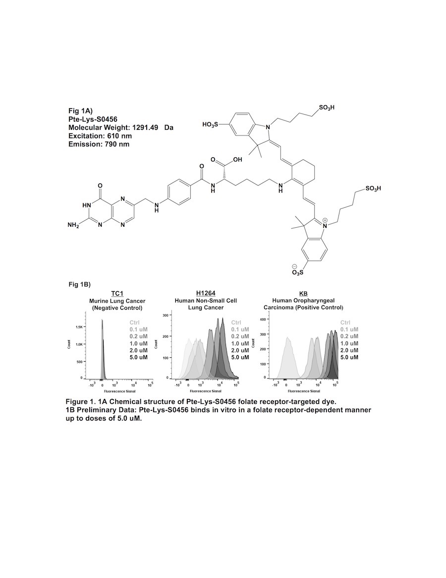
Previous attempts of folate receptor-targeted intraoperative molecular imaging (IMI) of pulmonary adenocarcinomas have been successful, but remain limited by photon-scatter and high levels of tissue reflection. One approach to overcome these limitations is by incorporating fluorophores displaying increased Stokes shifts (greater than 100 nm). In this study, we describe optical properties and preclinical data involving a third-generation folate receptor-targeted probe, Pte-Lys-S0456, that displays a Stokes shift measuring 180 nm.

Methods:

Excitation and emission spectra for Pte-Lys-S0456 were obtained with 10 nM aliquots measured with a luminometer. Next, several murine and human NSCLC models were co-cultured in variable concentrations of Pte-Lys-S0456 ranging from 100 nM to 10 uM for 2 hours. Following washout, fluorescence was assessed with flow cytometry and fluorescence microscopy. Fluorescent patterns were quantified and compared to previous folate receptor-targeted drugs (Folate-FITC and Folate-S0456). Following in vitro characterization, in vivo feasibility of systemc Pte-Lys-S0456 was tested using a small animal flank tumor model of cancer surgery (n=15). 

Results:

The peak excitation and emission wavelengths of Pte-Lys-S0456 were found to be 610 nm and 790 nm (Stokes shift of 180 nm). NSCLC models avidly bound Pte-Lys-S0456 in vitro in a folate receptor-dependent manner. Fluorescent signal of tumor cells co-cultured with Pte-Lys-S0456 resulted in increases in fluorescence up to a concentration of 5 uM (p < 0.001). Higher concentrations of Pte-Lys-S0456 did not improve tumor fluorescence. Upon fluorescent microscopic evaluation of tumor models, we observed strong cell membrane binding of Pte-Lys-S0456 in all groups. In cells exposed to the highest concentration, we observed internalization of Pte-Lys-S0456. When compared to Folate-FITC and Folate-S0456, we observed improved signal-to-background signal due to decreased tissue reflection and photon scatter.

Conclusion:

Pte-Lys-S0456 is a novel folate receptor-targeted optical contrast agent that binds NSCLC in a folate receptor-dependent manner. Improved optical properties associated with Pte-Lys-S0456 result in higher in vivo signal-to-background ratios, and thus may provide a more reliable agent for folate receptor-targeted IMI of human NSCLC.


1.11 Common Driver Mutation and Smoking History Affect Tumor Mutation Burden in Lung Adenocarcinoma

M. Nagahashi1, S. Sato2, K. Yuza1, Y. Shimada1, H. Ichikawa1, S. Watanabe3, S. Okuda4, K. Takabe5,6, M. Tsuchida2, T. Wakai1  1Niigata University Graduate School Of Medical And Dental Sciences,Division Of Digestive And General Surgeyr,Niigata City, NIIGATA, Japan 2Niigata University Graduate School Of Medical And Dental Sciences,Division Of Thoracic And Cardiovascular Surgery,Niigata City, NIIGATA, Japan 3Niigata University Graduate School Of Medical And Dental Sciences,Department Of Respiratory Medicine And Infectious Disease,Niigata City, NIIGATA, Japan 4Niigata University Graduate School Of Medical And Dental Sciences,Division Of Bioinformatics,Niigata City, NIIGATA, Japan 5Roswell Park Cancer Institute,Breast Surgery, Department Of Surgical Oncology,Buffalo, NY, USA 6University At Buffalo Jacobs School Of Medicine And Biosciences, The State University Of New York,Department Of Surgery,Buffalo, NY, USA

Introduction:
Although the expectation of immune checkpoint inhibitors, such as anti-PD-1 therapy, is tremendously high based on its dramatic efficacy, only a limited number of patients respond. Even worse, there is no definite biomarker to identify which tumor responds to the therapy. Recent progress in the genomic analysis using next-generation sequencing (NGS) technology has enabled comprehensive detection of mutations and tumor mutation burden (TMB) in cancer patients. The high TMB (TMB-H) tumor is defined as one with high somatic mutational rates, which correlates with the generation of neo-antigens and potential clinical response to immunotherapies. In this study, we analyzed the TMB in patients with lung adenocarcinoma utilizing NGS, and clarified the characteristics of patients with TMB-H in relation to common driver mutations of lung adenocarcinoma and smoking history.

Methods:
Genomic aberrations were determined in Japanese patients with lung adenocarcinoma (N=100) using NGS of 415 known cancer genes, and TMB was determined in each patient. High TMB was defined as 20 or more mutations per megabase of sequenced DNA. Common driver mutations, including ALK, EGFR, ERBB2, ROS, RET, MET, and smoking status were compared with TMB to examine their association.

Results:
The median TMB was 13.5 (range 5 – 33) per megabase among 100 patients with lung adenocarcinoma examined. Ten out of 100 (10%) patients showed TMB-H, and 90 (90%) patients showed low TMB (TMB-L) (Fig. 1). Only 2 out of 10 (20%) patients with TMB-H had one of common driver mutations (one had ALK, and the other had ERBB2 mutation), while 57 out of 90 (63%) patients with TMB-L had one of the driver mutations including ALK, EGFR, ERBB2, ROS, RET, MET (P<0.05) (Fig. 1). Of note, no EGFR mutation was observed in patients with TMB-H. Eight out of 10 (80%) patients with TMB-H had current smoking history, while 17 out of 90 (19%) patients with TMB-L had the history, respectively (P<0.001). Moreover, a group of current smokers without driver mutations had the highest TMB in our cohort.

Conclusion:
We analyzed the TMB in patients with lung adenocarcinoma utilizing NGS-based analysis. Our data indicates that the common driver mutations and smoking history are associated with TMB-H in lung adenocarcinoma, which may impact treatment strategies for these patients.
 

1.09 The Significance of Phospho-Sphingosine Kinase 1 in the Lymphatic Spread of Esophageal Carcinoma

H. Ichikawa1, M. Nagahashi1, M. Nemoto1, T. Hanyu1, T. Ishikawa1, Y. Kano1, Y. Muneoka1, Y. Hirose1, Y. Shimada1, J. Sakata1, T. Kobayashi1, H. Kameyama1, T. Kazuaki2,3, T. Wakai1  1Niigata University Graduate School Of Medical And Dental Sciences,Division Of Digestive And General Surgery,Niigata, NIIGATA, Japan 2Roswell Park Cancer Institute,Breast Surgery, Department Of Surgical Oncology,Buffalo, NY, USA 3University At Buffalo Jacobs School Of Medicine And Biomedical Sciences,The State University Of New York, Department Of Surgery,Buffalo, NY, USA

Introduction: Lymphatic invasion and lymph node metastasis are main mode of spread in esophageal squamous cell carcinoma (ESCC). Sphingosine-1-phosphate (S1P), a pleiotropic bioactive lipid mediator, is one of the important molecule in cancer progression. Sphingosine kinase 1 (SphK1) is activated by phosphorylation and produce S1P. Phospho-SphK1 (pSphK1) were also elucidated to confer to the lymphatic spread of cancer in previous studies. However, the significance of pSphK1 in the progression of ESCC have not been fully investigated to date.

Methods: We retrospectively analyzed the cases of 96 patients who underwent esophagectomy without preoperative therapy for ESCC from 2000 to 2008. Immunohistochemistry of the surgically resected specimens was conducted using the primary polyclonal antibody against pSphK1. Sixty-one of the 96 patients (63.5%) had tumors with high pSphK1 expression (pSphK1-high group), and the others had ones with low pSphK1 expression (pSphK1-low group). We compared clinicopathological factors and overall survival after esophagectomy between the two groups.

Results: Pathological N category according to TNM classification of UICC 7th edition was significantly higher in pSphK1-high group (P < 0.01). Median number of lymph node metastasis (pSphK1-high: 2 vs. pSphK1-low: 0; P < 0.01), the proportion of tumor with lymphatic invasion (67.2% vs. 17.1%; P < 0.01) and that with intramural metastasis (26.2% vs. 2.9%; P < 0.01) were significantly higher in pSphK1-high group than in pSphK1-low group. Multivariate analysis revealed that the presence of lymphatic invasion was independently associated with high expression of pSphK1 (Odds ratio = 8.45; P < 0.01). The 5-year overall survival rate after esophagectomy in the pSphK1-high group was significantly lower compared to the pSphK1-low group (50.8% vs. 67.3%; P = 0.01).

Conclusions: We provide the first evidence of the association between high expression of pSphK1 and lymphatic spread in ESCC. Our study demonstrated that S1P signaling pathway is worth investigating to further understand the molecular mechanism of lymphatic spread in ESCC.

1.10 Combined TLR/CD40 Stimulation Potentiates an Immunogenic Neoantigen Vaccine

T. Hoki1, T. Yamauchi1, C. A. Eppolito1, A. J. Francois1, K. Odunsi1,2,3, F. Ito1,4,5  1Roswell Park Cancer Institute,Center For Immunotherapy,Buffalo, NY, USA 2Roswell Park Cancer Institute,Department Of Gynecologic Oncology,Buffalo, NY, USA 3Roswell Park Cancer Institute,Department Of Immunology,Buffalo, NY, USA 4State University Of New York At Buffalo,Department Of Surgery, University At Buffalo Jacobs School Of Medicine And Biomedical Sciences,Buffalo, NY, USA 5Roswell Park Cancer Institute,Department Of Surgical Oncology,Buffalo, NY, USA

Introduction:
Cancer neoantigens are derived from nonsynonymous, tumor-specific mutations that create de novo epitopes for T cells, and bypass central thymic tolerance. Although they are highly immunogenic and induce immune responses in humans, the overall success of vaccination studies that target cancer neoantigens has so far been limited. To boost cell-mediated immunity against epithelial tumors, signaling through CD40 has been used with promising results. Toll-like receptor (TLR) agonists have also been implemented as adjuvants. Furthermore, combinatorial stimulation of TLRs and CD40 generates expansion of CD8+ T cells targeting nonmutated self-antigens compared with either agonist alone. However, therapeutic efficacy of combined TLR/CD40 stimulation in the setting of neoantigen vaccine remains elusive. 

Methods:
To this end, we used murine MC38 colon adenocarcinoma cells that harbor a single-epitope mutation within the Adpgk protein with the neo-epitope presented in MHC-I H-2Db molecules. C57BL/6 mice were inoculated subcutaneously with MC38 cells. MC38 tumor-bearing mice were treated with soluble Adpgk mutant epitope in combination with TLR agonist, anti-CD40 antibody or both. 

Results:
Therapeutic vaccination with the Adpgk mutant peptide combined with TLR agonist and an anti-CD40 antibody (TLR/CD40) significantly slowed tumor growth and improved survival. This was associated with expansion and terminal differentiation of CD8+ T cells in the periphery as well as in the tumor microenvironment. Significantly increased terminally differentiated neoantigen-specific CD8+ T cells in blood and spleen were identified using the tetramer staining assay.

Conclusion:
These studies provide the rational basis for the use of TLR and CD40 agonists together as essential adjuvants to optimize vaccines designed to elicit therapeutic immunity against cancer neoantigens.

1.07 Hsp-90 Inhibitors are Synergistic with Standard of Care Treatments in Resistant Head and Neck Cancer

K. J. Kovatch1, C. Subramanian2, M. E. Prince1, J. Sanchez3, M. S. Cohen2  1University Of Michigan,Department Of Otolaryngology-Head And Neck Surgery,Ann Arbor, MI, USA 2University Of Michigan,Department Of General Surgery,Ann Arbor, MI, USA 3University Of Michigan,Medical School,Ann Arbor, MI, USA

Introduction: Despite advancements in adjuvant treatments for advanced head and neck squamous cell carcinoma (HNSCC), survival has remained relatively unchanged. Current standard of care for HNSCC involves a combination of surgery, chemotherapy, and radiation; however, drug resistance is high and often leads to recurrent or persistent disease. Heat Shock protein 90 (Hsp90) inhibitors have shown promise in the past in HNSCC, but were abandoned due to dose-limiting toxicity (DLT) as a monotherapy. Here, we study a novel, potent Hsp90 inhibitor (that does not elicit the heat shock response and its associated DLT) in combination therapy with current standard of care as a promising new treatment strategy for resistant HNSCC.

Methods: Cisplatin- and cetuximab-resistant cell lines were generated for two HNSCC oral cavity cell lines (UMSCC-103 and UMSCC-108) using co-culture and incremental dosing with each respective drug. Resistance was confirmed by measuring IC50 values using MTS assay. Combination index (CI) was performed with simultaneous treatment of Hsp90 inhibitor (KU757, 0.58-5.0 uM) and either cisplatin (5-20 uM) or cetuximab (3.5-5 uM) for 72 hours. Synergistic effect was defined as CI < 1 using the Chou-Talaylay method. Intrinsic radioresistance of cell lines was characterized using clonogenic assay with treatment dosing of 0-8 Gy.

Results: UMSCC-103 and 108 cell lines were co-cultured with up to 5uM cisplatin and 5uM cetuximab. IC50 values for UMSCC-103 and 108 increased 4- to 6-fold for cisplatin, confirming drug resistance. A modest resistance effect was seen for cetuximab, with 3-fold increase for UMSCC-108 and only marginal increase in UMSCC-103.  Combination indices showed synergistic effects for all six cell lines (2 parent, 2 cisplatin-resistant, and 2 cetuximab-resistant) at the doses indicated in Table 1. These results were redemonstrated when cisplatin-resistant lines were treated with KU757 and cisplatin in series, suggesting a sensitizing effect of Hsp90 inhibition. Parent UMSCC-103 and UMSCC-108 cell lines showed radioresistance of 29% and 42% at 2 Gy, respectively. Clonogenic assay with KU757 pre-treatment showed radiosensitizing effect compared to control at 2-8 Gy.

Conclusion: Multidrug therapy inhibits multiple key regulatory pathways simultaneously, thereby lowering drug doses to improve toxicity profiles, and combat development of resistance. These results show for the first time ever synergistic treatment response of Hsp90 inhibition in combination with either cisplatin or cetuximab in both parent and drug-resistant cell lines. Further studies, including functional cellular studies and characterization of altered cellular pathways, are ongoing and may elucidate avenues to overcome mechanisms of resistance.


1.08 ARNT-related gene overexpression correlates with low immune signature and worse survival in melanoma

K. M. Leick1,2, J. M. Obeid2, S. Bekiranov2, C. L. Slingluff2  1University Of Iowa,Iowa City, IA, USA 2University Of Virginia,Charlottesville, VA, USA

Introduction:  We have identified a set of 8 genes expressed in human melanomas and associated with lack of T cell infiltrate and shortened overall survival. These include filaggrin (FLG) and 7 proteins that mediate mechanical barrier function in normal skin through cell-cell adhesions. These barrier molecule genes (BMGs) are concordantly expressed with genes of the epidermal differentiation complex (EDC), which is responsible for terminal differentiation of keratinocytes and is regulated by aryl hydrocarbon receptor (AHR) and AHR nuclear translocator (ARNT). Thus, we hypothesized that AHR/ARNT genes would be expressed concordantly with BMG and EDC genes and would also be negatively associated with immune signatures in melanoma.

Methods:  RNA-seq data from 471 patients with cutaneous melanoma were available from the Cancer Genome Atlas (TCGA). Our gene search included the EDC pathway, BMGs, and immune genes. Overexpression of selected genes was identified at z = 1.5, and heatmaps were generated using nonsupervised clustering. Associations of clusters with overall patient survival were assessed using Kaplan Meier curves and log-rank tests.

Results: Gene expression profiles divided tumors into 4 categories: (1) ImmuneHI, (2) BMG/EDCHI, (3) ARNTHI, and (4) Mixed (Figure 1A). ARNT-related genes clustered separately from BMG and EDC genes, which were concordantly expressed. ARNTHI demonstrated upregulation of ARNT-related genes with low immune signature and low BMG/EDC expression. BMG/EDCHI had low ARNT-related and immune gene expression in the presence of upregulated BMGs. Both the ARNTHI and BMG/EDCHI tumors had significantly shorter survival than tumors with high immune signatures (p=0.0003, Figure 1B).

Conclusion: Among patients lacking immune signatures, we have identified 3 different patient subsets: (a) ARNTHI, (b) BMG/EDCHI, and (c) mixed gene expression profiles. Overexpression of BMG/EDC genes are associated with worst survival overall. Despite the fact that AHR and ARNT induce BMG/EDG gene expression in keratinocytes, discordant expression of ARNT genes and BMG/EDC genes in melanomas suggests that BMG/EDC gene expression is controlled in an ARNT-independent manner in melanoma. ARNT HI tumors represent another subset of tumors, in addition to BMG/EDCHI tumors, where immune barriers exist. These findings raise the possibility that barriers to immune infiltrates may differ between EDC/BMG HI and ARNT HI groups. Future studies will define the regulation of EDC and BMG expression and how they may interfere with immune cell infiltration and survival of patients with melanoma and other cancers.


1.05 Triptolide Causes Melanoma Cell Death through DNA Damage-induced p53 Accumulation

V. Sethi1, B. Giri1, B. Garg1, M. Tarique1, S. Kurtom1, A. Farrantella1, S. Banerjee1, S. Ramakrishnan1, A. Saluja1, V. Dudeja1  1University Of Miami,Department Of Surgery,Miami, FLORIDA, USA

Introduction: Melanoma, especially when detected at an advanced stage, has a dismal prognosis. Only a few immunotherapeutic agents have demonstrated efficacy against this cancer, often only in a selected group of patients and accompanied with significant adverse effects. Triptolide, a biological derived from Tripterygium wilfordii, and its injectable prodrug, Minnelide have been shown by our group to have remarkable efficacy against pancreatic cancer and is undergoing Phase II clinical trial against advanced gastrointestinal malignancies. We aimed to do a preclinical investigation of triptolide in treating melanoma and to find a mechanistic basis for its action.


Methods: Melanoma cell lines B16-F10 and a cell line derived from genetically engineered mouse model Tyr::CreER; Braf V600E/+;Ptenlox5/lox5 were used to model skin primary and liver metastases. Minnelide (0.2 mg/kg/day) was given intraperitoneally at a dose that has been shown to be safe in human patients in a Phase I clnical trial. A dose- and time-dependent response of triptolide in determining melanoma cell viability, causing apoptosis, producing reactive oxygen species (ROS) was studied through relevant assays. qPCR and immunoblotting were used to study the effect of triptolide on various genes and proteins. Pifthrin α was used to inhibit the transactivation of p53 responsive genes.


Results: Minnelide treatment caused a significant decrease in melanoma burden in all three preclinical models and resulted in an increase in apoptosis as seen by greater cleaved caspase 3 staining inside tumors. In vitro, triptolide caused a dose- and time-dependent decrease in melanoma cell viability and a simultaneous increase in apoptosis. Triptolide caused ROS generation as early as 1 hour after treatment.  It also led to increased levels of γH2AX, suggesting DNA damage, and accumulation of p53. This accumulation was found to be due to decreased ubiquitination of p53 rather than change in the gene expression. Subsequently, inhibition of p53 led to a significant decrease in triptolide-induced apoptosis and ROS generation suggesting a role of p53-dependent apoptosis pathway.

Conclusion: Triptolide and its prodrug, Minnelide, show great promise in forming the basis of an effective chemotherapeutic regimen for treating melanoma. 


1.06 Stromal activation mediates metastatic outgrowth of pancreatic cancer cells in the liver

W. Lo1, E. Van Beek1, S. Sinha1, A. Ranjan1, G. Chakraborty2, J. Davis1, R. T. Ripley1, J. Hernandez1  1National Cancer Institute,Thoracic And GI Oncology Branch,Bethesda, MD, USA 2Memorial Sloan-Kettering Cancer Center,Department Of Medicine,New York, NY, USA

Introduction:  Recent studies have suggested that metastasis is initiated by a subpopulation of cancer stem-like cells or by cancer cells that revert to the stem-like fate upon invasion into the stroma of the target organ. Like normal adult stem cells, the metastasis-initiating cells appear to enter quiescence and undergo reactivation in response to niche signals that are part of the extracellular matrix, embedded in it, or exposed on the surface of adjacent stromal or epithelial cells. The signaling cascades that mediate metastatic reactivation from quiescence remain poorly defined.    

Methods:  In order to evaluate the signaling mechanisms that differentiate those cells capable of metastatic outgrowth, we designed and implemented an innovative negative selection method. Specifically, we isolated dormant variants of the KPC tumor cells (from Pdx-1-Cre, KrasG12DLSL, Trp53R172HLSL mice) that lack the ability to form liver metastases following splenic injection. We then compared the transcriptomes, secretomes, and exosomes of dormant and fully metastatic cells. Next, we serially examined mouse livers at defined time-points after splenic injection of cells using immunohistochemical and immunofluorescence techniques. Finally, sphere-forming assays were undertaken with and without the addition of extra-cellular matrix proteins. Statistical analysis was undertaken using GraphPad Prism where appropriate. 

Results: Comparison of the transcriptomes, secretomes, and exosomes of dormant and fully metastatic cells indicated that the metastatic cells produce and package into exosomes cytokines and matrix proteins able to activate TGF-β signaling. In addition, we have observed that nascent micrometastases coopt the periportal cells and undergo expansion in close apposition with the abluminal surface of blood vessel, which results in vascular occlusion reminiscent of external compression. Intriguingly, the nascent tumors activate local collagen-producing cells before the growing tumor mass egresses and invades/replaces normal hepatic parenchyma. Analysis of an isolated “escape” dormant clone (revertant phenotype), which acquired the capability of forming liver metastasis upon splenic injection, revealed secretion of collagen cross-linking enzymes Loxl-2 and Loxl-3 similar to fully metastatic cells. Accordingly, metastatic cells, but not the dormant cells, were able to augment sphere-forming capacity in the presence of collagen, but no other ECM proteins. 

Conclusion: Stromal activation appears to be one of the earliest steps in liver colonization for metastatic pancreatic cancer. Interrupting TGF-β signaling and/or matrix stiffening through collagen cross-linking may be a reasonable strategy for adjuvant therapy after resection of pancreatic cancer. 


1.03 A Novel Near-Infrared (NIR) Dye Can Accurately Measure Human Neuroendocrine Cancer Proliferation

B. R. Herring2, W. Jason2, S. Jang2, R. Jaskula-Sztul2, H. Chen2  2University Of Alabama at Birmingham,Department Of Surgery,Birmingham, Alabama, USA

Introduction:  Ex vivo patient-derived xenografts have the potential to test personalized therapies prior to their administration to the patient. However, techniques to measure cancer cell proliferation in these models are lacking. In this study, we investigated a novel cancer-specific near-infrared (NIR) fluorescent dye, IR-783. Specifically, we hypothesized that IR-783 accurately measures neuroendocrine (NE) cancer cell proliferation in vitro and in vivo.

Methods:  NE cancer cell lines (TT, H727, UMCII, MZ, and QGP1) and non-cancerous control cells (HEK293, WI-38 and 917) were plated in culture slides coated with fibronectin. Cells were then incubated with 20 uM IR-783 before fixation and images were acquired with confocal microscopy. Images of single cells were then obtained with an Imagestream Flow Cytometer, and their signal intensities were measured. Uptake of the dye in 2D culture was measured with an In Vivo Imaging System (IVIS) in 12 well plates containing increasing cell number, and the signal intensities were then compared to the results of the MTT assay. Furthermore, NE cancer cells stably transfected with Luciferase were subcutaneously injected into Nu/Nu mice, excised after 7wks of growth, and implanted into a 3D surrogate Bioreactor system for growth up to 20 days. IR-783 was added to the growth medium. The Bioreactor system was exposed to Luciferin before imaging with an IVIS for Luciferase activity and IR-783 uptake.

Results: IR-783 was retained to a higher degree in NE cancer cells compared to non-cancerous cells as detected by confocal microscopy and flow cytometry. NE cancer cells exhibited a mean maximum pixel intensity (mMPI) of 247 while non-cancerous control cells showed an mMPI of 103 (P=.015). In 2D culture, IR-783 signal intensity increased with increasing cancer cell density. This correlation was also shown in the 3D surrogate Bioreactor system (R2=0.49 and 0.96 for IR-783 signal and Luciferase activity, respectively)

Conclusion: As IR-783 is more avidly internalized by NE cancer cells compared to non-cancerous cells, it is a reliable indicator of changes in NE cancer cell number in both 2D culture and the 3D Bioreactor system. It could serve as a powerful tool for detecting the cytotoxic effects of drug candidates in the 3D Bioreactor system for NE cancer cells derived from patients.


1.04 Cancer- and Immune-Related Genes Expression in Peripheral Blood and Bone Marrow in Gastric Cancer

S. Ito1, T. Fukagawa2, T. Sato3, T. Masuda1, Y. Kuroda1, H. Eguchi1, M. Sasako4, K. Mimori1  1Kyushu University Beppu Hospital,Department Of Surgery,Beppu, OITA, Japan 2National Cancer Center Hospital,Gastric Surgery Division,Tokyo, , Japan 3Kyushu University,Medical Institute Of Bioregulation,Fukuoka, Fukuoka, Japan 4Hyogo College Of Medicine,Department Of Surgery,Nishinomiya, HYOGO, Japan

Introduction: Liquid biopsy, which is less invasive and simpler method than tissue biopsy, in cancer patients has received enormous attention because of its significant clinical implications. We have reported the relationship between various cancer-related genes expression in preoperative peripheral blood (PB) and bone marrow (BM), and patient prognosis in gastric cancer (GC) patients who underwent surgery. In the current study, we investigated which marker shows the strongest prognostic marker in GC patients.

Methods: mRNA levels of oncogenic genes (BMI-1, JUN, CDC42, PLD1, FOS, FOSB, S1PR1), tumor suppressor genes (FBXW7, MIR-34c), cancer stem cell marker (CD44), circulating tumor cell marker (PLS-3) and immune related genes (PD-1, PD-L1, CD8) in PB and BM samples from 415 GC patients before surgery were investigated by quantitative RT-PCR. For survival analysis, cases were divided into two groups using minimum P value approach based on each gene expression level. Flow cytometric analysis was performed to identify PD-1-expressed cells in peripheral blood mononuclear cells.

Results: Multivariate analysis showed that mRNA levels (high/low) of FOS (HR 0.52, P < 0.05), CD44 (HR 2.57, P < 0.05), PD-1 (HR 0.41, P < 0.001), PD-L1 (HR 1.91, P < 0.01) and CD8 (HR 0.54, P < 0.05) in PB sample, and mRNA levels (high/low) of BMI-1 (HR 0.37, P < 0.05), CDC42 (HR 4.12, P < 0.01), PLD1 (HR 0.50, P < 0.05), FOS (HR 0.51, P < 0.05), S1PR1 (HR 0.23, P < 0.01), FBXW7 (HR 0.49, P < 0.05), CD44 (HR 0.35, P < 0.01), PD-1 (HR 1.75, P < 0.01) and PD-L1 (HR 1.56, P < 0.05) in BM sample were independent factors for overall survival (OS). The strongest independent factor for OS was mRNA levels of PD-1 in PB. PD-1 mRNA levels in PB of GC patients were significantly higher than those of healthy volunteers (n=23); 4.2-fold increase was observed (P < 0.0001). PD-1 mRNA levels in PB of GC patients with neoadjuvant chemotherapy (NAC) (n=15) were significantly lower than those in GC patients without NAC (n=392) (P < 0.01). The proportion of CD3-positive cells (CD4+ and CD8+ T cells) in PD-1-positive cells was 95.4 ± 6.9% in GC patients, suggesting that most PD-1-expressed cells were T cells. Taken together, PD-1 mRNA levels were most associated with survival among cancer- and immune-related genes examined. Since PD-1 is reported to mainly express on activated CD4+ T cells and CD8+ T cells which are closely associated with immune response to tumor, our findings that PD-1 mRNA levels which mostly express on T cells in protein levels in PB were overexpressed in GC patients, and were decreased in patients with NAC, suggest that PD-1 mRNA levels in GC patients may reflect antitumor immune response and immunocompromised condition with NAC. 

Conclusions: The strongest prognostic factor was preoperative PD-1 mRNA levels in PB for GC patients who underwent surgery, and may reflect antitumor immune response.

1.02 High Expression of SLCO2B1 is Associated with Prostate Cancer Recurrence after Prostatectomy

T. TERAKAWA1,2, E. Katsuta3, M. Fujisawa2, K. Guru1, K. Takabe3  1Roswell Park Cancer Institute,Urology,Buffalo, NY, USA 2Kobe University,Urology,Kobe, , Japan 3Roswell Park Cancer Institute,Surgical Oncology,Buffalo, NY, USA

Introduction:
Solute carrier organic anion (SLCO) genes encode organic anion transport proteins, which is a family of transport proteins that influx number of substrates into the cells including androgens. Among them, high expression of SLCO2B1 has been shown to be associated with the resistance to androgen deprivation therapy and with the prognosis of the patients with hormone sensitive prostate cancer. Here, we hypothesized that the high expression of SLCO2B1 in human prostate cancer may increase influx of androgens to remaining undetectable dormant cancer cells thus is associated with worse disease-free survival after radical prostatectomy.

Methods:
Clinical and RNA-seq data were all obtained from the Cancer Genome Atlas (TCGA). Patients were classified as either high or low expression of SLCO2B1 by the mean value. Overall survival (OS) and disease-free survival (DFS) were compared between high and low expression group in whole cohort, as well as subgroups based upon Gleason score (GS≤6, =7 or ≥8). Gene set enrichment analysis (GSEA) were conducted between high and low expression group.

Results:
Of all patients, 193 and 305 patients were classified as SLCO2B1 high and low expression group, respectively. The patients with high expression of SLCO2B1 were found to have more aggressive cancer characteristics, including high Gleason score (p<0.001), higher T stage (p<0.001), and positive surgical margin (p=0.011). Among all patients, 5-year OS and DFS were 97.8% and 71.2%, respectively. High expression group showed significantly worse DFS after radical prostatectomy (5-year DFS rate: 60.3% vs 78.7%, p=0.026), whereas there was no significant difference in overall survival between these two groups (5-year OS rate: 99.3% vs 96.8%, p=0.923). The patients with higher Gleason score had significantly higher levels of SLCO2B1 expression (GS≤6, vs GS=7; p=0.045, GS=7 vs GS≥8; p=0.002). Significant difference in DFS between high and low expression groups were only observed in the patients with GS≥8 (5-year DFS rate: 38.6% vs 70%, p=0.005), and not in the patients with GS≤7 (GS≤6; p=0.640, GS=7; p=0.923). GSEA demonstrated that in the high expression group of SLCO2B1 enriched KRAS signaling, epithelial mesenchymal transition (EMT) and TGF beta signaling related genes.

Conclusion:
High expression of SLCO2B1 in the prostate cancer patients associated with the aggressive cancer characteristics and recurrence after radical prostatectomy. Furthermore, the higher recurrence rate of the patients with high expression of SLCO2B1 may be able to be explained by its metastatic potential with up-regulated EMT signaling by KRAS and TGF beta pathways.
 

1.01 Potential Cdk5 Targeted Preclinical Therapeutics in Pheochromocytoma

K. Strange1, P. Gupta1, A. Carter1, W. Howse1, C. Tan2, H. Ghayee3, K. Pacak4, A. Natarajan5, L. Meijer6, S. Reddy1, J. Bibb1  1University Of Alabama at Birmingham,Department Of Surgery,Birmingham, Alabama, USA 2University Of Texas Southwestern Medical Center,Department Of Psychiatry,Dallas, TX, USA 3University Of Florida & Malcom Randall VA Medical Center,Department Of Medicine, Division Of Endocrinology,Gainesville, FL, USA 4National Institute Of Health,National Institute For Child Health And Human Development,New York, NY, USA 5University Of Nebraska College Of Medicine,Omaha, NE, USA 6ManRos Therapeutics,Roscoff, FRANCE, France

Introduction:
Pheochromocytomas (PCC) are catecholamine-producing tumors arising from chromaffin cells in the adrenal medulla. Approximately 10% of PCC develop metastatic disease having 5-year survival rate <40%. No histopathological criteria exist to predict clinical behavior and current treatments for the malignant form of PCC are ineffective. Cdk5 is a non-traditional CDK family member activated by interaction with non-cyclin co-activators p35/p39. Emerging evidences indicates Cdk5 contributes to the pathophysiology of neuroendocrine (NE) cancers. However, the exact molecular mechanisms by which CDK5 causes tumorigenesis remains elusive. We discovered Cdk5 as a biomarker in different types of NE cancers including PCC. These findings gave rationale to test next generation Cdk5 inhibitors in cells derived from human PCC tumors and explore the mechanisms by which Cdk5 drives PCC cell neoplasia. Moreover, we have begun to model Cdk5-driven PCC in transgenic mice.

Methods:
Well characterized NE cell lines were used including hPheo1, PC12, TT, MTC-SK, SinJ, H1184 and H146. TCGA transcriptome data annotated in UALCAN portal was used to evaluate CDK5 transcript levels. The bitransgenic tetOp system was used to drive p25 expression and induce aberrant Cdk5 activity in mouse NE cells.

Results:
PCC were found to express significantly higher Cdk5 transcript levels compared to its normal counterparts. Protein expression analysis showed similarly elevated Cdk5/p35 in human progenitor PCC cell line (hPheo1) and positive immuno-histochemical staining in human derived tumor tissues. We tested four new analogues that selectively target Cdk5 across panel of human NE cancer cells and assayed the effects on cell viability. All derivatives arrested PCC cell growth with median effective concentrations of 0.02, 2.5, 0.3 and 0.11µM for the respective agents. IC50 values were several-fold lower than the non-selective Cdk5 inhibitor, Roscovitine (26 µM), indicating improved potency. To validate that the growth-inhibitory effects were Cdk5-dependent, we assessed the effect of these agents on novel phosphorylation sites on targets downstream of Cdk5 including pSUV, pH1B, pLARP6 and pRBL1. Quantitative immunoblotting with phosphorylation state-specific antibodies showed attenuation of all phosphosites in hPheo1 cells. Finally, we find that PCC arises in transgenic mice overexpressing aberrant Cdk5 activity, further supporting its role in this disease and suggesting new animal models of PCC will be available to use for the preclinical testing of drugs such as those used here.

Conclusion:
CDK5/p35 is overexpressed in human PCC. Novel Cdk5 inhibitors arrested PPC cell growth and reduced Cdk5-dependent phosphorylation of tumorigenic signaling mechanisms. Thus, these signaling mechanisms that may be critical to PCC initiation/progression.