14.04 Disparities in outcomes of pediatric fecal incontinence in specialized bowel management clinic

F. Hebal1, E. Port1, K. Barsness1,2, J. Grabowski1,2  1Ann and Robert H Lurie Children’s Hospital of Chicago,Pediatric Surgery,Chicago, IL, USA 2Feinberg School Of Medicine – Northwestern University,Surgery,Chicago, IL, USA

Introduction: Bowel management clinic outcomes are not traditionally classified based on diagnosis. A variety of factors associated with the etiology of incontinence, however, may influence symptom severity, treatment selection, and successful outcomes. We hypothesized that treatment results vary by underlying condition. The purpose of this study was to assess longitudinal continence results among a diverse population of children with fecal incontinence treated in a specialized bowel management clinic.Introduction:

Methods: We conducted an IRB approved retrospective review of children with fecal incontinence treated in the bowel management clinic from 6/2015 – 6/2018. Treatment success was defined as achieving full fecal continence. As urinary continence is often affected by stool burden, our secondary outcome was urinary continence. Because the majority of patients experience a plateau in treatment outcomes after 5 consecutive visits and graduate from clinic, only patients with complete data available for at least 5 consecutive visits were included in our study cohort. Chi-square was used for analysis (p<0.05 significant).

Results: Of 98 patients with at least five consecutive visits tracked in our longitudinal outpatient database since 2015, 67 having fecal incontinence were available for analysis. Diagnoses included anorectal malformations (ARM) (n=36), Hirschsprung’s disease (HD) (n=16), and idiopathic/constipation (n=15). Overall 30% (n=20) of our patients achieved full fecal continence by their fifth clinic visit. Patients with a diagnosis of HD or idiopathic constipation had a significantly better rate of achieving stool continence compared to those with an ARM (50%, 40%, and 17%, respectively; p<0.03). The entire cohort had improved urinary continence with similarly varied continence achieved (70%, 55%, and 16%, respectively; p<0.005).

Conclusion: Children who have a regimented bowel management program have an overall improvement in fecal and urinary continence. Patients with HD or idiopathic constipation demonstrate the most dramatic improvements over consecutive visits. These data suggest that there may be a fixed deficit in those patients with an ARM that is more complicated to manage compared to those with other diagnoses. This broad difference in outcomes supports an approach focusing on individualized treatment and data driven clinical decision making in long-term management of these patients. Future investigation will explore demographic and patient-specific factors that may impact successful outcomes.


14.03 Clinical features identified from the new classification about pancreaticobiliary maljunction in children

A. Sekioka1, K. Fukumoto1, M. Yamoto1, T. Takahashi1, K. Nakaya1, A. Nomura1, Y. Yamada1, N. Urushihara1  1Shizuoka Children’s Hospital,Pediatric Surgery,Shizuoka City, SHIZUOKA, Japan

Introduction: The classification of congenital choledochal cysts proposed by Alonso-Lej or Todani has been widely used. However, this classification did not include the concept of pancreaticobiliary maljunction (PBM). The Japanese Study Group on Pancreaticobiliary Maljunction (JSGPM) defined a new classification for PBM in 2015. Our aim is to identify the clinical features of the new PBM classification, including the diagnosis and surgical outcomes.

Methods: The hospital records were retrospectively reviewed. From 2001 to 2017, 91 children underwent excision of the extrahepatic duct for congenital choledochal cyst with PBM. According to the new classification, they were divided into 4 types (A, B, C, and D, shown as figure 1), based on the intraoperative cholangiography. The number of patients in each group was 42, 20, 26, and 3, respectively. Statistical test was performed between type A, B, and C about patients’ demographics and operative outcomes. Type D was excluded from the statistics, because the number was relatively small. Additionally, we evaluated the feasibility of the PBM diagnosis by multi-detector computed tomography (MD-CT) since 2008.

Results:There was no significant difference in the background among three groups (age, sex, body weight, and follow-up period). Prenatal diagnosis was only given in type A (36%, p<0.01). Amylase level in the bile was similar in three groups (91300-127000 IU/l, p=0.77). Abdominal pain or vomit was significantly often observed in type B or C (p<0.01). In contrast, jaundice or acholic stool was significantly found in type A (p=0.02, 0.03). Protein plug in the bile duct was more frequently observed in type C (70%, p<0.01). The rate of biliary perforation or impending perforation was also significantly high in type C (35%, p<0.01). There was no significant difference in the operative results (method, operative time, blood loss, postoperative length of stay, and short or long term complications). Regarding type D, 2 out of 3 patients had the comorbidity, such as annular pancreas and trisomy 21. The accuracy of PBM classification by MD-CT was 72% (36/50, between 2008 and 2017). In type A, 7 patients with the large choledochal cyst (45-170cm) could not reveal the PBM. Six patients had unclear CT images of the PBM. Type D (1 patient) could not be identified by MD-CT.

Conclusion:In the new classification, several characteristics were found in each type of PBM. This classification is simple and correlates with clinical features. In the present study, MD-CT is likely to play an important role to classify the PBM.

14.02 Non-neurologic Organ Dysfunction Plays Major Role In Outcomes After Pediatric Traumatic Brain Injury

K. Hanna1, M. Hamidi1, A. Tang1, E. Zakaria1, N. Kulvatunyou1, A. Northcutt1, L. Gries1, T. O’Keeffe1, B. Joseph1  1University Of Arizona,Trauma And Acute Care Surgery,Tucson, AZ, USA

Introduction:
Organ dysfunction commonly occurs after traumatic brain injury (TBI) and is associated with increased mortality. The aim of our study was to evaluate the prevalence of non-neurologic organ dysfunction and its association with outcomes in pediatric patients with isolated severe TBI.

Methods:
We performed a 4-year (2013-16) analysis of our prospectively maintained TBI database. All pediatric (age<18) trauma patients with a severe isolated-TBI (head-AIS≥3 and other body region-AIS<3) were included. Non-neurologic organ dysfunction was measured by the pediatric multiple organ dysfunction (P-MOD) score. Organ failure was defined as a P-MOD component score of >3 anytime during the patient stay in the hospital. Regression analysis was performed to control for confounders.

Results:
We analyzed 210 isolated TBI patients. Mean age was 11±6, 57% were male and the mortality rate was 18.1%. Median ICU and hospital stays were 12 [6-20] and 8 [2-9] respectively. A total of 96 organ system dysfunctions were identified in 74 patients. Single organ-system dysfunction was present in 51 patients (Table 1). The most common system involved was cardiovascular (16%) followed by respiratory (8.1%). On regression, the presence of any non-neurologic organ dysfunction was independently associated with in-hospital mortality (OR, 2.2[2.1-4.5]) (Figure 1).

Conclusion:
Non-neurologic organ dysfunction develops in one of every three severe TBI patients and is independently associated with adverse outcomes in pediatric patients. Identification of non-neurologic organ dysfunctions in isolated pediatric TBI and focusing on management of non-neurologic organ dysfunction may lead to improved outcomes.
 

14.01 The Impact of American College of Surgeons Verification on the Management of Pediatric Renal Trauma

D. R. Gurciullo1, N. Denning1,3, V. P. Vasudevan2,4, L. S. Palmer2,4, J. M. Prince1,3  1Cohen Children’s Medical Center at Northwell Health,Division Of Pediatric Surgery,New Hyde Park, NY, USA 2Cohen Children’s Medical Center at Northwell Health,Department Of Pediatric Urology,New Hyde Park, NY, USA 3Zucker School of Medicine at Hofstra/Northwell,Department Of Surgery,Manhasset, NY, USA 4Zucker School of Medicine at Hofstra/Northwell,Department Of Urology,Manhasset, NY, USA

Introduction:  In 2012, New York State began a process to transition from state recognition and accreditation of trauma centers to the American College of Surgeons Committee on Trauma (ACS-COT) system of trauma center verification. In 2015, our tertiary care children’s hospital transitioned from a NY State regional trauma center to an ACS-COT Level 1 pediatric trauma center. Improved outcomes for patients treated at designated trauma centers are well established. Recent literature has demonstrated that ACS-COT verification further improves outcomes for patients treated at adult trauma centers however, there is less data examining pediatric centers. In contrast to management of liver and spleen injuries, there is less standardization in pediatric renal trauma. As such, we sought to evaluate the impact of ACS-COT verification on management of renal injuries at a pediatric trauma center. 

Methods:  This is an IRB approved retrospective review of pediatric patients under 18 years of age admitted with a traumatic renal injury at our tertiary care children’s hospital from 2013 to 2017. Baseline demographics, including mechanism of injury, level of activation, and clinical status at presentation, were collected. In hospital and follow up data were recorded. Patient outcomes, treatment, and resource utilization were compared between patients treated before and after ACS-COT verification. 

Results: 26 patients with renal injury were identified; 14 pre-ACS-COT verification and 12 post-ACS-COT verification. The two groups were similar in regard to age, trauma mechanism, level of trauma activation, admission hemoglobin and creatinine, and average grade of kidney injury (2.7 ± 1.4 vs 2.5 ± 1.3). 88.5 % of patients required no renal intervention; there were no nephrectomies and three nephron-sparing procedures. Descriptive statistical analysis demonstrated there was a small decrease in percentage of PICU admissions (57% vs 50%) and percentage of patients receiving a transfusion (28.6% vs 25%), and notable decreases in hospital length of stay (5.8 ± 5.7 vs 3.8 ± 3.5 days), percentage of repeat CT scans (42.9% vs 25%), and phlebotomy (6.2 ± 6.2 vs 4.3 ±4.8 blood draws). There was a large increase in the number of patients receiving urologic consultation from 38.5% to 66.7% post-verification (p = 0.16).  There was a significant decrease in the percentage of patients who followed up with the trauma service post discharge, from 71.4% to 33.3% post-verification (p=0.05).   There were no mortalities in either group.  

Conclusion: This single institution observational study suggests improved patient outcomes and decreased resource utilization after ACS-COT verification. An increase in urology consultations post-verification demonstrates the recognition of the need for collaborative care. Interestingly, these potential inpatient improvements did not correlate with outpatient follow up.  Larger, multi-institutional studies are needed to verify these findings.    


13.20 Pelvic Fractures in Pediatric Patients: Are They Little Adults?

W. J. Yang1, M. B. Mulder1, D. Wietecha1, A. L. Cohen1, M. S. Sussman1, E. A. Perez1, J. E. Sola1, N. Namias1, K. G. Proctor1, C. M. Thorson1  1University Of Miami,Daughtry Department Of Surgery,Miami, FL, USA

Introduction:

Pediatric pelvic fractures are rare even at high volume centers. Associated injuries are ill defined, and management often differs from adults. The aim of this study was to compare pediatric (< 18y) and adult (≥ 18y) pelvic fractures to identify disparities in incidence, associated injuries, interventions and outcomes.


Methods:

All pediatric pelvic fractures at a level I trauma center from 1998-2017 were retrospectively reviewed and compared to adult pelvic fractures from 2013-2017. Descriptive statistics and multivariate regression were performed with significance defined at p≤ 0.05.


Results:

The incidence of pelvic fractures was 2% (n=168) in pediatrics and 8%(n=1211) in adults (p≤0.001). Children were more likely to be injured in auto accidents (41% v 29%, p=0.001), while adults more often sustained falls (17% v 5%, p≤0.001). Despite a similar Injury Severity Score (ISS) (22±16 v 23±17, p=0.39), associated injuries differed between groups, with higher rates of pediatric injuries involving the spleen (20% v 13%, p=0.01) and kidney (13% v 8%, p=0.04), and lower rates of spine injuries (28% v 39% p≤0.001) compared to adults. Adults were nearly twice as likely to require angiography (3% v 7%, p=0.03), transfusions (14% v 37%, p≤0.001), or operative interventions involving the abdomen or chest (20% v 29%, p=0.007). Embolization for pelvic bleeding was required in 40 (3%) adults but only 1 (0.6%) child, p=0.053. Many injury-related factors were all found to be independent predictors of emergent operative interventions on multivariate regression (Table 1; AUC=0.907). In terms of discharge disposition, pediatric patients were more often discharged directly home (73% v 57%, p=0.004). Mortality rates were similar in pediatric and adult patients (13% v 15%, p=0.331). An equal proportion of pediatric (3%) and adult (3%) patients died from pelvic exsanguination (p=0.823), accounting for 22% of total mortalities. Most mortalities (72%) occurred in the first 24hrs.


Conclusion:

Pediatric pelvic fractures are rare. Children present with more associated abdominal injuries, likely due to the high force required to fracture the growing pelvis. A high proportion of patients with pelvic fractures will require urgent non-orthopedic operative intervention. Despite children being less likely to require interventions, mortality remains high and similar to that of adults.


13.19 Mediastinal Lymphadenopathy in Children with Histoplasmosis

J. K. Staszak1, P. Zmijewski1, B. Bagga2, S. Arnold2, R. Williams1  1University of Tennessee Health Science Center,Pediatric Surgery,Memphis, TN, USA 2University of Tennessee Health Science Center,Pediatric Infectious Disease,Memphis, TN, USA

Introduction:   Mediastinal lymphadenopathy (ML) in children can arise from malignancy, infection, or rheumatic illness among others, and may be found incidentally on imaging or during work-up for a variety of symptoms.  Our aim was to describe the presentation and natural history of histoplasmosis in children who present to a tertiary care center with ML in an endemic area of the country.   We hypothesized there would be no evidence to support serial imaging or treatment with anti-fungal or other medications.

Methods:   After IRB approval, a retrospective study of all children (age < 21 years) presenting with proven (positive serologies) or suspected histoplasmosis (negative serologies, negative tuberculosis testing, benign outcomes in follow-up) over a 5-year period was done.  Seventy-four patients were tested; those with another diagnosis (n=6) or without ML (n=22) were excluded, for a cohort of 46 patients.  Demographics, clinical presentation, symptoms, laboratory data, treatment course, radiography studies, and in- and out-patient visits were examined.

Results:  Of the 46 patients with ML, 27 were proven and 19 were suspected to have histoplasmosis.  Median follow-up by imaging or clinical exam was 6.9 months (0.3 – 73.2 months), with 8 children lost to follow up after hospital discharge.  Sixteen patients received antifungal therapy with itraconazole and 15 patients received at least one course of steroids; 11 patients (24%) received both.  There was no difference in readmission rate (n=5 versus 2), p=0.7) or recurrence (n=2 versus 5, p=0.4) between patients who received an antifungal and those that did not.  Patients who received steroid therapy were more likely to have airway narrowing on their CT scan (p=0.003) and present with at least one respiratory symptom (n=14, p=0.004).  Receiving steroid therapy was associated with a higher readmission rate (6 patients versus 2 not treated with steroids, p=0.04), but not with disease recurrence.  Nine patients underwent biopsy of a lymph node; and pathology was non-diagnostic in all.  Twenty-seven patients had at least one positive confirmatory lab test for histoplasmosis; 23 complement fixation antibody titers, 14 immunodiffusion assays, 1 urine antigen and 2 serum antigen.  Only those with negative lab studies had complete resolution of ML on imaging (n=2).  Thirty-nine patients (84%) with a diagnosis of histoplasmosis (proven or suspected) were asymptomatic by 1-2 month follow up, with the remainder having intermittent chest pain or reactive airway disease.

Conclusion:  Mediastinal lymphadenopathy due to proven or suspected histoplasmosis is usually a self-limiting disease that can be managed with symptomatic treatment. Antifungals and steroids are of unclear benefit and may not alter the natural course of the disease. Biopsies are rarely diagnostic in the setting of mediastinal lymphadenopathy and invasive procedures should be avoided.  Associated lymphadenopathy is very slow to resolve even after symptoms improve. 


13.18 Pediatric Portal Hypertension: Surgical Outcomes in the Decade of Meso-Rex Bypass

F. Lopez-Verdugo2, A. S. Muñoz-Abraham4, J. Merola7, Z. Kastenberg2, A. Bertacco3, D. Alonso2, W. R. Hewitt5, M. Facciuto6, S. H. Emre7, M. I. Rodriguez-Davalos2  6Mount Sinai School Of Medicine,Department Of Transplantation,New York, NY, USA 7Yale University School Of Medicine,Department Of Surgery,New Haven, CT, USA 2Intermountain Primary Children’s Hospital,Department Of Surgery, Transplant And Hepatobiliary Surgery,Salt Lake City, UT, USA 3Università di Padova,Department Of Surgery, Hepatobiliary Surgery And Liver Transplantation,Padova, PD, Italy 4Saint Louis University School Of Medicine,Pediatric Surgery,St. Louis, MO, USA 5Mayo Clinic In Arizona,Department Of Surgery,Phoenix, AZ, USA

Introduction: Extrahepatic portal venous obstruction (EHPVO) as well as intrinsic liver disease (ILD) are major causes of portal hypertension in children often accompanied by development of cavernoma. Decompressive surgeries, including Meso-Rex bypass (MRB) and portosystemic shunts (PSS) are offered for definitive therapy in such cases. The study aims to review surgical outcomes in a cohort of patients who underwent MRB or PSS over the last decade.

Methods: Medical records from all children who received a MRB or PSS at across different institutions in the US between 2005 and 2018 were reviewed and described. Data collected included age, gender, diagnosis, clinical manifestations of PTH, type of procedure, and clinical outcomes. Patency and patient survival were assessed at 6-months and at the last follow-up visit.

Results: 34 surgeries were performed in 29 children. The mean age at the time of surgery was 8.1 years (range: 1.5-19),14 patients (48.3%) were male. Common causes of PTH were EHPVO (n=17) and ILD (n=9). PTH manifestations at presentation were GI bleeding, splenomegaly and hepatic encephalopathy (HE). There were 18 MRB and 8 selective PSS: 6 distal splenorenal and 2 spleno-caval. 4 proximal splenorenal, 2 mesorenal, one mesocaval, and one side-to-side splenorenal shunts. Nine patients (26.4%) had treatment failure before the 6-month follow-up, 4 of which happened on postoperative day 1. Treatment failure for MRB resulted from thrombosis (n=5), and collateral steal phenomenon (n=3) and was managed with thrombectomy (n=3), creation of an alternative PSS (n=2), or successful re-do MRB with occlusion of collaterals (n=2). 25 patients (73.5%) have been followed for more than 6 months (median: 28, range: 7-116) 23 are still alive and 2 died with a patent shunt. 18 procedures did not require further intervention, 2 required balloon angioplasty, 2 ILD needed LTx, and 1 patient required conversion of MRB to PSS.

Conclusion: In this series, MRB in patients without ILD was successful in 81.2%. Early shunt thrombosis may occur in the setting of an underdeveloped intrahepatic portal system though secondary patency rates of 60% were achieved without converting to PSS. These results support the notion that MRB should remain the standard of care for children with EHPVO or portal vein thrombosis after transplantation as it re-establishes hepatopetal flow; selective PSS should be the next line therapy for those patients with concurrent ILD or when MRB is not feasible due to technical or functional constraints. Creation of non selective shunts should be reserved for patients with massive splenomegaly or when the anatomy is not favorable, and HE is not a concern. Multidisciplinary work with interventional radiology is key to the successful treatment of these patients.

13.17 Pediatric Opiate Usage After Hernia Repair

K. Hope Wilkinson1, K. Somers2, M. Lingongo2, C. Calkins1,2  1Medical College Of Wisconsin,General Surgery,Milwaukee, WI, USA 2Children’s Hospital Of Wisconsin,General Surgery,Milwaukee, WI, USA

Introduction:
Pain is an expected result of surgery, but the ideal postoperative pain management regimen after outpatient pediatric surgical procedures is not well elucidated. Furthermore, the national “opioid epidemic” has led to concern about the routine use of postoperative opioids. The aim of this study was to examine the effects of postoperative oral opioids on outcomes of pediatric patients undergoing outpatient hernia repair. We hypothesized that the use of non-opioid medications alone would provide adequate postoperative analgesia compared to those patients receiving postoperative opioids after uncomplicated pediatric herniorrhaphy. 

Methods:
A retrospective chart review of all patients less than 18 years of age undergoing umbilical, inguinal and epigastric hernia repair at a single US free-standing children’s hospital during one calendar year (2017) was conducted. Patients were excluded if discharge did not occur within 24 hours of surgery. Individual physician practice dictated whether opioids were prescribed. Data extracted included age, sex, procedure, pain medications prescribed and follow-up visits within 30 days of surgery. The Wisconsin Electronic Prescription Drug Monitoring Program was queried for those patients who received an opioid prescription to determine if the prescriptions had been filled. Calls were conducted 24 to 72 hours after surgery to assess for pain control, nausea/emesis, and wound issues. 

Results:
564 children were included for analysis. 68% of children received prescriptions for opioid pain medications, of which 61% were filled. The average opioid prescription was for 6.4[1-29] doses. 59% of patients answered follow up calls, 97% reported pain as controlled. 7% of parents or guardians reported nausea/emesis. Children who received and filled an opioid script 9% reported nausea/emesis versus 1% of patients who received but did not fill an opioid script (p = 0.005). 11% of children had unscheduled visit to the general surgery clinic, their pediatrician, urgent care or emergency department; but the majority of visits were not related to the procedure (viral infections, otitis media, etc). 1% of children without an opioid prescription sought care for postoperative pain. 1% of children with an opioid prescription sought care for constipation, nausea or emesis. Two children who received opioid prescriptions required re-admission for respiratory distress compared to no children who did not receive opioid scripts.

Conclusion:
Non-opioid medications are effective in controlling postoperative pain after pediatric herniorrhaphy. Routine opioid administration does improve postoperative pain management in this population and is associated with a higher rate of medication related side effects.
 

13.16 Effect of Adult Opioid Reduction Interventions on Pediatric Patients Within the Same Institution

L. M. McGee1, A. Kolli1, C. M. Harbaugh2, R. A. Howard2, M. J. Englesbe2, C. M. Brummett3, J. F. Waljee2, S. K. Gadepalli4  1University Of Michigan,Medical School,Ann Arbor, MI, USA 2University Of Michigan,Department Of Surgery,Ann Arbor, MI, USA 3University Of Michigan,Department Of Anesthesia,Ann Arbor, MI, USA 4University Of Michigan,Section Of Pediatric Surgery, Department Of Surgery,Ann Arbor, MI, USA

Introduction: Procedure-specific prescribing guidelines and trainee education have reduced opioid overprescribing in adult surgical patients, but tailored interventions do not yet exist for children. Intervention effectiveness may be mediated through behavioral change in trainees, who perform the bulk of prescribing and rotate across adult and pediatric services. It is unknown what effect these adult interventions have had on postoperative opioid prescribing rates in children at the same institution.

Methods:  Opioid prescribing guidelines and trainee education were instituted for adult laparoscopic cholecystectomy in 11/2016. This retrospective study of patients aged 0–21 years undergoing Pediatric Surgery (PS), Pediatric Otolaryngology (ENT), or Pediatric Urology (URO) procedures at a single tertiary academic center assessed the opioid doses per patient before (1/1/2015–9/30/2016) and after (1/1/2016–3/31/2018) the intervention. Patient demographics, postoperative opioid prescribing, opioid refills, and emergency department (ED) visits <21 days after surgery were compared using chi-squared analyses and t-tests. Interrupted time series analyses (ITSA) assessed changes in the rate of opioid prescribing pre- and post- intervention for each subspecialty.

Results: There were 3,482 patients pre-intervention and 2,518 patients post-intervention. No significant differences existed in patient age, gender, or race between groups. After the intervention, fewer patients were prescribed an opioid (PS: 61% vs. 25%, p<0.001; ENT: 97% vs. 93%, p<0.001; URO: 98% vs. 93%, p<0.001) and fewer opioid doses were prescribed in each prescription (PS: 20.6±12.7 vs. 13.0±9.6 doses, p<0.001; ENT: 66.5±31.2 vs. 52.5±25.2 doses, p<0.001; URO: 33.7±23.6 vs. 22.1±11.4; p<0.001). There were no changes in opioid refill or ED visit rates post-intervention. There was a downward slope in ENT prescribing pre-intervention, with no significant change post-intervention (-2.0±1.0 vs. -3.6±0.7; p=0.24). Downward slopes in PS and URO prescribing significantly flattened post-intervention (PS: -2.0±0.1 vs. -0.5±0.1, p<0.001; URO: -4.2±0.2 vs. -2.4±0.5, p=0.005; Figure). 

Conclusion: Opioids are often overprescribed after surgery, increasing risk for accidental exposure and misuse of leftover medication. Opioid prescribing rates are decreasing, but adult interventions did not achieve greater reductions in pediatric opioid prescribing at the same institution. Development of evidence-based, procedure-specific prescribing guidelines that specifically address pediatric patients may be needed to effectively minimize opioid overprescribing in this population.
 

13.15 Computed Tomography Reduction in Pediatric Trauma Patients

E. Mahdi1, N. Toscano1, R. Dorman1, C. Davis2, A. Brayer2, J. Sanders3, M. Chess4, C. Gitzelmann1, W. Pegoli1, D. Wakeman1  1University Of Rochester,Department Of Surgery,Rochester, NY, USA 2University Of Rochester,Department Of Emergency Medicine,Rochester, NY, USA 3University Of Rochester,Department Of Orthopedics,Rochester, NY, USA 4University Of Rochester,Department Of Imaging Sciences,Rochester, NY, USA

Introduction:
Radiation poses significant risks to pediatric patients. We previously retrospectively validated algorithms to reduce unnecessary computed tomography (CT) scanning in pediatric blunt trauma patients. We hypothesized that prospective implementation of these algorithms would reduce unnecessary CTs.

Methods:
Imaging algorithms for the head, cervical spine, chest, and abdomen/pelvis were prospectively employed during pediatric trauma team activations starting July 2017. A historical control group from 07/2016–12/2016 was compared to the intervention group from 07/2017–12/2017. Chart review was performed and imaging practices were compared. Patients with penetrating mechanism, suspicion of non-accidental trauma, and CT imaging prior to arrival during the intervention group were excluded.

Results:
During the control period 63 CT scans were performed in 24 patients vs. 54 scans in 34 patients after intervention. The number of CTs performed per patient per month (mean 2.7 vs 1.6, median 2.75 vs 1.5), the number of scans not indicated by guidelines per month (mean 4.5 vs 2, median 5 vs 1.5), and the number of CTs not indicated by guidelines per patient per month (mean 1.2 vs 0.4, median 1.1 vs 0.4) all decreased after implementation of imaging guidelines. Injury severity score did not differ between groups. No clinically significant missed injuries were observed.

Conclusion:
Implementing imaging algorithms for traumatically injured children reduced non-indicated CT scans by 56% without compromising care. This practice reduces radiation exposure, health care costs, and time away from monitored care in a vulnerable patient population.  Development of electronic tools may simplify and increase algorithm usage.
 

13.14 Acute Chest Syndrome after Splenectomy in Children with Sickle Cell Disease

Y. El-Gohary1, A. Fleming2, T. Beazely2, A. Abdelhafeez1, J. Estepp3, J. Hankins3, A. Davidoff1, A. Murphy1  1St. Jude Children’s Research Hospital,Pediatric Surgery,Memphis, TN, USA 2University of Tennessee Health Sciences Center,Memphis, TN, USA 3St. Jude Children’s Research Hospital,Hematology,Memphis, TN, USA

Introduction:
Acute chest syndrome (ACS), defined by a new pulmonary infiltrate on chest radiograph and one or more of the following symptoms: chest pain, fever greater than 38.5 Celsius, respiratory symptoms, and/or relative hypoxemia, is a life-threatening complication in patients with sickle cell disease (SCD). Patients with SCD are at high-risk of developing ACS following anesthesia. Here we sought to determine the risk factors for developing ACS after splenectomy in children with SCD. 

Methods:
We retrospectively reviewed medical records of all children with SCD who underwent splenectomy at our institution between 1997-2017. Variables evaluated included age, SCD subtype, surgical method, operative time, splenic volume, total morphine equivalents administered, pain scale score (Face, Legs, Activity, Cry, Consolability (FLACC) scale), and peri-operative transfusions. Determination of the area under the curve (AUC) for FLACC score was calculated as a function of time. Categorical variables were compared using the Student’s t-test with a significance threshold of p<0.05

Results:
Sixty-six patients (n=50, HbSS; n=11, HbSC; and n=5 HbSβ -thalassemia) underwent splenectomy at a mean (SD) of 5.4 (±4.5) years of age. There were 36 males and 30 females. A laparoscopic approach was used in almost all cases (n=65, 98.5%). Of the laparoscopic procedures, 14 (21.5%) were single site, and two (3.1%) were converted to open partial splenectomy. One patient had an open partial splenectomy. Ten (15.2%) children developed ACS. No child died from postoperative ACS during this 20-year period. Children who developed postoperative ACS had similar operative times, perioperative RBC transfusions, preoperative hemoglobin levels and splenic volumes compared to those who did not develop ACS. Children who developed ACS had higher postoperative mean FLACC scores (6.2 ±3.0 versus 2.8 ±1.8, respectively; p <0.01), higher FLACC AUCs (117.9 ±56.1 versus 52.0 ± 33, respectively; p=0.01), and received more total morphine equivalents (1.6mg/kg ±1.5 versus 0.8 ±0.8, respectively; p = 0.01), compared to children who did not develop ACS

Conclusion:
The dogma of postoperative fever being due to atelectasis does not apply to SCD patients, and surgeons must be made aware of ACS as potentially fatal complication. Despite most of our patients undergoing minimally invasive surgery and receiving preoperative transfusions, our postoperative ACS rate is similar to the reported literature. In our cohort, ACS was associated with moderate to severe postoperative pain and narcotic use, supporting the investigation of non-narcotic pain control options in this patient population.
 

13.13 Trends of Opioid and Non-Opioid Utilization during Ambulatory Surgery in Children

Y. K. Rizeq1, B. T. Many1, I. Silver1, F. Hebal1, C. Stake1, J. C. Vacek1, F. Abdullah1, S. D. Goldstein1, K. Barsness1, M. V. Raval1  1Ann and Robert H. Lurie Children’s Hospital of Chicago,Division Of Pediatric Surgery,Chicago, IL, USA

Introduction: In the midst of the opioid crisis, recommendations have focused on judicious opioid prescribing practices through expanded use of non-opioid analgesic agents.  In 2010, the Food and Drug Administration approved intravenous (IV) acetaminophen as a safe, non-opioid analgesic. Longitudinal trends in the use of IV acetaminophen, oral acetaminophen, IV ketorolac, hydromorphone, oxycodone and morphine for pediatric patients in the ambulatory surgery setting have not been extensively explored. This study aims to characterize perioperative pain management patterns for pediatric surgical practices. 


Methods:  Using the Pediatric Health Information System (PHIS), 2010-2017 data was retrospectively reviewed for patients who received an opioid or non-opioid for ambulatory surgery. Inclusion criteria was less than 18 years and surgery by one of five subspecialties (otolaryngology, general pediatric, plastic/reconstructive, orthopedics and urology). Data from 49 tertiary care pediatric hospitals were used. Medications were identified via International Classification of Diseases (ICD) codes based on billing information for 18 commonly used analgesics, along with their specific route of administration during patient stay. The top six prescribed medications were further examined.


Results:  1,795,329 total patients across all specialties were identified, of which 87% received an opioid or non-opioid analgesic with a median age of 10 years.  Overall, opioid prescriptions in the perioperative setting for ambulatory procedures has decreased from 74.9% to 66.9% as a proportion of total prescriptions. Among opioids commonly used in this setting, morphine decreased the most from 19.8% to 15.4%, while hydromorphone and oxycodone use remained largely unchanged. Conversely, non-opiate prescriptions are on the rise. IV ketorolac use increased from 8.4% to 13.6%, while IV acetaminophen use increased from 0% to 8.5%. IV acetaminophen utilization more than doubled between 2013 and 2017, but this increase was accompanied by a significant decrease in oral acetaminophen use (14.4% to 9.3%). 


Conclusion:  In the ambulatory pediatric perioperative setting, overall opioid utilization appears to be decreasing in favor of non-opioid analgesic. In part, this may be attributed to increasing use of IV acetaminophen and ketorolac.  However, other trends such as a decrease in oral acetaminophen are specifically noted and raise issues related to route of delivery, costs, and resource utilization, which may be worthy of future analyses.

13.12 Cholestatic Hyperbilirubinemia in Infants with Congenital Diaphragmatic Hernia

C. A. Ikedionwu1, C. C. Style1, M. A. Verla1, T. C. Lee1, C. J. Fernandes2, S. G. Keswani1, A. M. Vogel1, O. O. Olutoye1  1Baylor College Of Medicine, Texas Childrens Hospital,Department Of Surgery, Division Of Pediatric Surgery,Houston, TX, USA 2Baylor College Of Medicine, Texas Childrens Hospital,Department Of Pediatrics, Newborn Section,Houston, TX, USA

Introduction:  Hyperbilirubinemia is often present in neonates with congenital anomalies, particularly congenital diaphragmatic hernia (CDH). Hence, we sought to identify risk factors for hyperbilirubinemia and the relationship with outcome in children with CDH.

Methods: An IRB approved retrospective review of all infants admitted with CDH at a single institution from January 2012 to December 2017 was performed. The cohort was categorized by the presence or absence of hyperbilirubinemia defined as either a total serum bilirubin (TSB) of >5mg/dL, direct bilirubin (DB) >1.5mg/dL or DB >20% of TSB. Patients were further stratified into physiologic vs cholestatic (conjugated) hyperbilirubinemia based on normograms for bilirubin levels and neonatal age. Data analyzed included demographics, prenatal imaging features, and postnatal clinical outcomes. Data were analyzed using chi-square, Mann-Whitney U test and Student’s t-test, and logistic regression as appropriate; a p-value of <0.05 was considered significant.

Results: A total of 122 CDH patients were identified, of which 75% were left-sided, 53% were male, and hyperbilirubinemia (HB) developed in 69% (84). The primary HB subtype, physiologic hyperbilirubinemia (PHB) was noted in 35% (43/122), while cholestatic HB (CHB) accounted for the remaining 34% (41/122). The majority of those with HB development had “liver up” morphology (81% (68/84), p=0.012) and all received total parenteral nutrition (TPN). Compared to infants with PHB, infants with CHB had prenatal findings with smaller lung volumes (O/E TFLV of 28% vs 38% p=0.007), albeit without significant difference in percent liver herniation. Postnatally, those with CHB were more likely to have had a patch repair, longer duration of TPN, developed sepsis and needed ECMO when compared to both infants with PHB and those without evidence of HB (p<0.05, Table 1). Of these variables, logistic regression revealed that ECMO was the foremost risk factor for the development of CHB (OR 0.161; CI:0.31-0.84].  Infants with CHB had a longer length of hospital stay and lower survival compared to those with PHB. However, there was no difference in the survival between those with CHB and those with no jaundice. In spite of the high incidence of CHB in the cohort, only one patient had findings of hepatic encephalopathy.

Conclusion: CDH infants are high risk for developing clinically significant hyperbilirubinemia. Cholestatic hyperbilirubinemia is associated with increased severity of CDH and portends a worse prognosis. ECMO appears to be a major factor associated with parenteral nutrition associated liver disease (PNALD) in CDH.  Patients with CDH whgo undergo ECMO should be considered for management strategies to reduce PNALD.


13.11 Gastrostomy Tube Placement Before First Cardiac Surgery is Associated with Increased Complications

H. Osei2, A. S. Abraham2, A. Martino1, S. Kazmi1, K. Bates1, J. S. Kim1, J. Myint1, K. Chatoorgoon1,2, F. Colleen1,2, G. A. Villalona1,2, J. Greenspon1,2  1Saint Louis University,School Of Medicine,St. Louis, MO, USA 2SSM Cardinal Glennon Children’s Hospital,Pediatric Surgery,St. Louis, MO, USA

Introduction:

Gastrostomy tube placement in pediatric patients has gained wide acceptance for long-term enteral nutrition support. We aimed to determine whether type of congenital heart disease (CHD) affects outcomes of patients undergoing gastrostomies.

Methods:

A retrospective review of all gastrostomy tube placement at our institution from 2010 to 2017 was performed. Data collected included demographics, type of congenital heart disease (CHD), timing of cardiac surgery, outcomes and 90-day complications. We divided the patients into patients with non-cyanotic CHD (Group A) and patients with cyanotic CHD (Group B).  Chi-square test and independent t-test analysis were performed as appropriate to compare the two groups. Multivariable analysis with logistic regression was performed for concurrent evaluation of predictive factors for complications. Outcomes with two-sided p ≤ 0.05 were considered significant.

Results:

Eighty two patients met criteria, Group A consisted of 62 patients (75.6%)  and Group B had 20 patients (24.4%). Forty-one patients (50%) were male. Mean age for patients with CHD was 19.6 months (SD 47.3 months). There were no differences in demographics, procedure (laparoscopy or open), mean operative time or type of tube placed between the two groups. Group A patients had more complications (69% vs 40%, p=0.019), both in univariate and multivariable analysis (adjusted odds ratio, 4.30 [1.34 – 13.79], p=0.014). However, cyanotic CHD patients who had their gastrostomies placed prior to initial cardiac surgery had significantly higher complications when compared to those after initial cardiac surgery (53.3% vs 0%, p=0.035).

Conclusion:

For patients with cardiac disease undergoing gastrostomy, non-cyanotic congenital heart disease have higher complications. On the other hand, patients with cyanotic congenital heart disease that underwent gastrostomy before initial cardiac surgery had increased complications. Gastrostomy tube placement should likely be delayed until after first cardiac surgery is performed.

13.10 To Button or Not To Button? Primary Gastrostomy Tubes offer No Significant Advantage over Buttons

H. Osei1,2, A. S. Abraham1,2, S. Kazmi2, A. Martino2, K. Bates2, J. S. Kim2, J. Myint2, K. Chatoorgoon1,2, J. Greenspon1,2, F. Colleen1,2, G. A. Villalona1,2  1Cardinal Glennon Children’s Hospital,Pediatric Surgery,St. Louis, MO, USA 2St. Louis University,School Of Medicine,St. Louis, MO, USA

Introduction:
Outcomes between primary gastrostomy tubes and buttons have not been established in pediatric patients. We hypothesized that primary gastrostomy tubes have decreased complications when compared to buttons.

Methods:
A retrospective review of all surgeon-placed gastrostomy devices from 2010 to 2017 was performed. Data collected included demographics, outcomes and 90 day complications. We divided the patients into primary gastrostomy tubes (Group A) and primary gastrostomy buttons (Group B). We excluded patients who underwent standard open Stamm and PEG procedures. Primary outcome was postoperative major complication defined as need for NPO/TPN or surgery due to external leaks, and need for surgery due to granulation tissue or dislodgement. Groups were compared using chi-square test for categorical data and independent t-test for continuous data. Multivariable analysis using binary logistic regression was performed for simultaneous assessment of predictive factors for major complication. All data evaluation were performed using SPSS Version 24 (Armonk, NY: IBM Corp).  Outcomes with p ≤ 0.05 (two-sided) were considered significant.

Results:
Of the 265 patients, 142 (53.6%) were male. Median age and weight at the time of surgery were 7 months (IQR, 2 – 44 months) and 6.70 kg (IQR, 3.98 – 14.15 kg) respectively. Group A (G-tube) had 80 patients (30.2%), whiles Group B (G-button) had 185 patients (69.8%). There were 153 complications within 90 days. These were categorized into minor (n= 142 {92.8%}) and major (n= 11{7.2%}) complications. There was no significant difference in overall complications between groups (Group A, 63.8% vs Group B, 55.7%, p= 0.192). More importantly, there were no significant difference in major complications among the two groups (Group A, 5% vs Group B, 3.8%, p= 0.455). However, when adjusted for type of gastrostomy device placed, the independent predictive factors for major complication were preoperative antibiotics (adjusted odds ratio [aOR], 14.82 {CI: 2.60 – 84.34}, p= 0.002) and open procedure (aOR, 6.14 {1.01 – 37.24}, p= 0.049).

Conclusion:
Primary gastrostomy tubes have no significant difference in overall or major complications, when compared to primary buttons. However, preoperative antibiotics and open procedures may be independent predictive factors for major complication when adjusted for type of device placed.
 

13.09 To Sew Or Not To Sew: Surgeon Choices In Gastroschisis Management

J. Shelton1, K. J. Keck2, M. Alexander2, D. Van Der Heide2, J. Liao2, J. Shilyansky1  1University Of Iowa,Division Of Pediatric Surgery,Iowa City, IA, USA 2University Of Iowa,Department Of Surgery,Iowa City, IA, USA

Introduction:

Despite an increasing incidence of gastroschisis, controversy remains regarding ideal surgical management for these patients. Both sutured closure (SC) and sutureless plastic closure (PC) are commonly used techniques but little information exists regarding current surgeon practices.  The purpose of this study was to query pediatric surgeons regarding preferred closure techniques, location of closure, and need for general anesthesia with the goal of developing a clinical trial to assess gastroschisis closure techniques and outcomes. 

Methods:

A survey was developed using an iterative process and was focus group tested prior to circulation. An electronic version was distributed to 1351 active members of the American Pediatric Surgical Association. Responses were analyzed using Stata 15.0 (StataCorp, College Station, Texas). 

Results:

A total of 366 responses were received (response rate of 27.1%). The majority of responses were from practicing surgeons (93.2%) affiliated with academic medical centers (70.8%). Since the introduction of this technique in 2004, more than half of respondents (54.7%) report using sutureless closure methods. Those who choose PC cite ease of procedure, decreased anesthetic requirements, avoidance of the OR, and cosmetic result as the basis for their preference. PC use is affected by temporizing methods (silos) with only 29.2% using PC after silo placement while 69.3% elect SC. Regarding location of closure, 63.5% of surgeons prefer the OR, noting equipment availability, lighting, and need for anesthesia and ancillary staff as reasons for this preference, however, 94.4% of PCs occur in the NICU. PC reduces sedation use with 17% and 31.3% of surgeons reporting no or limited use of sedation, respectively.

Conclusion:

Pediatric surgeons are increasingly familiar with PC for gastroschisis. Lack of consensus among pediatric surgeons, even in academic settings, remains. These results should foster the development of clinical trials addressing optimal surgical management of this increasingly common congenital abdominal wall defect.
 

13.08 Does Hirschsprung associated enterocolitis differ in children with and without Trisomy 21?

D. R. Halleran1,2, H. Ahmad1, E. Maloof1, M. M. Paradiso1, H. Lehmkuhl1, J. Wu1, P. C. Minneci2, M. A. Levitt1, R. J. Wood1  1Nationwide Children’s Hospital,Center For Colorectal And Pelvic Reconstruction,Columbus, OH, USA 2Nationwide Children’s Hospital,Division Of Pediatric Surgery,Columbus, OH, USA

Introduction:  Hirschsprung associated enterocolitis (HAEC) is a potentially life-threatening condition seen in patients with Hirschsprung disease (HD). Although children with Trisomy 21 (T21) are reported in the literature to experience a higher incidence of HAEC, whether their individual episodes are more severe has not been previously evaluated. The objective of this study was to compare episodes of HAEC between these groups.

Methods:  We performed a single institution retrospective cohort study of patients with HD admitted to the hospital with HAEC between 2009 and 2017. Patients were divided into two groups based on an associated diagnosis of T21. HAEC was defined as episodes in which patients presented with fever from a presumed colonic source, abdominal distension, or diarrhea and received antibiotic treatment. Patient characteristics, HAEC score, and clinical outcomes were compared using t-tests for continuous variables and Chi-squared or Fisher exact tests for categorical variables.

Results: Of 86 admissions for enterocolitis over the study period, 12 (14%) were for patients with T21. Patients with and without T21 and enterocolitis were similar in terms of age (3.0 vs. 2.6 years, p=0.72), male gender (58% vs. 77%, p=0.17), proportion with disease proximal to the left colon (9% vs. 12%, p=0.70) history of 1 or more episodes of enterocolitis (67% vs. 53%, p=0.39), and proportion of patients whose HAEC episode occurred after their pull through (92% vs. 86%, p=0.62) (Table 1). At presentation, the presence of fever (17% vs. 12%, p=0.67), abdominal distention (75% vs. 76%, p=0.96), diarrhea (58% vs. 57%, p=0.86), and WBC count (10.0 vs. 12.2, p=0.18) were similar between both groups. The HAEC score was higher in patients with T21 (7.1 vs. 5.6, p=0.05). Patients with T21 were more likely to require ICU admission (58% vs. 12%, p=0.006) and receive a longer course of antibiotics (13 days vs. 7 days, p=0.02). The length of stay was similar between both groups (5.5 vs. 4.8 days, p=0.34). There were no deaths related to HAEC over the study period.

Conclusion: Patients with T21 experience more severe episodes of HAEC. Based on these results, patients with T21 and Hirschsprung disease should receive targeted education and more aggressive care plans to avoid and treat HAEC.


13.07 Predictors of Enteral Autonomy in Pediatric Intestinal Failure

G. Yan1,4, K. T. Anderson1,4, J. K. Chica1,4, D. M. Ferguson1,4, G. E. Hatton1,4, J. C. Chapman3,5, E. M. Imseis3,6, A. Kawaguchi1,3,4, A. L. Speer1,3,4  1McGovern Medical School at UTHealth and Children’s Memorial Hermann Hospital,Department Of Pediatric Surgery,Houston, TEXAS, USA 3McGovern Medical School at UTHealth and Children’s Memorial Hermann Hospital,Short Bowel Syndrome Therapy And Rehabilitation (STAR) Team,Houston, TEXAS, USA 4McGovern Medical School at UTHealth and Children’s Memorial Hermann Hospital,Center For Surgical Trials And Evidence-based Practice (C-STEP),Houston, TEXAS, USA 5McGovern Medical School at UTHealth and Children’s Memorial Hermann Hospital,Department Of Pediatrics, Division Of Neonatology,Houston, TEXAS, USA 6McGovern Medical School at UTHealth and Children’s Memorial Hermann Hospital,Department Of Pediatrics, Division Of Gastroenterology,Houston, TEXAS, USA

Introduction:
Although achievement of enteral autonomy (EA) after pediatric intestinal failure (IF) is known to improve survival, the reported rates of and contributing factors to EA are highly variable in the literature. The aim of our study was to determine the incidence and predictors of EA in pediatric IF patients treated in a tertiary referral intestinal rehabilitation program.

Methods:
We conducted a single-center retrospective cohort study of pediatric (<18 years) IF patients (2013-2018). IF was defined as either a bowel resection or gastrointestinal motility disorder diagnosed at <1 year old requiring parenteral nutrition (PN) for ≥60 of 74 consecutive days. EA was defined as discontinuation of PN for >3 consecutive months with maintenance of growth variables. Demographics, clinical characteristics, and operative details were collected. Descriptive statistics, Wilcoxon-rank sum, chi2, and multiple logistic regression were used for analysis.

Results:
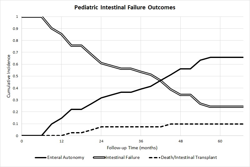
Forty-one patients met inclusion criteria. The majority were male (56%), other race (51%), non-Hispanic (58%), and Medicaid funded (90%). Median age at study inclusion was 85 days (IQR 76-102). Median gestational age was 30 weeks (IQR 25-34) and birth weight was 1170 grams (IQR 725-2140). The most common cause of IF was necrotizing enterocolitis (63%). EA was achieved in 27 patients (66%) at a median of 123 days (IQR 101-184). Of the 14 (34%) that did not achieve EA, 8 (20%) remained PN dependent, 4 (10%) died, and 2 (5%) weaned off PN but did not meet EA criteria. No patient underwent intestinal transplant (Figure). The median follow-up was 20 months (IQR 11-30). On univariate analysis, EA was associated with preserved ileocecal valves (ICV) (85 vs 36%, p=0.00), longer residual small bowel length (SBL) (60 vs 33 cm, p=0.01), and higher percent of expected SBL (42 vs 17%, p=0.01). Additionally, the following variables were identified as potential contributing factors to EA and used in our multiple logistic regression model: lower direct bilirubin (2.6 vs 3.9, p=0.14), lower aspartate aminotransferase to platelet ratio index (1.0 vs 1.7, p=0.08), decreased cholestasis (59 vs 86%, p=0.08) and increased restoration of intestinal continuity (91 vs 71%, p=0.14).  Residual SBL and percent of expected SBL were omitted on multiple logistic regression analysis, due to incomplete data (n=21).  Only a preserved ICV predicted achievement of EA (OR 5.65, CI 1.06-30.21).

Conclusion:
An EA rate of 66% was comparable to prior studies and was best predicted by ICV preservation. Efforts to preserve the ICV and SBL are paramount to the achievement of EA and, ultimately, to the survival of this vulnerable patient population.
 

13.06 Open vs. Laparoscopic Right Hemicolectomies in Pediatric Patients with Crohn's Disease

H. J. Quiroz1, E. A. Perez1, C. M. Thorson1, A. H. Langshaw2, J. E. Sola1  1University of Miami – Miller School of Medicine,Surgery,Miami, FL, USA 2University of Miami – Miller School of Medicine,Pediatrics,Miami, FL, USA

Introduction: Surgical intervention remains an important treatment modality for the management of Crohn’s disease in the pediatric population. The objective of this study was to perform a comparative analysis of open right hemicolectomy and laparoscopic right hemicolectomy for management of Crohn’s disease in pediatric patients.

Methods: The Kids’ Inpatient Database (KID) was analyzed for the years 2009 through 2012 utilizing ICD-9 procedure codes for open right hemicolectomy (45.73) and laparoscopic right hemicolectomy (17.33) in patients with Crohn’s disease (ICD-9 codes: 555.0, 555.1, 555.2, 555.9). Open and laparoscopic procedures were compared using propensity score-matched analysis (PSMA) of 38 variables, including risk adjustment with Elixheuser comorbidities.   

Results:  A total of 889 patients were identified and 448 open right hemicolectomy and 441 laparoscopic right hemicolectomy patients were propensity score matched for our analysis. 821 cases were included in the cohort (median age=17 years, male=58%, Caucasian=73%). There were zero in-hospital deaths for all patients (0/821) included in the study. Following propensity score matching, open right hemicolectomy (n=380) patients were more likely to have septicemia, respiratory compromise, pneumonia, perforation and/or laceration, complications and require blood transfusions (all, p<0.05). Although laparoscopic right hemicolectomy patients (n=441) were more likely to develop post-operative nausea/vomiting/diarrhea (p<0.0001), they had a shorter hospital length-of-stay (p<0.0001) and lower overall hospital charges and cost (p<0.001).

Conclusion: Open and laparoscopic right hemicolectomies in KID have similar low in-hospital mortality for pediatric patients with Crohn’s disease. However, open procedures are associated with higher morbidity including an increased risk for respiratory complications, surgical complications, need for blood transfusions, and increased resource utilization compared to patients who had laparoscopic procedures.

13.05 The Value of Prophylactic Chest Tubes for Tracheoesophageal Fistula Repair

M. Nguyen1, P. Delaplain1, C. Gayer1  1Children’s Hospital Los Angeles,Pediatric Surgery,Los Angeles, CA, USA

Introduction: Intraoperative chest tubes (IOCTs) can be placed at the time of esophageal atresia/tracheoesophageal fistula (EA/TEF) repair, in part to detect air and fluid leaks early. An IOCT may also prevent a second anesthesia if postoperative drainage is required. However, indwelling chest tubes come with risk, including pain, limited movement, and repeated imaging. As a result, IOCT placement is not standard and is somewhat controversial, with recent literature suggesting that it is unnecessary. We examined whether IOCT placement improved postoperative outcomes in a pediatric tertiary referral center. We hypothesized that the postoperative course of patients who received IOCTs would not differ from that of patients without IOCTs. 

Methods: We performed a five-year retrospective review of all patients with EA/TEF. Data collection included patient demographics (birth weight, EA/TEF type, hospital length of stay (LOS)), comorbidities, operative details (approach, gap length, IOCT placement), postoperative management (chest X-rays, esophagrams, postoperative cehst tube (POCT) placement), and patient outcomes (mortality, indications for reoperation, and complications). Descriptive statistical analysis was performed using GraphPad Prism version 7. Comparison between groups was performed using t-test, chi-square, or Fischer's exact test for continuous and categorical variables respectively. 

Results: Seventy-eight patients were included in the final analysis. Type C EA/TEF was most common (92%). Seventy patients underwent esophageal anastomosis; five type E patients did not require it, and three type C patients died prior to repair. No type A or E patients received IOCTs. Patients who received IOCTs had nearly two-times higher LOS (53.8±62.18 vs. 24±25.22, p=0.0195) and higher rate of reoperation for stricture (17 (34%) vs. 2 (7.1%), p=0.0083). The IOCT group also had increased incidence of POCT placement, however this was not significant. In a subgroup comparison, low birthweight (LBW) patients had increased incidence of pneumothorax (5 (17%) vs. 1 (2.2%), p=0.0229), more POCTs despite IOCTs (8 (28%) vs. 3 (12%), p=0.0416), and higher mortality (6 (20.7%) vs. 0, p=0.0026) versus normal birthweight (NBW) patients. There was no difference in rates of extra- versus intrapleural dissection between these groups.

Conclusion: EA/TEF patients who received IOCTs during repair had significantly increased hospital LOS and reoperation for stricture, suggesting that prophylactic IOCT placement is associated with worse postoperative course. Though LBW patients required more POCTs, this was despite IOCT placement, suggesting that prophylactic IOCTs are not beneficial even in this more fragile patient population.