16.11 How the Other Half Dies: Characterization of Mortalities 30 to 90 days after Complex Cancer Surgery

B. J. Resio1, J. Hoag1, A. Monsalve1, J. Blasberg1, D. J. Boffa1  1Yale University School Of Medicine,New Haven, CT, USA

Introduction: The vast majority of research to reduce surgical mortalities has focused on the first 30 days after surgery. Unfortunately, nearly half of the patients who die after complex surgery, do so beyond the traditional 30-day window.  Data from the surveillance, epidemiology and end results (SEER) database linked to Medicare claims was evaluated in an effort to increase the understanding of the events surrounding deaths occurring between 30 and 90 days after complex cancer surgery.

Methods: Patients who underwent lung, colon or esophageal resection for non-metastatic cancer and died within 90 days of surgery were identified in SEER-Medicare. Cause of death (COD) was grouped based on ICD10 diagnosis codes. Place of death was determined using the last place of discharge before death.

Results: A total of 1,480 patients died between 30-90 days of complex cancer surgery (“later” mortality cohort). Readmission was strongly linked with later mortality as 78% of patients were readmitted at least once before dying within 30-90 days.  The COD was listed as “cancer” in 57% of the late mortalities, which seems unlikely within 90 days of surgery. Of the patients with COD other than cancer, the most common were acute cardiac disease (23%), chronic lung disease (12%), chronic heart disease (11%), sepsis/shock (8%), pneumonia (5%) and stroke (4%). Sixty-one percent of late mortality patients died in the hospital (8% during the index hospitalization), 10% were last discharged to home, 14% to a nursing facility and 16% to hospice.


The “later” mortality cohort was compared to an “earlier” cohort of 1,985 patients who died within 30 days of surgery. The 30-day and 90-day mortality rates varied by surgery type (colectomy 6.2% vs 11.0%; lobectomy 3.1% vs 6.0%; pneumonectomy 10.5% vs 18.9%; esophagectomy 6.3% vs 14.1%). Age, sex, race and stage were similar between earlier and later mortalities. Compared to earlier mortalities, later mortalities were less likely to occur during a hospital admission (61% vs. 85% p<0.01) and less likely during the initial hospitalization during which surgery was performed (8% vs. 51%, p<0.01). In terms of COD, cancer was listed for a similar proportion of early and late patients (56.8% vs. 57.0%). While there were similarities in noncancer COD, death from acute GI disease (bowel ischemia, bleed, perforation, leak, peritonitis etc.) was less common in the later cohort compared to the earlier cohort (4% vs. 14% of non-cancer COD, p<0.01).

Conclusions: Among elderly Medicare beneficiaries, nearly half the perioperative mortalities occur after 30 days. Most patients who die beyond 30 days were readmitted at least once, suggesting a failure to rescue from a postoperative complication.  Surgical teams should minimize the over-listing of “cancer” as the cause of death in the perioperative period, as this may impede the identification of opportunities to increase rescue.   


13.18 Pediatric Portal Hypertension: Surgical Outcomes in the Decade of Meso-Rex Bypass

F. Lopez-Verdugo2, A. S. Muñoz-Abraham4, J. Merola7, Z. Kastenberg2, A. Bertacco3, D. Alonso2, W. R. Hewitt5, M. Facciuto6, S. H. Emre7, M. I. Rodriguez-Davalos2  6Mount Sinai School Of Medicine,Department Of Transplantation,New York, NY, USA 7Yale University School Of Medicine,Department Of Surgery,New Haven, CT, USA 2Intermountain Primary Children’s Hospital,Department Of Surgery, Transplant And Hepatobiliary Surgery,Salt Lake City, UT, USA 3UniversitĂ  di Padova,Department Of Surgery, Hepatobiliary Surgery And Liver Transplantation,Padova, PD, Italy 4Saint Louis University School Of Medicine,Pediatric Surgery,St. Louis, MO, USA 5Mayo Clinic In Arizona,Department Of Surgery,Phoenix, AZ, USA

Introduction: Extrahepatic portal venous obstruction (EHPVO) as well as intrinsic liver disease (ILD) are major causes of portal hypertension in children often accompanied by development of cavernoma. Decompressive surgeries, including Meso-Rex bypass (MRB) and portosystemic shunts (PSS) are offered for definitive therapy in such cases. The study aims to review surgical outcomes in a cohort of patients who underwent MRB or PSS over the last decade.

Methods: Medical records from all children who received a MRB or PSS at across different institutions in the US between 2005 and 2018 were reviewed and described. Data collected included age, gender, diagnosis, clinical manifestations of PTH, type of procedure, and clinical outcomes. Patency and patient survival were assessed at 6-months and at the last follow-up visit.

Results: 34 surgeries were performed in 29 children. The mean age at the time of surgery was 8.1 years (range: 1.5-19),14 patients (48.3%) were male. Common causes of PTH were EHPVO (n=17) and ILD (n=9). PTH manifestations at presentation were GI bleeding, splenomegaly and hepatic encephalopathy (HE). There were 18 MRB and 8 selective PSS: 6 distal splenorenal and 2 spleno-caval. 4 proximal splenorenal, 2 mesorenal, one mesocaval, and one side-to-side splenorenal shunts. Nine patients (26.4%) had treatment failure before the 6-month follow-up, 4 of which happened on postoperative day 1. Treatment failure for MRB resulted from thrombosis (n=5), and collateral steal phenomenon (n=3) and was managed with thrombectomy (n=3), creation of an alternative PSS (n=2), or successful re-do MRB with occlusion of collaterals (n=2). 25 patients (73.5%) have been followed for more than 6 months (median: 28, range: 7-116) 23 are still alive and 2 died with a patent shunt. 18 procedures did not require further intervention, 2 required balloon angioplasty, 2 ILD needed LTx, and 1 patient required conversion of MRB to PSS.

Conclusion: In this series, MRB in patients without ILD was successful in 81.2%. Early shunt thrombosis may occur in the setting of an underdeveloped intrahepatic portal system though secondary patency rates of 60% were achieved without converting to PSS. These results support the notion that MRB should remain the standard of care for children with EHPVO or portal vein thrombosis after transplantation as it re-establishes hepatopetal flow; selective PSS should be the next line therapy for those patients with concurrent ILD or when MRB is not feasible due to technical or functional constraints. Creation of non selective shunts should be reserved for patients with massive splenomegaly or when the anatomy is not favorable, and HE is not a concern. Multidisciplinary work with interventional radiology is key to the successful treatment of these patients.

104.11 Do Words Matter? The "Chair" Title and Gender Gaps in Academic Surgery

C. Peck1, S. J. Schmidt2, D. A. Latimore1, M. I. O’Connor1  1Yale University School Of Medicine,New Haven, CT, USA 2Yale Law School,New Haven, CT, USA

Introduction:  Gender-marked titles have shown to be exclusionary of women in a variety of professional settings. The purpose of this study was to analyze and compare the use of the words "chair" and "chairman" on academic websites for both surgical and non-surgical departments in the US. 

Methods:
Orthopedics, Neurosurgery, Obstetrics and Gynecology, and Pediatrics departments from 139 US allopathic medical schools were reviewed. Official websites were screened for use of the word chair or chairman. Any use of the word chairman was classified according to type of use and location on the website. Patterns of chair use were compared by specialty, region, and gender of the current chair. 

Results:
Overall, 59.8% of all academic departments used only the gender-neutral term chair. In surgical specialties, this number was significantly lower (p<.001)—40.3% in Orthopedics and 42.5% in Neurosurgery, compared to 70.1% and 64.0% in Obstetrics/Gynecology and Pediatrics respectively. Departments with female chairs used gender-neutral titles 89% of the time, compared to 53.1% in departments led by males. Gender-neutral title use was highest in the West across specialties (p<.01). The proportion of female chairs was highest in Obstetrics and Gynecology at 32.5%, compared to 26.0% in Pediatrics, 4.3% in Neurosurgery, and 0.8% in Orthopedics. Use of the word chair correlated with a 437% increase in the likelihood of having a female chair (p<.01).

Conclusion:
Our studies show persistence of the gender-marked title "chair" across academic specialties—particularly in surgical specialties—and suggest a association between title use and overall gender diversity. Increasing the use of gender-neutral titles may be a simple way to promote gender parity in academic surgery. 

103.05 Impact of Temporal Artery Biopsy on Clinical Management of Suspected Giant Cell Arteritis

C. Deyholos1, M. Systek1, S. Smith1, J. Cardella1, K. C. Orion1  1Yale University School Of Medicine,Section Of Vascular Surgery, Department Of Surgery,New Haven, CT, USA

Introduction: Temporal arteritis (TA) or giant cell arteritis (GCA) is a systemic inflammatory vasculitis of unclear etiology that affects medium sized vessels. The gold standard for diagnosis has traditionally been histological by TA biopsy.  Due to the risk of permanent vision loss if the disease is left untreated, standard of care is to begin steroid therapy prior to confirming the diagnosis.  In up to one third of GCA patients, the temporal arteries are not involved and there has been reported facial nerve injury during TA biopsy. Improved imaging modalities such as color duplex, PET CT or MRI have been increasingly used to aid diagnosis and are  recommended in the newest 2018 European (EULAR) Guidelines.  We hypothesize that a negative TA biopsy result does not change management in patients for whom temporal arteritis is strongly suspected and that duplex ultrasound can be successfully used as a screening tool.

Methods: A retrospective review of patients undergoing TA biopsy between May 1, 2012 and December 31, 2015. We reviewed patient's age, gender, co-morbidities, symptoms, histology, and whether patients were prescribed steroids prior to or following biopsy. We also began small prospective series of 3 patients where ultrasound of the bilateral temporal arteries was performed prior to biopsy, using a high frequency linear transducer to evaluate for wall thickening. Radiology report and pathology report were then reviewed.

Results: Within period of study, 171 temporal artery biopsies were performed. 7.6% positive (n=13) 92.4% negative (n=158) for acute GCA.  Patients with positive biopsy result had mean age 80± 6 (Range 69-88). Patients with negative biopsy had mean age of 72± 11 (Range 17-95). We also performed subgroup analysis on patients with negative biopsies (n=158). Cases in which there was no documentation of steroids prior to or after biopsy were excluded (n=15). 20% of patients who had negative biopsies were not on steroids prior to the procedure (n=28). 31% of patients with negative biopsies continued on steroids despite the negative result (n=45).  In series of 3 ultrasounds, all 3 correlated with subsequent biopsy histology. 1 was positive, and 2 were negative.

Conclusion:  Our results suggest that the yield of temporal artery biopsy is low, and a negative biopsy alone often does not lead to termination of steroid therapy. Ultrasound may present a viable diagnostic tool to reduce number of unnecessary temporal artery biopsies performed.

10.14 Intraoperative Cholangiogram – an Analysis of Trends and Outcomes for Management of Cholecystitis.

K. Zhang1, V. Natkha1, J. Mccauley1, M. L. Warren1, J. Luo3, Y. Zhang2,3, K. Y. Pei1  1Texas Tech University Health Sciences Center,Surgery,Lubbock, TX, USA 2Yale School of Medicine,Section Of Surgical Outcomes And Epidemiology,New Haven, CT, USA 3Yale School of Public Health,Environmental Health Sciences,New Haven, CT, USA

Introduction:


Common bile duct injury and retained stones continue to be rare, but potentially catastrophic outcomes of laparoscopic cholecystectomy.  Although still controversial and unsettled, there is some evidence that intraoperative cholangiography during laparoscopic cholecystectomy may mitigate such complications.  Despite multiple national practice management guidelines espousing liberal use of intraoperative cholangiography, it is unknown practice patterns among US surgeons.


Methods:


The ACS NSQIP database was queried for patients undergoing laparoscopic cholecystectomy with (CPT code 47563) and without (CPT code 47562) intraoperative cholangiography for diagnosis of cholecystitis (identified by ICD 9 and ICD 10 codes) from 2005 to 2016. Patients undergoing cholangiogram for known common bile duct stones were excluded.  Trends and practice patterns were evaluated as percentages of total procedures performed from NSQIP participating hospitals.  Standard descriptive statistics were analyzed using student t test, chi-squared as indicated.  Multivariable logistic regression was utilized to compare outcomes of interest including complications, mortality or reoperation.


Results:


A total of 19,636 procedures (80.3% without cholangiography) were included for analysis.  There were no significant differences among patient characteristics between the 2 groups.  Among NSQIP participating hospitals, majority of surgeons do not perform intraoperative cholangiogram and there appears to be an increasing trend to forgo cholangiography during the study period (Figure 1).  After adjusting for patient characteristics, there were no differences in overall complications [OR 0.86 95% CI (0.74-1.00)], 30-day mortality [OR 0.95 95% CI (0.59-1.52)],or reoperation [OR 1.16 95% CI (0.31-4.35)].

Conclusion:


Most surgeons do not perform intraoperative cholangiography during laparoscopic cholecystectomy for cholecystitis.  There were no significant differences in overall complications, mortality, or reoperative risk.

10.13 Transfusion Rates in Emergency General Surgery – High but Modifiable

A. J. Medvecz1, A. Bernard3, C. Hamilton3, K. Schuster2, O. Guillamondegui1, D. Davenport3  1Vanderbilt University Medical Center,Nashville, TN, USA 2Yale University School Of Medicine,New Haven, CT, USA 3University Of Kentucky,Lexington, KY, USA

Introduction:  Transfusion of red blood cells (RBC) increases risk-adjusted morbidity and mortality in surgical patients. Blood management programs have been developed to reduce rates of transfusion. However, emergency general surgery cases are at inherent risk for transfusion given case complexity and patient acuity. We sought to examine the rate of transfusion in emergency general surgery and whether it has changed over time. 

Methods:  This is a retrospective review of ACS NSQIP data from 3 academic medical centers that are also Level I Trauma Centers. Operations performed by general surgeons on adults (age ≥ 18 years) were selected. Data were analyzed from two periods, calendar years 2011 to 2013 and 2014 to 2016. We grouped procedures by the first four digits of the primary procedure CPT code. Groups with fewer than 100 cases and fewer than 10 total transfusions were combined into an “Other” group resulting in 40 procedure groups. Transfusion is defined as any transfusion of RBC during or < 72 hours after the operation. Composite morbidity was defined as any ACSNSQIP complication within 30 days of the operation that was not present prior to the operation.

Results: Between period 1 and 2 overall general surgery transfusion rates decreased from 6.4% to 4.8% of cases (emergent: 16.6% to 11.5%; non-emergent 4.9% to 3.7%; Fisher’s exact p’s < 0.001), Table). Among patients transfused, the number of units received also decreased slightly (median 2 U [IQR 2-3] to median 2 U [IQR 1-3], Mann-Whitney U p = 0.005). Morbidity decreased over the same period from 13.8% to 12.3% (p = 0.001); with emergent cases decreasing from 26.3% to 20.6%, p < 0.001. Mortality did not change by period. (emergent 6.6%, 6.7%; non-emergent 1.4%; 1.5%).

Conclusion: Rates of RBC transfusion have decreased over time. EGS requires twice the rate of transfusion of elective general surgery. However, efforts to reduce transfusion have also been successful in the EGS population. Morbidity improved over the same time period while mortality was unchanged.

10.04 Modern Management of Perforated Peptic Ulcers – a Decade Long ACS NSQIP Analysis.

R. Dev1, J. Mccauley1, T. Wyatt1, V. Natkha1, J. Luo3, Y. Zhang2,3, K. Y. Pei1  1Texas Tech University Health Sciences Center,Surgery,Lubbock, TX, USA 2Yale University School Of Medicine,Surgical Outcomes And Epidemiology,New Haven, CT, USA 3Yale School of Public Health,Environmental Health Sciences,New Haven, CT, USA

Introduction:

Decades after the introduction of acid reducers and the recognition of endemic helicobacter pylori infection as the cause of peptic ulcers, medical management has largely replaced surgical therapy.  Nevertheless, a portion of patients will require operative intervention for perforated ulcers.  With the increasing adoption of advance laparoscopic techniques, it is uncertain the practice patterns among US surgeons during the last decade.  This study evaluates the trends and outcomes in management of perforated peptic ulcers.

Methods:

The ACS NSQIP database was queried for patients with diagnosis of peptic ulcer perforation (ICD 10 codes K26.5, K27.2, K26.1 K27.1, and K27.5 and ICD 9 codes 532.1, 532.5, 533.2, 533.5, 533.10)from 2005 to 2016.  Only the index operation identified by the current procedural codes (CPT) was included for analysis.  Based on CPT codes, operations were divided into 3 major groups including: simple open suture repair, laparoscopic repairs, and open suture repair open suture repair with omental patch). Trends and practice patterns were evaluated as percentages of total procedures performed from NSQIP participating hospitals. Standard descriptive statistics were analyzed using student t test and chi-squared as indicated.  Outcomes including complications, mortality or reoperation were evaluated by procedural group.


Results:

A total of 2,603 procedures were included for analysis.  There were no significant differences among patient characteristics between the 3 groups. Among NSQIP participating hospitals, majority of surgeons perform simple open suture repair of perforated ulcers (50.0% open suture repair only, 7.8% laparoscopic repairs, 42.2% open suture repair with omental patch) and there appears to be a plateauing of laparoscopic implementation (Figure 1).  Patients undergoing simple open suture repair only had the highest mortality rate (12.25%) whereas laparoscopic repairs had the lowest mortality (7.35%).  Other outcomes of including any complication, reoperation, and length of stay were similar.  Of note, no historical procedures including vagotomy and drainage operations were reported during this study period.

Conclusion:
 

The national experience in management of perforated peptic ulcer is rare and only a small minority of surgeons perform laparoscopic patch repairs.  Surprisingly, the majority of surgeons performed simple open suture repair without patch.  Barriers to implementation of laparoscopic techniques warrants further study.

02.14 Penicillin Protects the Small Bowel from Ischemia: Applications in Intestinal Transplantation

V. M. Baratta1, T. M. Gisinger1,2, M. J. Barahona1, J. Ollodart1, D. Mulligan1, J. P. Geibel1,3  1Yale University School Of Medicine,Department Of Surgery,New Haven, CT, USA 2Paracelsus Medical University,Department Of Medicine,Salzburg, SALZBURG, Austria 3Yale University School of Medicine,Department Of Cellular And Molecular Physiology,New Haven, CT, USA

Introduction: Intestinal failure is the inability to maintain nutritional autonomy due to dysfunction or loss of functional native intestine. In both pediatric and adult populations, a heterogenous list of disorders may lead to intestinal failure and necessitate either total parenteral nutrition (TPN) or intestinal transplantation. Unfortunately, intestinal transplantation remains technically challenging. One of the barriers to successful intestinal transplantation is the progression of ischemia from tissue harvest to implantation. Finding ways to abate small bowel ischemic injury will help prolong graft function and viability during and after procurement. In this study, we demonstrate how exposure of the small intestine to Penicillin G can protect from ischemic injury in a rat model.

Methods: Distal and middle small bowel segments were harvested and attached to an ex-vivo intestinal perfusion device. Each segment was maintained at 37°C and perfused both from the luminal and basolateral side. FITC-Inulin (fluorescein isothiocyanate-inulin), was used to assess the ischemic conditions of the intestinal grafts in real-time, as previously described. A decrease in fluorescence is reflective of the onset of cellular ischemic injury. The perfused bowel segments were bathed in a 4-(2-hydroxyethyl)-1-piperazineethanesulfonic acid), HEPES-Ringer solution. The extraluminal buffer solutions were bubbled with 100% nitrogen to mimic intestinal ischemia. The experimental bowel segments were exposed to intraluminal 5 mM Penicillin G, while the control segments were perfused without Penicillin G.

Results: Control (without Penicillin G) small bowel samples showed significantly greater ischemic injury than experimental segments (with Penicillin G), as reflected by differences in FITC-Inulin fluorescence concentrations (i.e. decreased ischemic injury) compared with experimental segments (47.37 ±  0.7288 μM FITC-Inulin vs. 35.76 ± 4.469 μM FITC-Inulin, p 0.0248, Figure 1).

Conclusion: Penicillin G-exposed small intestines are more resistant to ischemic injury than segments without antibiotic exposure. This was demonstrated by the drop in fluorescent concentrations over time, which reflects cellular fluid secretion and apoptosis. This finding may have numerous applications in the field of transplant surgery, such as prolonging organ viability following perfusion with Penicillin G.

02.10 Ex Vivo Assessment of Marginal Organs Using Dynamic Computed Tomography

M. K. Harris1, J. DiRito1,3, A. Feizi4, N. Boutagy2,5, A. Feher2,5, P. Yoo1, D. Haakinson1, D. Mulligan1, A. J. Sinusas2,5, G. Tietjen1  1Yale University School Of Medicine,Department Of Surgery,New Haven, CT, USA 2Yale University School Of Medicine,Department Of Internal Medicine, Section Of Cardiovascular Medicine,New Haven, CT, USA 3University of Cambridge,Department Of Surgery,Cambridge, CAMBRIDGESHIRE, United Kingdom 4Yale University School Of Medicine,New Haven, CT, USA 5Yale University School Of Medicine,Yale Translational Research Imaging Center,New Haven, CT, USA

Introduction:
Despite a high transplant waiting list mortality, many marginal organs are discarded based on incomplete and subjective data. Organ viability assessment relies on risk stratification of donor data and limited organ evaluation by visualization and frozen biopsy. Dynamic computed tomography (CT) is used in vivo to assess microvascular tissue perfusion and ischemia, factors related to damage sustained by transplanted organs. Dynamic CT has potential to provide objective, quantifiable data to improve marginal organ utilization. We adapted in vivo dynamic CT methods to an ex vivo setting to measure individual organ perfusion variability.

Methods:
CT was performed at the Yale Translational Research Imaging Center on a GE Medical Systems clinical CT scanner. 10 kidneys and 5 livers were obtained from a local pig slaughterhouse. Organs were dissected, flushed with cold Custodial HTK preservation solution, and stored on ice until imaging. DICOM images were reconstructed in Horos and analyzed using custom MATLAB code to measure rates of contrast enhancement and clearance throughout the organs over time.


Results:

Marginal organs are known to sustain microvascular changes. We expect that these microvascular obstructions impair flushing and promote tissue injury. We adapted dynamic CT for use with ex vivo organs by performing sequential scans during contrast administration with iohexol and during subsequent continuous flush. Using custom MATLAB code, we measured enhancement of two regions of cortex equidistant from the right and left sides of each organ and measured peak enhancement, rates of enhancement, and rates of clearance.

Figure 1A includes representative images of two kidneys. Region of interest (ROI) 1 and ROI 2 are measured in the cortex on the left and right sides of each organ and demonstrate varied contrast kinetics within organs. Kidney 3L had a slower rate of contrast accumulation, lower total enhancement, and faster rate of decay in the tissue (Figure 1B and 1C). These preliminary data may suggest that less microvasculature was engaged and that contrast primarily remained in the larger vessels.

Conclusion:
Dynamic CT of ex vivo organs can be used to directly quantify perfusion deficits and microvascular changes. Its use could be extended to assess marginal organs to more accurately identify viable organs and improve utilization. This technique also creates opportunities to potentiate other tools for assessing viability and to evaluate the effects of emerging techniques in ex vivo organ preservation, recovery, and treatment.


01.04 Morphometric Parameters Vary with Age in the Adolescent and Adult Murine Small Intestine

C. J. Park1, M. P. Shaughnessy1, R. A. Cowles1  1Yale University School Of Medicine,Pediatric Surgery,New Haven, CT, USA

Introduction: Animal models allow researchers to study the effects of pharmacological and surgical interventions on the gastrointestinal (GI) mucosa. The benefits of using mice in the laboratory include low cost, ease of maintenance, and the applicability of a wide range of molecular tools. While the typical laboratory mouse reaches sexual maturity at 6-8 weeks, maturation ensues and mice are considered mature adults at 12-24 weeks. Meanwhile, the ages and genders of mice used for experiments reported in the literature vary drastically. We hypothesized that there is age-, but no gender-related variation in intestinal morphometric parameters, necessitating thoughtful experimental design.

Methods:  With IACUC approval, C56Bl/6J mice of varying ages (6, 12, 18, 24 weeks; n=4/group) were euthanized and the small intestine was isolated from the ligament of Treitz to the ileocecal valve. 2 cm segments of proximal, middle and distal small intestine were harvested and morphometric parameters assessed with microscopy. The distal segments were also stained for Ki67 to determine the crypt proliferation index (CPI). Secondary analysis comparing males and females (n=4/group) of matched ages was performed. Means were compared with Student’s t-test and variance of proportions was assessed with the Chi-squared test to a significance of p<0.05.  

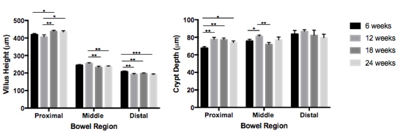
Results: There was variation in the measured morphometric parameters (Figure). In the proximal small intestine, 18-week old mice had taller villi compared to 6- and 12-week old mice and 24-week old mice had taller villi compared to 12-week old mice. In the middle segments, 12-week old mice had taller villi compared to 18- and 24-week old mice. In the distal segments, 6-week old mice had taller villi compared to all other age groups. When comparing crypt depth, 6-week old mice had shallower crypts compared to all other groups in the proximal region and 12-week old mice had deeper crypts compared to 6- and 18-week old mice in the middle segments. CPI was statistically lower only in 12-week old mice compared to all other age groups. When comparing age-matched males and females, there was no difference in villus height or crypt depth except for the middle small intestine where villus height was greater in females compared to males. There was no difference in CPI between genders.

Conclusion: Small, but statistically significant, differences in villus height, crypt depth and crypt proliferation are present in mice of different ages, while fewer differences exist between male and female mice of the same age. Investigators studying the GI mucosa should be aware of these differences and aim for consistent age-matching of experimental animals in order to avoid errors and allow direct comparison between studies.


01.02 Exome Sequencing of Syndromic Adrenocortical Cancer Reveals Distinct Genetics from Sporadic ACC

N. G. Nicolson1, J. M. Healy1,2, R. Korah1, T. Carling1  1Yale University School Of Medicine,Yale Endocrine Neoplasia Laboratory, Department Of Surgery,New Haven, CT, USA 2Connecticut Children’s Medical Center,Hartford, CONNECTICUT, USA

Introduction:  Next-generation sequencing has provided detailed genetic profiles of sporadic adrenocortical carcinoma (ACC). However, the genetic landscapes of ACC developing in patients with tumor predisposition syndromes are not well-characterized, as they are excluded from large-scale studies. Understanding the somatic genomic events complementing the background of germline syndromes is critical for designing personalized therapeutics for these unique patients, and may shed light on the molecular underpinnings of both sporadic and syndromic ACC.

Methods:  ACC tissue and matched normal adrenal from a pediatric patient with clinically characterized Beckwith-Wiedemann syndrome were subjected to whole-exome sequencing (WES). Using multi-layered bioinformatics analysis, the WES data was compared to 21 sporadic ACCs which had also undergone WES. Single nucleotide variants (SNVs) of interest were subjected to damage prediction protocols to identify potential ACC drivers, and somatic copy number variations (CNVs) were also investigated.

Results: WES analysis revealed 8 relevant germline mutations in the index case, including a TP53 mutation previously associated with Li-Fraumeni syndrome. 25 potentially damaging somatic SNVs including multiple unique novel gene variants were identified. A damaging somatic mutation was identified in tumor suppressor CACNA2D3, a calcium channel gene in the same family as those previously reported to be mutated in some adrenocortical tumors. Although the mutation burden in the index case was similar to the sporadic ACC cohort, the total absence of CNVs was distinct from sporadic cases, all of which carried CNVs.

Conclusion: This study characterizes for the first time the genomic landscape of syndromic adrenocortical carcinoma of a patient with markers of both Beckwith-Wiedemann and Li-Fraumeni syndromes. The unique SNV and CNV profiles demonstrate that syndromic adrenocortical tumors may represent a genetically distinct entity from sporadic tumors.