44.07 CNP-miR146a Alters Macrophage Phenotype in Diabetic Wounds

L. Dewberry1, C. Zgheib1, M. M. Hodges1, S. A. Hilton1, J. Hu1, J. Xu1, K. W. Liechty1  1University Of Colorado Denver,Department Of Surgery,Aurora, CO, USA

Introduction:

Diabetic wounds are characterized by chronic inflammation. Persistent, proinflammatory macrophage polarization (M1) has been implicated in this pathophysiology. Previous work in our lab has demonstrated that miR-146a downregulates inflammation in the wound, and that the delivery of miR-146a conjugated to cerium oxide nanoparticles (CNP-miR146a) improves diabetic wound healing. We hypothesize one of the mechanisms of this improved wound healing is through the promotion of macrophage polarization from M1 to M2, which leads to resolution of inflammation.

Methods:
Using a murine model, two 8mm dorsal wounds were created using eleven-week-old male mice homozygous for the Leprdb mutation (db/db) and age-matched male nondiabetic heterozygous littermates (db/+). At the time of wounding, one group (n=5) was injected with 10micromolar CNP-146a in each wound. This was compared to the db/db group injected with PBS (n=5) and the db/+ group (n=5). Wounds were harvested at day 10, and macrophages were isolated from with wound homogenate using magnetic separation. Genetic expression was evaluated with real-time quantitative PCR.

Results:
At day 10, diabetic wounds treated with CNP-146a demonstrated decreased expression of the markers associated with the M1 phenotype, TNF, STAT-1, and IL-1B, compared to diabetic wounds treated with PBS, with the level of expression similar to those seen in the non-diabetic wounds.

Conclusion:
Administration of CNP-146a to a diabetic wound returns the level of inflammation in macrophages to that of a non-diabetic wound and supports the hypothesis that administration of CNP-146a may restore the transition of macrophages from M1 to M2 to facilitate normal wound healing.

44.06 Activation of the Vacuolar H+-ATPase via CaSR Activation in the Rat

M. J. Barahona1, J. Ollodart1, T. M. Gisinger1,3, V. M. Baratta1, Y. W. Stroehl1,2, D. Mulligan1, J. P. Geibel1,4  1Yale University, School of Medicine,Department Surgery,New Haven, CT, USA 2Charité University Medicine Berlin,Faculty Of Medicine,Berlin, BERLIN, Germany 3Paracelsus Medical University,Department Of Medicine,Salzburg, SALZBURG, Austria 4Yale University School Of Medicine,Department Of Cellular And Molecular Physiology,New Haven, CT, USA

Introduction:  Gastroesophageal reflux disease is a prevalent chronic disorder, yet symptom management is often difficult to achieve or maintain. Traditional treatments involve proton pump inhibitors and H2-receptor antagonists, though many patients have persistent symptomatology. Recently, an unaccounted apical vacuolar H+-ATPase was identified and found to be activated in the absence of H,KATPase activity. This H+-ATPase is activated by the calcium-sensing receptor (CaSR), which is in turn modulated by the calcium-activated chloride channel (CaCC). Production of gastric acid via this mechanism can be inhibited by blocking gastric CaCC. Tannic acid has been shown to block the CaCC in other organ systems. Here, we demonstrate that modulation of CaSR via the calcimimetic R568 and CaCC, via tannic acid can indirectly influence acid secretion through the vacuolar H+-ATPase.

Methods:  Gastric glands from rats were isolated via a hand dissection technique. The individual glands were then perfused in vitro with a K+-free HEPES (4-(2-hydroxyethyl)-1-piperazineethanesulfonic acid) to suppress H,KATPase activity. The control group was perfused with 400 nM R568 hydrochloride, a positive allosteric modulator and agonist of CaSR, while the experimental group was perfused with 400 nM R568 hydrochloride and 200 µM tannic acid. Next, the extracellular calcium concentration was elevated to 5.0 mM in the presence and absence of R568 and tannic acid. The H+-ATPase activity was selectively monitored with the rate of proton extrusion (ΔpH/min) on individual parietal cells by observing a change and recovery rate of intracellular pH after acid loading the cells with various solutions, leaving only the H+-ATPase as an active proton secretory pathway.

Results: After exposure to R568 in glands from fasted animals there was increased vacuolar H+-ATPase activity with normal extracellular (1.0 mM) Ca2+ with a rate of proton extrusion increase of 0.01255 ± 0.00120 ΔpH/min. Elevations in Ca2+ concentration (1.0 mM to 5.0 mM) caused a further increase in K+-independent H+ secretion 0.01621 ± 0.00087 ΔpH/min. When tannic acid, an inhibitor of CaCC was added to the perfusates with 1.0 mM Ca2+ and 5.0 mM Ca2+there was a significant inhibition of proton secretion under all conditions with a lower proton extrusion relative to the control 0.00296 ± 0.00027 ΔpH/min (p<0.0001) and  0.00409 ± 0.00053 ΔpH/min (p<0.0001), respectively.

Conclusion: An increase in extracellular calcium concentration leads to CaSR stimulation and elevated H+-ATPase activity. In this study, we demonstrate tannic acid-induced inhibition of the H+-ATPase through inactivation of the CaCC. The inhibition of H+-ATPase activity along with H,KATPase activity theoretically may lead to complete blockade of gastric acid production under resting and stimulated conditions. The CaSR along with CaCC can be important new pharmacologic targets to suppress acid secretion.
 

44.05 A Thermogelling Hyaluronic Acid Vaginal Stent to Reduce Postoperative Vaginal Fibrosis

J. Hakim1, O. Wyman2, S. Keswani1  1Texas Children’s Hospital,Pediatric Adolescent Gynecology,Houston, TX, USA 2Baylor College Of Medicine,Houston, TX, USA

Introduction:  There is a need for new therapies to prevent vaginal fibrosis. Up to 50,000 girls and 213,000 adult women yearly in the United States require vaginal reconstructive surgery. There is a high rate (up to 73%) of vaginal fibrosis after these surgeries. Sequelae from vaginal fibrosis can be life-long and lead to significant reductions in quality of life. Currently, treatments are limited to conjugated estrogen (CEE) creams in combination with vaginal stents. Unfortunately, current CEE creams cannot assess delivered estrogen due to cream egress from the vagina and the cumbersome nature of vaginal stents leads to early discontinuation. We have created a novel hydrogel that utilizes “click” chemistry to make a thermogelling hyaluronic acid (HA) drug eluting hydrogel. We sought to compare local estrogen delivery with Premarin cream to sustained estrogen delivery with a novel CEE-eluding NorbHA hydrogel on vaginal wound healing.

Methods:  Norbornene-functionalized hyaluronic acid (NorbHA) and tetrazine-functionalized hyaluronic acid (TetHA) was synthesized. Hydrogel was composed of a 1:1 ratio of NorbHA:TetHA with various concentrations of estradiol (E2), 1.5 MDa HA, or a combination of both. The vaginal stents were placed into a full-thickness 1 mm murine vaginal excisional injury model. Animals were sacrificed at days 0, 2, 3, 7, 10. Macrophage inhibiting factor 1 (MIF1), transforming growth factor-β3 (TGFβ3) expression and vascular endothelial growth factor (VEGF) expression were analysed as markers of inflammation and angiogenesis respectively. Histology and immunohistochemistry were used to assess wound resolution and estrogen receptor (ER) density.

Results: Dissolution parameters demonstrated that drug release was still possible until 72h in the thermogelling hydrogel compared to CEE cream (12h). There was a burst release of E2 over the first 12h period followed by a steady-state release while HA had a burst release at 24 h. A statistically significant decrease in MIF1 expression was found in the group containing both HA and E2 compared to the CEE cream between the day 0 and day 3 timepoints. Greater expression of TGFβ3 and VEGF were found in the groups containing E2, or E2/HA compared to the estrogen cream by day 2. ER density increases with E2 delivery but appears to be dependent on circulating estrogen levels. 

Conclusion: Vaginal tissue healing is enhanced through a novel thermogelling vaginal hydrogel with sustained estrogen and HA release compared to local exogenous estrogen cream. Improvements in both the anti-inflammatory and pro-angiogenic effects of sustained estrogen delivery included reduction in neutrophil and macrophage infiltration, modulating ECM degradation and stabilizing collagen. Further development of this platform may provide a substantial increase in efficacy of E2 delivery to the vaginal tissue and reduce post-operative vaginal fibrosis.


44.04 NOD2 polymorphism is associated with stricutring phentotype in Crohn's disease

J. Hallion1, S. O’Brien1, C. Fiechter1, M. B. Ekman1, J. Burton1, M. Eichenberger1, S. Galandiuk1  1Price Institute of Surgical Research,University Of Louisville,Louisville, KENTUCKY, USA

Introduction: Crohn’s disease is a common inflammatory disorder of the gastrointestinal tract. It has a multifactorial etiology with immunological, environmental, and genetic factors. Phenotypically, Crohn’s disease was classified (Montreal Classification) using age of onset, disease location, and disease behavior. Single nucleotide polymorphisms (SNPs) are common single base pair genetic variants between individuals and are associated with disease phenotype in a number of diseases. Recent genome wide exploratory studies have identified SNPs that are associated with a stricturing phenotype in Crohn’s disease. The aim of this study was to determine if SNP allele frequencies truly correlate with stricturing Crohn’s disease phenotype in a mid-western Caucasian United States population.


Methods:  Patients with Crohn’s disease were selected from a prospectively maintained university surgical digestive practice database. Blood samples were drawn during clinic appointments. Patients with stricturing (Montreal B2) and non-stricturing (Montreal B1) phenotypes were eligible for inclusion. Clinical data were extracted from an electronic database and patients were matched for gender and race. Genomic DNA was extracted from the blood sample, and 4 SNPs (NOD2 rs2066844 R702W, FUT2 rs601338, IL23-R rs1004819, ATG16L1 rs2241880 T300A) were assessed using a TaqMan genotyping assay.

Results: One hundred and sixty-three patients with Crohn’s disease were included in this study; 65% (106/163) were female. Age of onset (Montreal A) was equally distributed between the stricturing and non-stricturing groups (p=0.408). There was no difference in disease location (p=0.814), or the presence of upper GI disease (p=0.274) between the groups. There was no difference in allele frequency or genotype frequency between the stricturing and non-stricturing groups for FUT2, ATG16L1, or IL23-R (Table 1). The T allele (p=0.004) and the TT genotype (p=0.035) in the NOD2 R702W SNP were significantly associated with a non-stricturing phenotype.

Conclusion: We confirmed a significant association between a non-stricturing phenotype and the TT genotype and the T allele in the NOD2 R702W SNP. NOD2 is a well characterized gene that is involved in Crohn’s disease pathophysiology and the results of this study validate the results of large genome wide association studies in a United States population. Further study is required to determine if the NOD2 R702W T allele is a protective factor against stricturing disease.
 

44.03 The Other Side of the Sword: The Role of Autophagy in Survival and Tumor Cell Growth

B. L. Rademacher1, T. Steeno1, K. A. Matkowskyj2, A. Auyeung1, E. H. Carchman1  1University Of Wisconsin,Surgery,Madison, WI, USA 2University Of Wisconsin,Pathology And Laboratory Medicine,Madison, WI, USA

Introduction:  Autophagy is thought to have a dual role in cancer; protective against carcinogenesis, while an important pro-survival mechanism for tumor cells, especially in the setting of treatment (chemotherapy or radiation). We have previously been shown, both pharmacologically and genetically, that autophagy is important in preventing anal carcinogenesis. However, the role of autophagy in already established cancer, in terms of tumor growth and “patient” survival, has yet to be examined. We hypothesized that autophagy is a protective mechanism used by tumor cells to address their increased metabolic needs and that by knocking out autophagy we would decrease tumor growth rates and improve survival. 

Methods:  We generated a conditional, genetic knockout mouse model for the essential autophagic protein Atg7. The generated mice carry Atg7f/f gene globally. They also carry an inducible CreER transgene is expressed from an epithelial-specific K14 transcriptional promotor, and its activity is induced by topical exposure to 4-OH tamoxifen (4-OH TAM), resulting in the knockout of the essential autophagic protein Atg7. For this study, the anuses of the mice were treated with the carcinogen, 7,12 dimethylbenz[a]anthracene (DMBA), until anal cancer developed (2-5 mm in diameter upon entry into the study). Fourteen of the mice were then treated with 4-OH TAM to knockout autophagy locally while twelve were not. Tumor sizes were then measured weekly until euthanasia requirements were met or the mouse died. Tumor volumes were then calculated and normalized to day 0 (date of treatment with 4-OH TAM or not). These tumor volumes were then log transformed, then regressed linearly against time in days, resulting in a slope that equates to the tumor growth rate. Differences in tumor growth rates and mouse survival between the two treatment groups were then examined via independent samples t-test and Kaplan-Meier statistics with Log-Rank comparison, respectively. 

Results: Mice treated with 4-OH TAM had a statistically significant longer mean survival compared to control mice not treated with tamoxifen (98 days vs 49 days, p-value 0.018). In terms of tumor growth rates, there was a trend to decreased mean tumor growth rates in the 4-OH Tam treated group compared to the control group (0.00167 mm/day +/- 0.003 vs 0.00129 mm/day+/-, p-value 0.725).

Conclusion: The localized knockout of autophagy in anal tumors resulted in a trend in decreased growth rates with a significant increase in mouse survival. Increased sample sizes are needed to reduce the variance in tumor growth rates to further elucidate the role of autophagy in tumor growth. 


44.02 Farnesoid-X Receptor Inhibition Protects the Intestinal Barrier by Upregulating Cell Junction Genes

M. Nguyen1, F. Li2, L. Hung2, O. Escobar1, C. Gayer1  1Children’s Hospital Los Angeles,Pediatric Surgery,Los Angeles, CA, USA 2Children’s Hospital Los Angeles,Single Cell, Sequencing, CyTOF Core Laboratory,Los Angeles, CA, USA

Introduction: Farnesoid-X receptor (FXR) is a nuclear receptor involved in bile acid homeostasis. Our lab has shown that FXR activation interferes with the epidermal growth factor receptor (EGFR) pathway, decreasing cell proliferation. Furthermore, we have shown that the intestinal epithelial barrier of FXR knockout (FXRKO) mice is protected from lipopolysaccharide (LPS)-induced injury compared to that of wild-type (WT) mice, as measured by serum fluorescence after oral gavage with FIT-C dextran. We hypothesized that gene expression of molecules in the EGFR pathway and tight junctional proteins would be increased in FXRKO compared to WT mice.

Methods: WT and FXRKO mice were treated with intraperitoneal injections of either normal saline or LPS (n=4-6 in all treatment groups). After sixteen hours, mice were sacrificed, and RNA was extracted from mucosal scrapings of terminal ileal samples. Gene expression profiling was perfomed by RNA-seq, and differentially expressed genes were identified by DESeq2. Genes demonstrating statistically significant (adjusted p<0.05, Benjamini-Hochberg FDR) differential gene expression were cross-referenced with the NIH DAVID Bioinformatics Resources 6.8 database for molecular function and pathway analysis.

Results: Principal component analysis revealed distinct clustering by genotype, indicating a baseline difference between FXRKO and WT mice. We also found distinct clustering by treatment group. A unique LPS response was observed in FXRKO versus WT mice (Figure 1, each row is one of 494 genes with adjusted p=<0.05). Of note, expression of EGFR was upregulated by LPS treatment in FXRKO mice compared to WT mice, as was expression of Pik3ca and Pic3c2a, members of the PI3K family, and Map3k1, a member of the MAPK family. Other genes demonstrating unique LPS response in FXRKO mice included Ddx3x, Yes1, and Arhgap5, which are involved in pathways related to cadherin binding, adherens junctions, and focal adhesions, respectively.

Conclusion: Acute intestinal injury as modelled by LPS injection significantly alters the gene expression profile of FXRKO mice in comparison to WT mice, specifically affecting pathways involved in intestinal barrier integrity and restitution. These data provide avenues for investigation into specific genes and molecular pathways to understand the mechanism through which the intestinal barrier is protected from acute injury in FXRKO mice. Understanding the role of FXR in acute injury will help identify therapeutic targets for the treatment of diseases such as necrotizing enterocolitis and inflammatory bowel disease.


44.01 Upregulation of miR-146a Suppresses Inflammatory Responses in Bleomycin-induced Acute Lung Injury

C. Zgheib1, S. A. Hilton1, L. C. Dewberry1, J. Hu1, J. Xu1, S. Seal3, L. Hernandez-Lagunas2, E. Nozik-Grayck2, K. W. Liechty1  1Laboratory for Fetal and Regenerative Biology, Department of Surgery, University of Colorado Denver School of Medicine and Children’s Hospital Colorado,Aurora, CO, USA 2Developmental Lung Biology and Cardiovascular Pulmonary Research Laboratories, University of Colorado,Aurora, CO, USA 3Department of Material Science Engineering, AMPAC, Nanoscience Technology Center, College of Medicine, University of Central Florida,Orlando, FL, USA

Introduction:

Acute lung injury is characterized by an initial inflammatory response accompanied by acute respiratory distress and surfactant deficiency, which can progress to chronic pulmonary fibrosis. Following lung injury, the respiratory epithelium releases pro-inflammatory mediators which promote the recruitment of neutrophils and subsequently macrophages into the injury sites, further increasing cytokine production and modulation of the extracellular matrix, including collagen. We have previously shown that following injury, delivery of miR146a – an anti-inflammatory microRNA – via conjugation with cerium oxide nanoparticles (CNP), significantly reduced inflammation and promoted healing of impaired diabetic wounds. Thus, we hypothesized that intra-tracheal delivery of CNP-miR146a at the time of bleomycin injury may reduce pulmonary inflammation and subsequent fibrosis.


Methods:

Using an established murine model, juvenile (10 week) male C57BL/6 mice, underwent intra-tracheal instillation of bleomycin (3 units/kg), PBS (n = 5), or bleomycin (n = 6) and a single dose of CNP-miR146a (100 ng, n = 6). Animals were euthanized 14 days post-injury for lung inflation and tissue harvest. Tissue was homogenized and total RNA was extracted. QPCR analysis was used to measure the gene expression of the following inflammatory markers IL-6, IL-1b, and TNFα.


Results:

Our data revealed that bleomycin-induced lung injury in mice resulted in significant upregulation of pro-inflammatory cytokine tumor necrosis factor-alpha (TNF-α), IL-6, and IL-1β, compared to PBS treated controls. Interestingly, intra-tracheal instillation of CNP-miR146a at the time of bleomycin injury showed decreased expression of these pro-inflammatory markers. This data is in agreement with our previous findings that demonstrated less mucosal sloughing/hemorrhage, decreased CD45+inflammatory cells, and markedly decreased lung fibrosis in the CNP-miR146a-treated bleomycin group compared to the bleomycin treatment alone.


Conclusions:

This study results’ indicate that CNP-miR146a decreases the inflammation associated with the bleomycin induced lung injury. CNP-miR146a is a promising therapeutic that could limit the consequences of acute lung injury and warrants further evaluation.

24.10 Dietary Aryl Hydrocarbon Receptor Ligands Shape the Composition and Function of the Microbiota

K. M. Brawner1, W. Van Der Pol2, L. Duck3, V. Yeramilli1, L. Smythies3, C. Morrow4, C. Elson3, C. Martin1  1University Of Alabama at Birmingham,Surgery,Birmingham, Alabama, USA 2University Of Alabama at Birmingham,Center For Clinical And Translational Science,Birmingham, Alabama, USA 3University Of Alabama at Birmingham,Medicine,Birmingham, Alabama, USA 4University Of Alabama at Birmingham,Cell, Developmental, And Integrative Biology,Birmingham, Alabama, USA

Introduction: Proper immune responses to gut microbes are mediated through host-cell receptor signaling. The aryl hydrocarbon receptor (Ahr) is engaged by microbial-derived tryptophan (Trp) derivatives to contribute to the development of a functional intestinal barrier.  We have shown that removal of dietary sources of Ahr ligands results in decreased fecal immunoglobulin (Ig) A levels. The effect of dietary Ahr ligand manipulation on intestinal microbiome composition and function is largely unknown. 

Methods: We randomly assigned mouse littermates to a conventional diet or an Ahr ligand-free diet.  DNA from fecal samples was isolated for 16S rRNA gene sequencing or fecal samples were cultured anaerobically. Faecalibaculum rodentium was grown anaerobically in broth with or without carbohydrates and supplemented or not with Trp.  Ahr ligands in F. rodentium –conditioned supernatants were measured by luciferase assay.   An in vitro model of IgA transcytosis was used to determine if fecal microbiota from Ahr ligand-free diet mice and F. rodentium-conditioned supernatants are capable of degrading IgA.  For gene expression analysis, RNA was harvested from primary epithelial cells used in IgA transcytosis assays.

Results: We observed a global alteration of fecal microbiota upon dietary Ahr ligand deprivation.  Mice on the Ahr ligand-free diet harbored a microbiota that was less diverse compared to mice on the conventional diet. The most common DNA sequence from the combined dataset of both groups of mice belonged to the genus Allobaculum. This genus was enriched in the feces of mice on the Ahr ligand-free diet.  The Allobaculum sequences in our dataset were most closely related to F. rodentium. Compared with non-conditioned broth, F. rodentium-conditioned supernatant contained lower amounts of Ahr ligands.  Cultured fecal bacteria from mice on the Ahr ligand-free diet, but not the conventional diet, were able to degrade IgA.  Finally, F. rodentium-conditioned supernatant was also capable of IgA degradation without affecting IgA transcytosis.

Conclusion: Here we show that removal of dietary Ahr ligands alters the composition and function of intestinal microbiota.  The ability of F. rodentium-conditioned supernatants to degrade Ahr ligands suggests Allobaculum thrives in an environment in which Ahr is inactive.  Our IgA transcytosis experiments indicate a cause and effect relationship between altered microbiota and reduced fecal IgA on the Ahr ligand-free diet.  We also show that F. rodentium is capable of IgA degradation, recapitulating our results from cultures of whole fecal samples.  To our knowledge, this is the first time a specific commensal species has been implicated in Ahr ligand and IgA degradation.        


24.09 Intra-tumor Heterogeneity is Associated with Immune Response and Improved Survival in Colon Cancer

A. L. Butash1, T. Kawaguchi2, L. Yan3, Q. Qi3, X. Peng3, M. Asaoka1, G. Mann1, E. Otsuji2, K. Takabe1  1Roswell Park Cancer Institute,Department Of Surgery,Buffalo, NY, USA 2Kyoto Prefectural University of Medicine,Department of Surgery,Kyoto, KYOTO, Japan 3Roswell Park Cancer Institute,Department Of Bioinformatics And Biostatistics,Buffalo, NY, USA

Introduction:
Tumor heterogeneity arises from differences among cancer cells that are inherited during cell division.  It implies the coexistence of sub-populations of cancer cells that differ in their genetic, phenotypic, or behavioral characteristics. Several studies have documented a role for intra-tumor heterogeneity (ITH) in driving tumor progression and treatment resistance in colon cancer. However, ITH, especially related to intra-tumor immune microenvironment, is not well examined in colon cancers.  In this study, we aim to examine the association of ITH and immune gene signatures and its clinical relevance in colon cancers.

Methods:
Integrated genomic and clinical data was collected from colon cancer patients in The Cancer Genome Atlas (TCGA). ITH was estimated by calculating Mutant Allele Tumor Heterogeneity (MATH) using computational analyses. Intra-tumor immune signatures and tumor infiltrating immune cell composition were calculated using integrated transcriptomics and CIBERSORT or TIMER, respectively. Clinical relevance was assessed by Kaplan-Meier survival curve. 

Results:
Colorectal tumors with high ITH were found to have fewer activated CD4 T-cells (p<0.0074) and CD8 T-cells (p<0.0084) when compared to tumors with low ITH. A similar trend was seen with decreased expression of immune checkpoint molecules; such as PD-1, PDL-1, CTLA4, and LAG3 (p<0.01, p<0.0067, p<0.023, p<0.005 respectively) in tumors with high ITH, reflecting the overall decreased presence of immune cells. Expression of cytolytic molecules; PRF1 (p<0.0046) and GZMA (p<0.00059), as well as the immune cytolytic activity (p<0.0023) were also lower in tumors with high ITH. We also retrieved a genomic classifier created of >200,000 microsatellite loci to identify microsatellite instability (MSI), and found that MSI-high tumors showed lower ITH, suggesting that genomic instability could be associated with intra-tumor heterogeneity. MSI-high tumors were associated with low ITH (p<0.0001). Gene Set Enrichment Analysis (GSEA) demonstrated that enrichment of immune-response gene sets was observed in the lower ITH tumors. Low MATH was associated with improved overall survival (OS) while high MATH demonstrated worse survival (p<0.029), as well as disease-free survival (p<0.0093). In right sided colorectal tumors, low ITH was associated with improved survival (p<0.0023), but this trend was not observed with left sided colorectal tumors in this patient population.

Conclusion:
We found that low ITH was associated with enhanced intra-tumor immunogenicity or immune response and is prognostic of improved survival in colon cancer. Our study is in agreement with the notion that ITH is determined by tumor immune microenvironment including anti-tumor immunity, which is expected to have future implications for clinical application. 
 

24.08 Effects of Plant Based Enteral Nutrition Formula on Colonic Gene Expression in a Murine Colitis Model

R. G. Ramos1, M. Rogers1,2, A. Yeh1,2, J. Tian1,2, B. Firek1,2, E. Novak1,2, K. Mollen1,2, M. Morowitz1,2  1University Of Pittsburg,Surgery,Pittsburgh, PA, USA 2Children’s Hospital Of Pittsburgh Of UPMC,Pediatric Surgery,Pittsburgh, PA, USA

Introduction:

The beneficial effects of a high fiber diet on epithelial integrity, immune reactivity, and bacterial diversity have been well documented in the medical literature. Previously we have shown that when compared to standard mouse chow or conventional enteral nutrition (CEN), plant based enteral nutrition (PBEN) formulas significantly decrease disease activity in murine colitis models. To define the mechanisms underlying this beneficial effect, we utilize RNA Seq to compare colonic gene expression in mice with colitis on different nutritional regimens.

Methods:

Four groups of four C57BL6 male mice each were fed regular mouse chow, PBEN (Liquid Hope), CEN1 (PediaSure), or CEN2 (Vital) for 7 days. On day 8, 2% dextran sodium sulfate (DSS) was started. On day 12, mice were sacrificed and colon was preserved for RNA-seq analysis. Colonic gene expression was characterized with RNA-seq and reference matched with KEGG. R software with vegan package was used for analysis. False discovery rate (FDR) was used to adjust for multiple hypothesis testing, logarithmic fold change (LFC) was used to measure effect size, statistical significance was defined as a p value < .05.

Results:

As we have seen previously, weight loss and disease activity scores were significantly lower in PBEN animals (see figure). RNA Seq of whole colon tissue from each dietary group demonstrated a statistically significant change in transcription of the following pathways in the PBEN colitis group: interleukin 17 (IL-17) and tumor necrosis factor α  (TNF-α ) signaling, vitamin A metabolism, nucleotide binding oligomerization domain domain-like receptors (NOD), and inflammatory bowel disease (IBD) associated pathways. A twofold change in expression levels was identified in the following pathways with LFC, upregulated genes include: Gasdermin-C4 (Gsdmc4), antigen peptide transporter 1 (Tap1), aldehyde dehydrogenase family 1 subfamily A2 (Ald1a2), UDP-Glucuronosyltransferase-2B7 (UGT2B7), members of major histocompatibility complex II (MHC), signal transducer and activator of transcription 1 (STAT1), capase 1 (Casp1), caspase 4 (Casp4), and mitofusin 2 (Mfn2). Downregulated genes include: TNF ligand superfamily member 11 and 14 (RANKL and LIGHT), IL-17 receptor A (IL17RA), interleukin 1 beta (IL-1β ) and interleukin 6 (IL-6).

Conclusion:

The downregulated TNF-α  signaling (RANKL and LIGHT) and IL-17 signaling (IL17RA, IL-1β  and IL-6) pathways, along with the upregulated vitamin A metabolism (Ald1a2 and UGT2B7), IBD (MHC II and STAT1) and NOD (Casp1, Casp4, and Mfn2) pathways offer a biologically plausible explanation of the molecular mechanisms underlying PBEN’s beneficial effects on epithelial integrity and inflammation.

24.07 TAK228 Enhances Antitumor Activity of Eribulin in Triple Negative Breast Cancer

N. Owusu-Brackett1, K. W. Evans1, A. Akcakanat1, E. Yuca1, E. Ileana Dumbrava1, F. Janku1, F. Meric-Bernstam1  1University Of Texas MD Anderson Cancer Center,Houston, TX, USA

Introduction: Phosphatase and tensin homologue (PTEN) negatively regulates the phosphatidylinositol 3-kinase (PI3K)/AKT/mTOR pathway. Triple negative breast cancers (TNBC) are often PTEN-deficient, making mTOR a compelling target. We evaluated the efficacy of catalytic mTOR inhibitor TAK228 alone and in combination with eribulin in triple negative breast cancers (TNBC) with and without PTEN loss.

Methods: We tested TAK228 in combination with eribulin in a panel of TNBC cell lines with cell proliferation assays. Western blot analysis was performed to assess PTEN status and PI3K pathway inhibition. In vivo, antitumor efficacy of TAK228 as a single agent as well as in combination with eribulin was evaluated.

Results: Five of eight triple negative breast cell lines were sensitive to TAK228, independent of PIK3CA/PTEN status. Western blotting demonstrated inhibition of mTORC1/2 signaling as demonstrated by decreased phosho-AKT, phospho-S6 and phospho-4EBP1. In vitro, TAK228 was synergistic with eribulin in all eight TNBC cell lines. The combination of TAK228 and eribulin did not enhance apoptosis but increased G2/M growth arrest (p<0.001). In vivo, TAK228 led to modest growth inhibition in TNBC derived xenografts with no tumor regression observed. In two TNBC PDXs with PTEN loss, one with intrinsic eribulin sensitivity, another eribulin resistance, TAK228 in combination with eribulin did not enhance in vivo efficacy. In a third PTEN-negative TNBC model, eribulin alone achieved disease stabilization, but the combination of TAK228 and eribulin led to significantly smaller tumor volumes compared to eribulin alone (p<0.001).

Conclusion: TAK228 enhances the antitumor efficacy of eribulin in TNBC models in vitro, and enhanced in vivo activity in selected models. Further study is needed to determine the potential of this combination, and optimal patient selection strategies.


24.06 An innovative approach to protecting immune function in breast cancer through Hsp90 inhibition

T. Wang1, C. Subramanian1, M. Cohen1  1University Of Michigan,General Surgery,Ann Arbor, MI, USA

Introduction: Breast cancer is the second leading cause of cancer-related death in women in the US. To date, use of immunotherapy has been difficult due to decreased mutational burden in breast cancer compared to other cancers such as melanoma. Hsp90 has been an exciting target for the development of anti-tumor agents, as multiple cancer signaling pathways can be disrupted simultaneously through Hsp90 inhibition; however, current Hsp90 inhibitors have significant immunosuppressive effects by decreasing dendritic cell maturation, antigen uptake, and tumor recognition by T cells. Recently our group has developed novel Hsp90 inhibitors that target only the carboxy-teminus (KU758) or are selective to the beta-isoform of Hsp90 (KUNB31). We hypothesize that these novel approaches to Hsp90 inhibition will have decreased immunosuppressive effects (due to their improved selectivity for client proteins) compared to traditional N-terminal inhibitors (17-AAG) and would be more ideal drugs for combination with immunotherapy.

Methods: Validated MCF7 (ER+) and MDA-MB-231 (triple neg) breast cancer cells were treated with 17-AAG, KU758, and KUNB31 for 30 hr. Dendritic cells (DC) were isolated from murine bone marrow and plated on a 96-well plate. The supernatant from the treated cells was then transferred to DCs for 24 hours. IL-6, TNA-α, and TGF-β secretion was measured by ELISA assay. SPSS software was used for statistical analysis and all experiments were repeated in triplicate.

Results: IL-6 secretion from DCs was decreased by exposure to 17-AAG treated MCF7 cells compared to untreated cells by 45.9% (p=0.05). Similar results were observed in exposure to 17-AAG treated MDA-MB-231 cells (52.6% decrease, p=0.001). In contrast, IL-6 levels were not significantly changed with exposure of DCs to both cell types treated with KU758 or KUNB31, except slightly at high concentrations >10uM of KUNB31 (decreased by 15.2%, p=0.13). In DCs exposed to Hsp90 inhibitor-treated MCF7 cells, there was decreased TNF-α expression with all drugs, but the greatest effect was noted with 17-AAG (decreased by 39.6%, p=0.07). TNF-α expression was significantly decreased by 81.9% (p=0.01) from DCs exposed to 17-AAG treated MDA-MB-231 cells. In contrast, no significant decrease in either TNF-α expression or TGF-β secretion was seen with KU758 and KUNB31 treatment. See Figure 1.

Conclusion: Compared to N-terminal inhibitors, our novel approach to Hsp90 inhibition may be better targets for combination with immunotherapy agents due to their lack of immunosuppressive effects. Additional studies in translational models are warranted to further validate this significant opportunity and support future clinical applications.

24.05 Myofibroblast-Derived Angiogenin Regulates Tumor Growth in a Mouse Model of Colorectal Cancer

R. Plummer1, T. Liu1, N. Ciomek2, J. Yoo1  1Tufts Medical Center,Surgery,Boston, MA, USA 2Tufts Medical Center,Pathology,Boston, MA, USA

Introduction:   The myofibroblast (MFB) is an inportant stromal cell of the GI tract that has been implicated in the pathophysiology of colorectal cancer (CRC).  Angiogenin (ANG), a secreted 14-kDa member of the ribonuclease superfamily, enhances cell growth and survival.  We recently reported that ANG regulates cell signaling events within the MFB, but the impact of MFB-derived ANG on CRC has not been previously studied.  Our goal was to evaluate the role of MFB-derived ANG on CRC growth in a novel mouse model involving orthotopic implantation of primary CRC and MFB cells via murine colonoscopy.  

Methods:   After isoflurane anesthesia, a colonoscopy was performed with endoscopic microinjection of a cell suspension that contained genetically defined (ApcΔ /Δ ;Kras G12D/+;Trp53Δ /Δ ) primary mouse CRC cells (1×10⁴-4×10⁴) into 8-10 week-old wild-type (WT) and ANG-knockout (ANG-KO) C57BL/6 mice.  In a second set of experiments, CRC cells (4×10⁴) were injected with primary syngeneic WT or ANG-KO MFB (2×10⁵) into WT and ANG-KO C57BL/6 mice.  A follow-up colonoscopy was performed weekly for 4 weeks with photo and video documentation.  Tumor size was graded based on tumor diameter relative to colon circumference (grade 1=just detectable; grade2=1/8; grade3=1/4; grade 4=1/2).  Endoscopic data was correlated with anatomic/histologic findings.  

Results:  A total of 37 mice were injected with a survival rate of 94%.  Injected CRC cells successfully implanted in colon submucosa of WT immune-competent mice and grew in a dose-dependent fashion (Week 1: 5×10³ – grade 0, 1×10⁴ – grade 1, 4×10⁴ – grade 1.4, n=17 total) that persisted over 4 weeks.  However, injected CRC cells at all concentrations failed to grow in ANG-KO mice over 4 weeks (grade 0, n=8).  Compared to injection of CRC cells alone, co-injection of CRC with WT MFB in WT mice led to enhanced tumor growth (Week 1: grade 3.3, n=4).  Tumor growth was unchanged following co-injection of CRC with WT MFB in ANG-KO mice (Week 1: grade 3.1, n=4).  However, there was significantly diminished tumor growth following co-injection of CRC with ANG-KO MFB in ANG-KO mice (Week 1: grade 1.5, n=4).

Conclusion:  Orthotopic implantation of CRC leads to dose-dependent tumor growth in WT mice but no growth in ANG-KO mice, suggesting that ANG is required for tumor growth.  WT MFB enhances tumor growth when orthotopically co-injected with primary CRC in WT mice.  This effect can be sustained when host ANG is absent (ANG-KO mice) but MFB-derived ANG (WT MFB) is still present.  However, the loss of both host ANG (ANG-KO mice) and MFB-derived ANG (ANG-KO MFB) resulted in markedly reduced tumor formation and growth. These results suggest that MFB enhances CRC growth through a mechanism that involves MFB-derived ANG.

24.04 The First HSP90β Inhibitor Differentially Regulate Wild & Mutant p53 Expressing Breast Cancer Cells

C. Subramanian1, N. Zhang1, B. B. Blagg2, M. S. Cohen1  1University of Michigan,General Surgery,Ann Arbor, MI, USA 2University of Notre Dame,Department Of Chemistry And Biochemistry,Notre Dame, INDIANA, USA

Introduction: Heat shock protein 90 (Hsp90) is a molecular chaperone that is critical for several cellular processes including the folding, maturation and degradation of client proteins involved in the development of cancers (including the breast cancer) and their metastatic spread. Current Hsp90 inhibitors in clinical trials non-selectively target all four isoforms of Hsp90 and have shown detrimental toxicities. Through an innovative structure guided design, our collaborative group has recently developed the first Hsp90β isoform specific inhibitors. During the initial screens with our lead β  specific Hsp90 inhibitor, KUNB105, we noted that cancer cells with wild type (wt) p53 were more sensitive to the drug than p53 mutant (mt) cells. Therefore, we have hypothesized that it is the differential effect in p53 pathway proteins to Hsp90β  selective inhibition that leads to this selective effect on p53 wt breast cancer (BC) cells compared to mt p53 cell lines.  

Methods: Validated BC cell lines MCF7 (p53 wt) and MDA-MB-231 (p53 mt) were treated with KUNB105 and viability of cells were measured using cell TiterGlo. Changes in expression levels of Hsp90 clients were analyzed by western. Apoptosis was measured using RealTime-Glo Annexin V Apoptosis and Necrosis Assay. Changes in cancer pathway genes was analyzed after treatment of cells with KUNB105 using Qiagen Cancer Pathway Profiler assay. Prism software was used for statistical analysis and all experiments were repeated in triplicate.

Results: Viability of MDA-MB-231 and MCF7 cells after treatment with KUNB105 showed an IC50 value of 20.5 & 8.2 μ M respectively. Analysis of p53-pathway by Western showed dose dependent increase in p53 (3-5 fold) and mdm2 (2-3 fold) starting from 10  μ M KUNB105 treatment for MCF7 cells whereas the levels of p53 did not show observable change even at the highest concentration of 30 μ M KUNB105 (p<0.001) for MDA-MB-231. By contrast, the cell cycle dependent protein p21 showed dose dependent upregulation starting at 1μ M KUNB105 (2- fold, p<0.01) in both cell lines indicating that KUNB105 treatment leads to cell cycle arrest. Evaluation of the effect of KUNB105 treatment on apoptosis showed increase in luminescence starting from 2 h (30000-33000 compared to around 12000 for the control, p<0.001) and peaked at 10 h (50,000-54,000, p<0.001) for both the cells indicating significant induction of apoptosis. Evaluation of cancer pathway after KUNB105 treatment of BC cells showed differential expression of genes in cell cycle (AURKA, CCND2, E2F4, MK167, WEE1, STM1), DNA damage and repair (ERCC5 and GADD45G) as well as apoptosis (BCL2L11).

Conclusions: Our results indicate that KUNB105 has selectivity to p53 wt BC and induces apoptosis through p53 pathway-specific mechanistic differences between wt and mt BC cell lines. Further studies related to this unique targeting effect are needed for translational applications of this promising and novel Hsp90β  inhibitor.

24.03 Orthotopic implantation develop better triple negative breast cancer Patient-Derived Xenograft

M. Okano1, M. Oshi1, K. Takabe1  1Roswell Park Cancer Institute,Surgical Oncology,Buffalo, NY, USA

Introduction: Patient-Derived Xenograft (PDX) has come into the limelight of breast cancer research to be used for pre-clinical studies. Some of its weaknesses are its poor engraftment rates and slow growths, which often limits its use as an avatar of the donor patient. Therefore, improvement of the models especially in engraftment and tumor growth are in urgent need. We hypothesized that orthotopically implanted tumors (Ortho) engraft better, grow faster and larger compared from subcutaneously implanted PDX (SQ), which is the standard model. 

Methods: NSG mice were used to generate PDX. 2 tumors were derived from brain metastasis (B-met), and the others were from primary breast cancers. 3 tumors were ER(+)HER2(-) and 7 tumors were triple negative (TN). Both of B-met tumors are ER(+)HER2(-). Tumor “engraftment” was defined as tumorigenesis of palpable tumor after implantation regardless of time it took.

Results:The overall engraftment rate was significantly better in Ortho than SQ (77.8% (n=137/176) vs. 50.7% (n=79/156), p<0.01). Ortho tumors grew remarkably larger than SQ tumors. The mean tumor weight was significantly heavier in Ortho than SQ (0.75g vs 0.14g, p<0.01). Ortho tumors demonstrated more abundant mitotic figures compared with SQ tumors (19.2 vs 7.9, p<0.01). Ortho tumors had more Ki-67 positive cells than SQ tumors (31.5 vs 21.8, p=0.015). The tumor weight was significantly larger when implantation was made to the 2nd or to the 4th mammary fat pad (0.73g vs 0.96g, p=0.02). Tumor engraftment of 1st generation was low (24.8% (n=32/129)), but the rate of 2nd (82.2% (n=46/56)) and 3rd (80.6% (n=58/72)) generation was significantly increased (p<0.001). The time it took for the 1st generation to grow was the longest between 3 generations (1st; 152days, 2nd; 66days, 3rd; 63days, p<0.01). The mean tumor weight was significantly higher in Ortho sites among all generations (1st -3rd) of TN cancer (0.2g, 1.1g vs 0.8g, respectively, p<0.01). ER positive cancer xenograft revealed significantly lower engraftment rate (26.7% (n=12/45) vs 65.1% (n=216/332), p<0.01), slower tumor growth, and lighter tumor weight (0.18g vs 0.47g, p<0.001) than TN xenograft. The xenograft from brain-metastasized breast cancer also showed higher engraftment rate in MP than SQ (94.4% (n=68/72) vs. 69.4% (n=50/72), p<0.01) although was not the organ that brain metastasis originally grew. The brain metastasis tumors also demonstrated higher tumor weight in MP than SQ (0.28g vs 0.54g, p<0.001). The brain metastasis tumors grew faster than primary tumor (52days vs 95days, p<0.01). 

Conclusion:Orthotopical implantation showed better take rate, greater tumor size and weight than heterotopic implantation, regardless of the cancer subtypes and their sources.


24.02 Plasminogen, collagenolytic bacteria and anastomotic leak: prevention via tranexamic acid enema

R. A. Jacobson1,2, A. Williamson2, S. Gaines2, K. Wienholts3, B. D. Shogan2, O. Zaborina2, J. C. Alverdy2  1Rush University Medical Center,Surgery,Chicago, IL, USA 2University Of Chicago,Surgery,Chicago, IL, USA 3Radboud University,Nijmegen, Netherlands

Introduction: The protease plasminogen (PLG) and its activator urokinase play a critical role in regulating collagen remodeling during gastrointestinal healing. We have previously shown that colonization of anastomotic tissue by collagenolytic Enterococcus faecalis can cause anastomotic leak (AL). We have recently discovered that E. faecalis can cause overactivation of PLG. The aims of this study were to: 1) determine the mechanism of PLG activation in bacterial-mediated AL pathogenesis, and 2) analyze the ability of tranexamic acid (TXA), a suppressor of PLG activation, to prevent AL caused by bacterial pathogens.

Methods: Activation of PLG and pro-urokinase (puPA) by collagenoltyic E. faecalis V583 and mutant strains lacking the collagenase genes gelE and sprE was measured using fluorogenic assays.  In an established model of colorectal AL caused by collagenolytic bacteria, mice received either TXA or vehicle control on postoperative days 1, 2 and 3 via enema. Anastomoses were evaluated on postoperative day 8 using anastomotic healing score (AHS – 0: perfectly healed; 1 – flimsy adhesions; 2 – dense adhesions; 3 – abscess; 4 gross disruption).  Fluorescence microscopy was used to demonstrate the temporospatial dynamics of PLG deposition and the impact of TXA on this process.

Results: Wild type E. faecalis activated puPA to a greater extent than mutants deficient in GelE, SprE, or both; activation was partially rescued when mutant strains were complemented with their deficient genes (Fig 1A). In the presence of puPA, the parent strain activated more PLG than mutants (Fig 1B). The presence of TXA inhibited the ability of E. faecalis to bind (flow cytometric data not shown) and activate PLG (497.8±20.1 RFU/s 0 TXA vs 272.1±7.3 10mM TXA, p<.05). When applied to our mouse model of E. faecalis-induced AL, TXA reduced the incidence of leak compared to vehicle (AHS 2.7±0.7 vehicle vs 1.4±0.5 TXA). Rescue of AL with TXA was redemonstrated in our model of P. aeruginosa-induced leak (AHS 1.6±0.5 vs 3.8±0.4, p<.05). Fluorescence microscopy demonstrated qualitatively less PLG at the anastomotic site when mice received TXA compared to vehicle control (Fig 1C).

Conclusion: To our knowledge this is the first mechanistic description of PLG activation by E. faecalis and its involvement in AL. Our data demonstrate microbial PLG activation is a novel, plausible and generalizable mechanism for pathogen-mediated AL. TXA limits the ability of pathogens to activate PLG and disrupt the anastomosis. TXA is inexpensive and FDA approved for use in elective surgery; its local delivery to the anastomotic site may be a promising approach to prevent AL in high risk anastomoses.

01.20 Novel Murine Thyroid Cancer Model – Is Surgical Resection Possible? A Feasibility Study.

K. K. Rossfeld1, D. Huk2, S. E. Justiniano2, M. Saji2, L. A. Shirley1, M. D. Ringel2, L. S. Kirschner2, J. E. Phay1  1Ohio State University,Surgical Oncology,Columbus, OH, USA 2Ohio State University,Endocrinology, Diabetes, And Metabolism,Columbus, OH, USA

Introduction:   Patients with locally aggressive and metastatic thyroid cancer have limited treatment options. Preclinical animal modeling is an important step in developing new therapies for these patients. Existing thyroid cancer mouse models promote tumor growth either through orthotopic injection of cancer cells or via genetic alterations. Unfortunately, tumor growth in the thyroid gland in these models often causes local compression or invasion requiring early euthanasia.  We sought to determine whether surgical resection of the thyroid without the demise of the animal was feasible. 

Methods:  Mice with thyroid-specific deletion of Pten develop follicular adenomas, and mice with thyroid-specific deletion of Prkar1a develop follicular thyroid cancer (FTC).  Mice with combination Pten and Prkar1a thyroid deletion develop vascular FTC; half develop pulmonary metastases.  We have recently described a medullary thyroid cancer (MTC) orthotopic xenograft model. Five mice were selected for surgical resection, one with Pten deletion, one with Prkar1a deletion, and one with an MTC orthotopic xenograft, along with two mice with double Prkar1a/Pten deletion.  

Results:  Three of the five mice survived surgical resection of their tumors.  The Pten knockout mouse survived 6 months postoperatively.  The Prkar1a knockout mouse had a local recurrence and required euthanasia one month following surgical resection.  The mouse with the MTC orthotopic xenograft survived resection but also developed local recurrence and required sacrifice after three months.  Both Prkar1a/Pten knockout mice suffered perioperative mortality.?

Conclusion:  Survival after surgical resection of a thyroid neoplasm in the murine model was demonstrated in three of four models.  The mortalities seen in Prkar1a/Pten knockout mice were secondary to difficulty with vascular control as well as hyperthyroidism. Interestingly, local recurrence of the resected tumor was observed in two cancer models, which is often a clinical challenge in patients with thyroid cancer.  These models may allow for further investigation of molecular mechanisms underlying local recurrence, novel surgical techniques, such as image-guided surgery, and novel chemotherapies for metastatic disease. ?


01.19 Antiproliferative effects of trichothecene and cytochalasin mycotoxins on neuroendocrine tumor cells

J. Whitt1, A. Aune2, N. E. Avalon3, B. J. Baker3, H. Chen1, R. Jaskula-Sztul1  1University Of Alabama at Birmingham,Surgery,Birmingham, Alabama, USA 2Auburn University,Pharmacy,Auburn, ALABAMA, USA 3University of South Florida,Chemistry,Tampa, FLORIDA, USA

Introduction:
Neuroendocrine tumors (NETs) may arise from neuroendocrine cells located throughout the body, but usually occur in the gastrointestinal tract, lungs, or thyroid. Clinical trials have demonstrated a response rate of only 20% for single agent chemotherapy, leaving surgery as the only cure. However, the disease has usually metastasized by the time of diagnosis and widespread metastases make complete surgical resection impossible. Thus, there is a need to identify new therapeutics that will reduce NET development and progression, while improving patient quality of life. Recent experiments from our group and others have demonstrated a tumor suppressor function of the Notch pathway in NETs. Natural compounds belonging to the trichothecene and cytochalasin families have previously demonstrated anti-proliferative effects against various cancer types. We investigated the potential therapeutic effect of these compounds via the reduction of cell proliferation and activation of the Notch pathway in NETs.

Methods:
Fourteen compounds isolated from Myrothecium verrucaria and other fungal species were received from the Chemistry Department at the University of South Florida. NET cell lines BON, H727, TT, and MZ cell lines were treated with several concentrations of the compounds. For comparison, the non-cancerous WI-38 cell line was treated with the same concentrations of the compounds. Cell viability and cell cycle was assessed using MTT and flow cytometry, respectively. RNA was isolated from treated and untreated cells and RT-PCR was used to analyze changes in gene expression.

Results:
Two compounds significantly reduced cell viability in a dose-dependent manner. These compounds, Cytochalasin D and Roridin E, exhibited low micromolar IC50 values in BON, H727, MZ, and TT cell lines. Both Roridin E and Cytochalasin D increased Notch1 expression within 24 hrs, which was followed by a decrease in cell viability. Roridin E also decreased the expression of ASCL1, a transcription factor important for the development of NETs. 

Conclusion:
Two compounds were identified as transcriptional activators of Notch1 signaling. This is the first time that a specific effect on Notch signaling has been identified for these compounds. Our findings support the development of these compound classes for the treatment and palliation of patients with NETs, which can significantly enhance the therapeutic outcome of NE cancer therapy while minimizing undesirable side-effects.
 

01.18 The negative impact of androgen receptor expression on overall survival in ER positive breast cancer

T. Takeshita1, M. Okano1, E. Katsuta1, X. Peng2, L. Yan2, K. Takabe1  1Roswell Park Cancer Institute,Surgical Oncology,Buffalo, NY, USA 2Roswell Park Cancer Institute,Biostatistics & Bioinformatics,Buffalo, NY, USA

Introduction: The androgen receptor (AR) is one of the members of the steroid nuclear receptor family, which includes estrogen receptor (ER) and progesterone receptor (PR). AR is expressed in 50–90 % of breast cancers. The role of AR in breast cancer is mechanistically complex and remains controversial. Some in vitro data have shown that androgen and AR have a role in proliferation of normal and malignant breast tissues. High AR expression also demonstrated resistance to tamoxifen and aromatase inhibitors in both in vitro and in vivo systems. The possible mechanism of this resistance was that breast cancer tumor cells could be changed from ER-dependent to AR-dependent. Further, it has been demonstrated that AR supports estradiol-mediated ER activity in ER/AR both positive breast cancer cells. In this study, we investigated the association of AR mRNA and protein expression and patient survival using gene and protein expression data of the publically available large cohort.

Methods:  Clinical, gene and protein expression data were obtained from The Cancer Genome Atlas (TCGA) and METABRIC through cBioPortal. Disease free survival (DFS), overall survival (OS) , gene set enrichment analysis (GSEA) and CIBERSORT analysis were conducted comparing high and low AR expression groups, which were defined as lower quartile based upon previous publications.

Results: AR high and low expression group were 817 and 272 patients in TCGA whole cohort and 1068 and 356 patients in METABRIC whole cohort, respectively. AR expression was significantly higher in ER positive tumors compared to ER negative tumors (p<0.001) in both cohorts. The high expression AR group showed significantly worse OS in ER positive patients in TCGA cohort (p=0.007). In METABRIC cohort, AR high group showed significantly worse OS in Luminal B patients (p=0.007). No significant difference in survival was observed by AR protein expression in TCGA cohort. To explore the mechanism of these results, GSEA was conducted. As expected, it was demonstrated that androgen response related gene set was significantly enriched with AR mRNA high expression (Normalized enrichment score; NES=1.75, p=0.003). Protein secretion related gene set (NES=1.76, p=0.01) and estrogen response related gene set (NES=1.67, p=0.02) were also significantly enriched with high AR. On the other hand, DNA repair related gene sets was significantly enriched in AR low expressed tumors in ER positive tumors (NES=-1.75, p=0.01). In CIBERSORT analysis, AR high tumors were negatively associated with immune-eliminating cells, such as CD8 T-cells, Gamma-Delta T-cells and memory B-cells (p>0.01).

Conclusion: High expression of AR showed worse progress in ER positive breast cancer. High AR expression tumor was enriched estrogen response related gene expression that might associate with worse OS in ER positive patients.
 

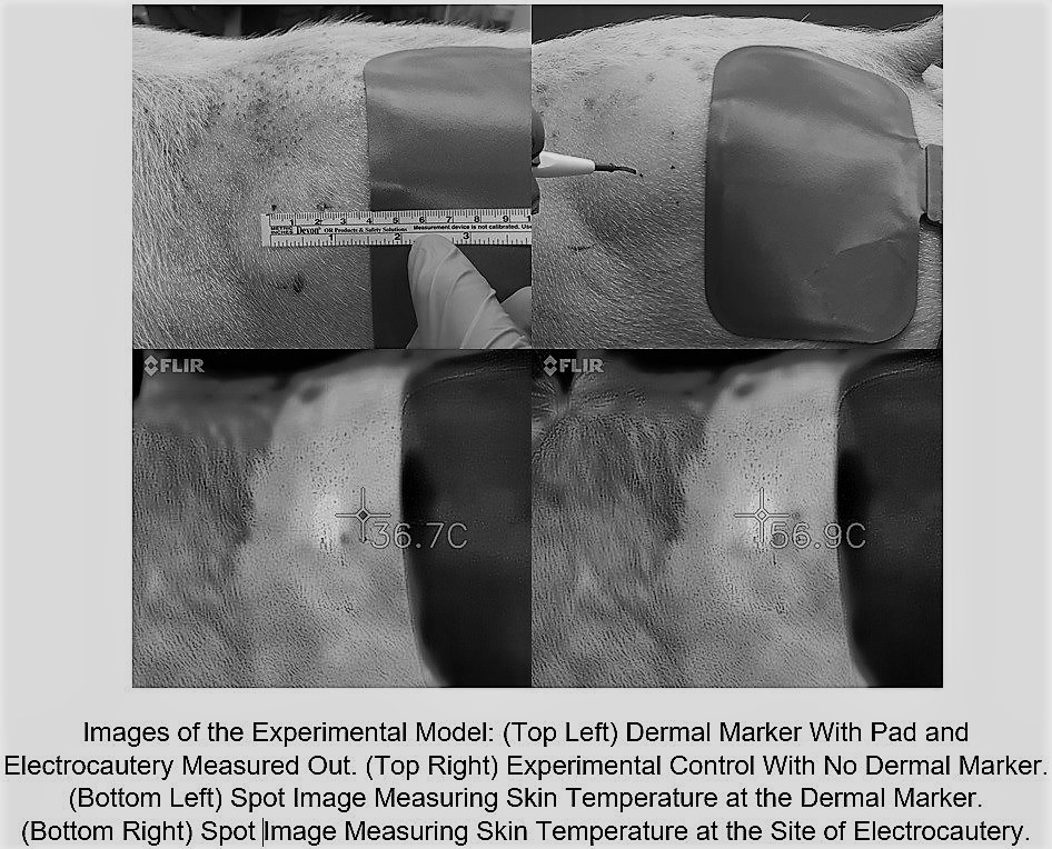
01.17 Microdermal Implants Show No Effect On Surrounding Tissue During Surgery With Electrocautery

R. R. Sheldon1, M. Loughren1, C. Marenco1, K. Morte1, W. S. Do1, D. T. Lammers1, J. B. Weiss1, D. M. Forte1, V. Y. Sohn1, M. J. Martin1, M. J. Eckert1, R. O. Burney1, S. T. Marko1, J. Bingham1  1Madigan Army Medical Center,Tacoma, WA, USA

Introduction:   Microdermal implants are an increasingly popular form of body jewelry in which the device is surgically implanted beneath the skin and held in place by scar tissue that forms around the dermal anchor. The potential for electrical conduction burn at the site of metal jewelry left in situ during electrosurgery prompted surgical societies to recommend routine removal. However, routine removal of microdermal implants can be complicated, requiring destruction of the device or local excision. Given the paucity of evidence regarding electrosurgical burn risk associated with microdermal implants, we assessed in vivo thermal effect and tissue damage at implants during electrocautery use.

Methods:   Stainless steel microdermal anchors (3 mm) were surgically implanted into 4 adult swine.  Implants were placed into the bilateral flank and one of two randomized hindquarters. After 4 days to allow for initial healing and scar tissue formation, the bilateral flank implants were evaluated and excised for histologic examination. A Bovie electrocautery grounding pad was placed 2 cm caudal to the microdermal implant.  Continuous electrocautery (coagulation/ 30 watts) for 30 seconds was applied to the skin at a point 2 cm cranial to the dermal implant.  Surface skin temperature was recorded during electocautery using thermal imaging.  Tissue damage was assessed by both gross examination and histologic evaluation of tissue immediately surrounding the excised microdermal implant. The same procedure was then performed to the contralateral non-implanted side as a sham control.

Results:  Electrocoagulation for 30 continuous seconds raised the skin temperature around the electrocautery tip 49.9° F to an average of 148.7° F.  Skin around the dermal implant only rose 3.24° F to an average of 101.7° F whereas the control skin without the dermal implant rose 3.65° F to 102.3° F (P=0.627). Skin temperatures at the dermal implant at 5 second intervals throughout the coagulation interval showed no statistical difference to those taken from the sham side at any time point.  Histologic review of excised tissue samples showed no evidence of thermal injury.

Conclusion:  Dermal implants appear to have no effect on skin temperature change during the use of electrocautery even when they are in close proximity to both the grounding pad and site of electocautery use.  This suggests that microdermal implants may be safe and aggressive steps to ensure their removal before surgery are unnecessary.