01.12 Actinomyces is a Potential Cause of Perforated and Gangrenous Appendicitis

R. Patel1, S. Misra1, S. Joseph1 1Texas Tech University Health Sciences,Department Of Surgery,Odessa, TX, USA

Introduction:

Acute appendicitis can present with a wide range of symptoms from an indolent course to severe peritonitis and overwhelming sepsis. The patients clinical status often indicates the severity of the disease and if perforation and diffuse peritonitis is present. While there have been many attempts to identify patients who may develop sepsis and peritonitis, little has been found as to the cause of the most severe cases.

Actinomyces, a gram positive facultative anaerobe, is part of the normal flora of the mouth, GI tract, and female vagina, however when pathologic it causes severe inflammation, gangrene, perforation, and obstruction. Actinomyces can easily be identified on pathologic examination by the formation of sulfur granules. We hypothesized that Actinomyces could be a causative agent in patients who present with perforated or gangrenous appendicitis.

Methods:

We did a retrospective review of the last 102 appendix specimens removed for acute appendicitis at our institution. Pathologic specimens were reviewed by a single pathologist to identify sulfur granules or actinomyces species within the specimen. Demographic data, lab value, and radiology were reviewed for all patients in the review. Appendiceal specimens were categorized as either gangrenous/perforated or acute inflammation.

Results:

There were a total of 20 cases of perforated/gangrenous appendicitis amongst our 102 cases (19.6%). For patients with gangrene/perforation (n=20) there were 7 cases of Actinomyces identified (35%). For the 82 patients with acute inflammation, Actinomyces was identified in 6 specimens (7%). p=0.0008

The average WBC level for all patients in our sample was 14.5. No clinically significant difference was found in WBC levels between gangrenous/perforated and acutely inflamed (14.4/ 14.5). However, patients with Actinomyces had a slightly higher average WBC of 15.3.

Conclusion:

In patients with gangrenous/perforated appendicitis there is a 35% chance that Actinomyces is present in the appendix. While in patients without perforation/gangrene there is a 7% chance of identifying Actinomyces. Patients with Actinomyces were found to have a slightly higher WBC level then the patients without, however this was not statically significant.

This data suggests that Actinomyces may be a cause of gangrenous/perforated appendicitis. Since Actinomyces is best treated with a long course of Penicillin based antibiotics, patients with gangrene/perforation may benefit from penicillin based treatment for longer than the standard 2 week course. Finally, Actinomyces may account for failure of non-operative management in selected patients.
01.13 Combination Cancer Treatment: Interferon-Expressing Adenovirus and 5FU Chemotherapy

J. Sell1, A. Oliviera1, E. Jensen1, E. Greeno1, M. Yamamoto1, J. Davydova1 1University Of Minnesota,Department Of Surgery,Minneapolis, MN, USA

Introduction: Interferon-α (IFN) is a cytokine known to have direct and indirect antitumor effects. Previous studies have shown that IFN-based chemoradiation therapy (CRT) can improve survival after resection of pancreas cancer. However, its clinical utility to this point has been limited due to the severe toxicity related to its use. Our aim in this study is to evaluate our group’s novel oncolytic adenovirus (OAd) which allows targeting IFN treatment to cancer cells while sparing healthy tissue. We have previously shown the ability of IFN to sensitize cancer cells to chemotherapy as well as demonstrated an increased therapeutic effect of the drug in immunocompetent models. This study was conducted to analyze the combination of 5FU chemotherapy and our OAd in vitro in order to assess the interaction of treatments and determine the optimum combined treatment regimen.

Methods: Treatments were analyzed in two pancreatic cancer and one esophageal adenocarcinoma cell lines: Panc1, S2013, and OE19. Recombinant OAds expressing luciferase rather than IFN were used to isolate the combination of 5FU and the virus. Two viral models were evaluated. Our therapeutic virus (Cox2) selectively replicative in Cox2 expressing cancer cells and a nearly identical but universally replicative virus (wild type) were compared. Cells were treated with 0, 1 or 10 viral particles per cell and 0, 5, 10, or 20 uM 5FU. Three timing regimens were used: simultaneous administration, 5FU 48 hours before virus, and virus 48 hours before 5FU. Crystal Violet and MTS Assays were used to measure cell death. Viral Copy number to assess viral replication was measured using qPCR.

Results: Cell death analysis showed time dependent killing of each virus, with a 2 day delay for the Cox2 virus. 5FU and each virus produced dose dependent cell death independently. There was a nearly full additive effect seen in cell death from combining treatments only with simultaneously given 5uM 5FU and virus. 10 and 20 uM concentrations and virus produced less than fully additive cell death. There was little additive effect seen in treatments of virus 48 hours before 5FU, across all concentrations. Patterns observed were similar for both S2013 and OE19. Studies with Panc1 are in progress. Viral copy experiments are in progress. Treatments with 5FU 48 hours before virus are in progress.

Conclusion: Our Cox2 OAd shows a killing effect similar to wild type in multiple cancer cell lines. When combined with 5FU treatment the expected addition in overall cell death from the independent treatments diminishes, more so as 5FU concentration increases. This may suggest an inhibition of the virus by 5FU. The killing ability and interaction of the combination treatments appears different when the timing of treatments is varied, suggesting the possibility of a treatment regimen with optimal therapeutic effect. Further studies investigating different chemotherapeutic drugs should be conducted to examine these trends.

01.14 Violet 405-Nm Light: A Novel Therapeutic Agent Against Common Pathogenic Bacteria

M. Barneck2,3, M. De La Presa1, A. Poursaid1, M. Nourian1, M. A. Firpo1, J. T. Langell1,2 1University Of Utah,School Of Medicine, Department Of Surgery,Salt Lake City, UT, USA 2University Of Utah,Engineering,Salt Lake City, UT, USA 3Oregon Health And Science University,School Of Medicine,Portland, OR, USA

Introduction: Hospital Acquired infections (HAIs) are the most frequent adverse event in healthcare delivery worldwide, occuring in 5 -10% of acute care hospital admissions, costing the healthcare system an estimated $28-$45 billion annually. Increasing rates of antimicrobial resistant bacteria have increased the morbidity, mortality and cost of treating these infections.

Visible light sterilization (VLS) using high intensity LEDs and lasers have been adapted into several medical applications and have shown bactericidal activity. The inactivation mechanism is believed to be oxygen dependent photoexcitation of bacterial endogenous porphyrins, which leads to the production of a cytotoxic singlet oxygen species. While some research has been done in this field, an in depth comparison of common clinically pathogenic bacteria in a single study has not been published. Here we demonstrate a comprehensive evaluation of common gram-positive and gram-negative species at varying treatment times, intensities, bacterial concentrations, and photokinetic doses.

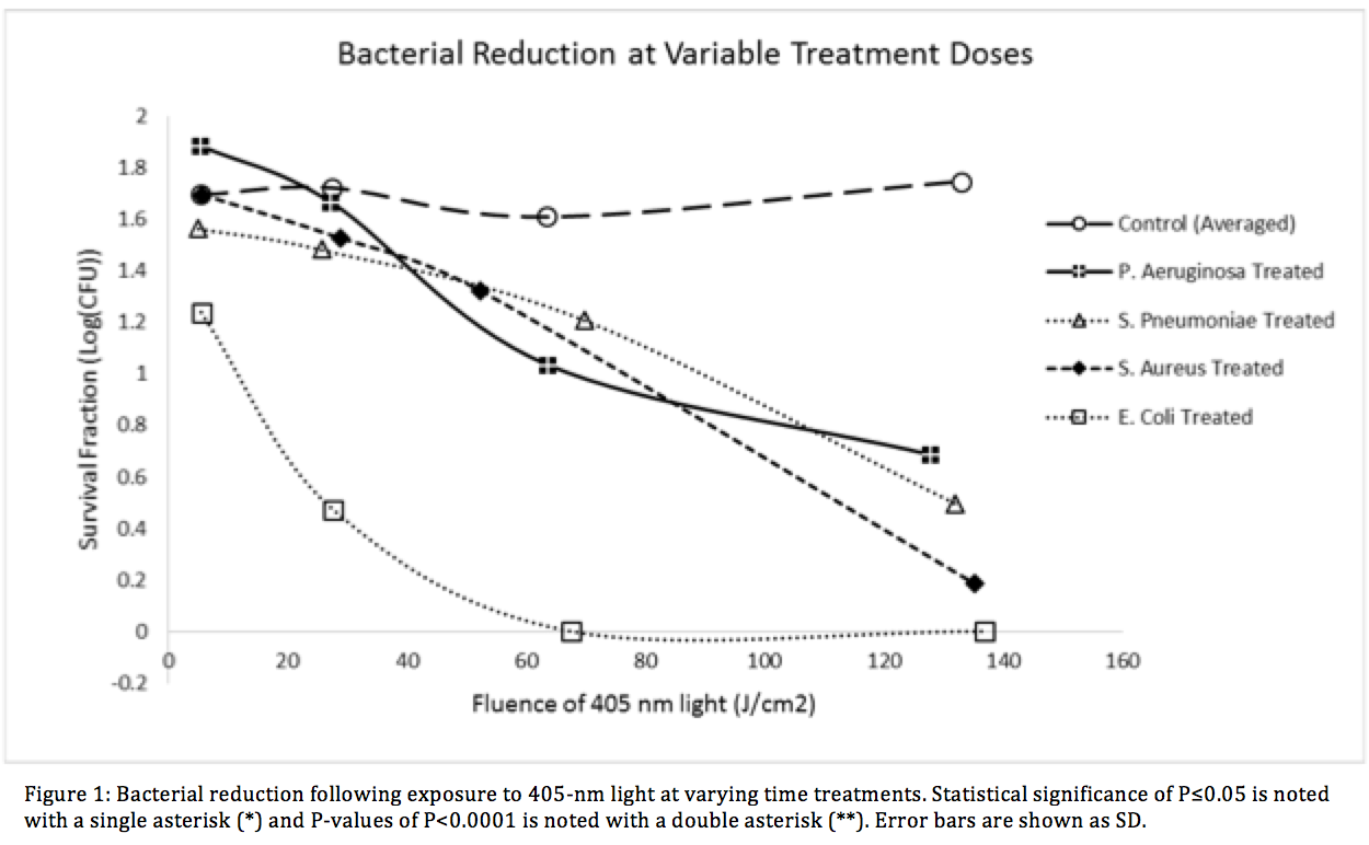
Methods: Visible light emitting diodes centered at 403.0-nm ± 10.8 were used to illuminate a panel of four relevant HAI facultative anaerobic bacteria, Escherichia coli, Staphylococcus Aureus (ATCC 29213), Pseudomonas Aeruginosa (ATCC 27853), and Streptococcus Pneumoniae (ATCC 49619). The E. coli K-12 strain transformed with the pCIG mammalian expression vector conferring ampicillin resistance via expression of the β-lactamase gene. Each bacteria was tested against the VLS with varying treatment durations, light intensities and colony forming unit (CFU) density. Treatment times ranged from 10 minutes to 16 hours, irradiance levels between 2.4mW/cm2 ± 0.3 and 8.9mW/cm2 ± 0.6, and fluence levels up to 132.98 ± 6.69 J/cm2.

Results: Dose dependent effects were visible in visible in all species. The largest inactivation occurred in β -lactam resistant E. coli at 6.27 ± 0.54 log for 250 minutes. Statistically significant results (<0.0001**) were found at each time point.

Conclusion: We have successfully demonstrated high efficacy bacterial reduction using a visible light sterilization system. The VLS showed statistical significance against both gram positive and gram-negative species within the given treatment times. The β-lactam antibiotic-resistant E. coli showed very significant reduction levels which suggests that light therapy could a suitable option for sterilization in drug resistant species of bacteria. Our research illustrates the potential of using VLS in treating clinically relevant bacterial infections in vivo.

01.09 Inhibition of ECM Protein Expression in Hepatic Stellate Cells by JHC0123, an Inhibitor of STAT3

F. J. Bohanon1, X. Wang1, O. A. Nunez Lopez1, A. Kandathiparampil1, N. Ye1, S. J. Vasudevan1, H. Chen1, J. Zhou1, R. S. Radhakrishnan1 1University Of Texas Medical Branch,Galveston, TX, USA

Introduction: STAT3 plays an important role in many physiologic and pathologic processes by regulating cell proliferation, differentiation, and metabolism. It has been reported that the STAT3 pathway is involved in hepatic fibrogenesis. Activated hepatic stellate cells (HSC) are the major effector cells for liver fibrosis, and HSC activation is characterized by increased proliferation and over-expression of α-smooth muscle actin, and extracellular matrix (ECM) proteins. Recently, our team developed a series of novel small-molecule inhibitors of STAT3 and demonstrated their potent anti-tumor effects in breast cancer cells. In the present study, we tested their anti-fibrogenic effects using activated HSC.

Methods: The proliferation of activated human and rat HSC cell lines LX-2 and HSC-T6 were measured by Alamar Blue. Cellular proteins were determined by Western blots and immunofluorescence. Flow cytometry was conducted for cell cycle study. Yo-pro-1 staining was used for apoptosis.

Results:HJC0123 treatment significantly inhibited LX-2 and HSC-T6 cell growth and attenuated HSC activation marker α-smooth muscle actin expression. The expression of endogenous ECM collagen type I and fibronectin was suppressed by HJC0123 in a dose- and time-dependent manner. TGFβ has been identified as the most potent stimulator for ECM. Our data show that in LX-2 cells, TGFβ significantly up-regulated collagen type I and fibronectin expression, and pretreatment with HJC0123 prevented TGFβ-stimulated ECM production. Furthermore, pretreatment with HJC0123 impaired TGFβ-induced pSmad2/3 expression and nuclear translocation, indicating that the TGFβ/pSmad pathway plays a role in ECM regulation by HJC0123.

Conclusion:HJC0123 inhibits HSC proliferation, suppresses endogenous and TGFβ-induced ECMs expression. HJC0123 is a promising anti-hepatic fibrogenic agent.

01.10 Depletion of Bacteria Protects from Chemo-Induced Enteritis Independent of Epithelial Signaling

J. Carr1, S. King1, C. Dekaney1 1University Of North Carolina At Chapel Hill,Chapel Hill, NC, USA

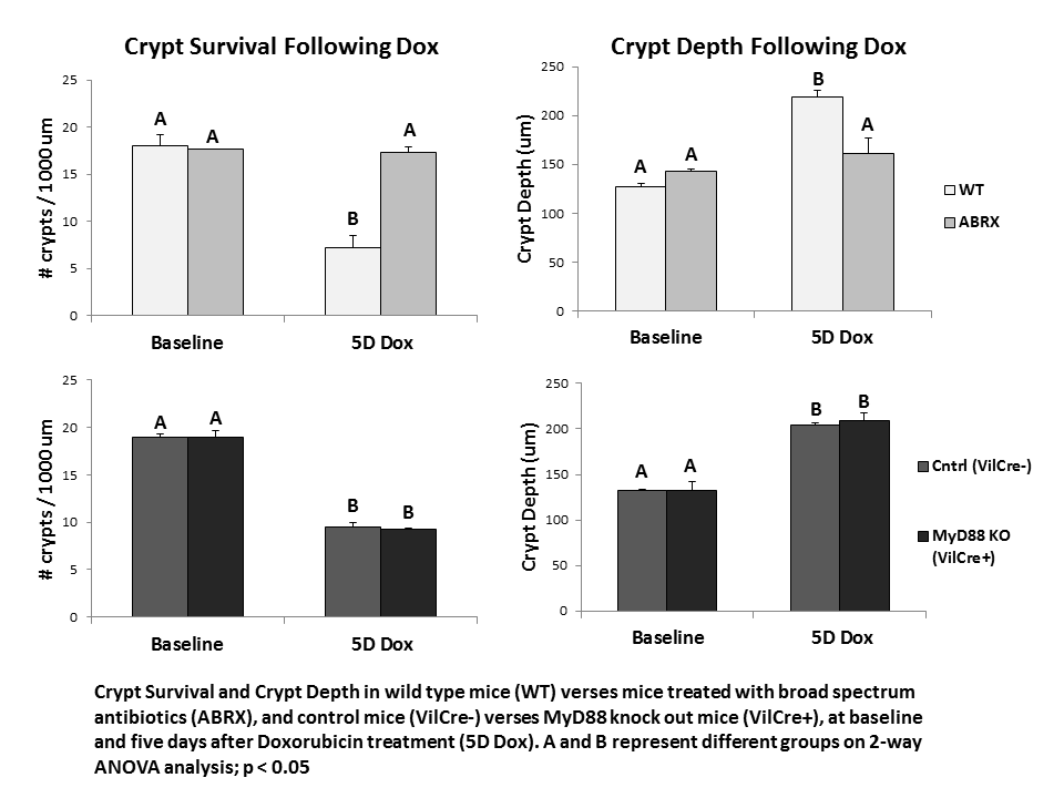
Introduction: Enteritis is a significant side effect of chemotherapy, and is a potential dose-limiting complication. Pharmacotherapy can diminish symptoms, but there are no effective therapies to prevent or treat the intestinal epithelial injury associated with enteritis, most likely because the mechanisms guiding epithelial damage and repair are unknown. Our preliminary studies suggest that enteric bacteria play a critical role in precipitating mucosal damage after treatment with doxorubicin (Dox), a common chemotherapeutic drug. Following Dox, mice treated with broad-spectrum antibiotics (ABRX) do not show the small intestine crypt loss, increased crypt depth, or alterations in proliferation demonstrated in wild type mice (WT). Bacteria and bacterial products signal via distinct toll like receptors (TLRs), while MyD88 is a key adapter molecule utilized by nearly all TLRs to activate downstream signaling pathways. ‘Knocking out’ MyD88 in the intestinal epithelial cell (IEC) specifically eliminates IEC bacterial signaling, and therefore allows an evaluation of the role of IEC bacterial signaling in mediating the bacterial contribution to enteritis. In the current study, we utilize this knock out model to determine whether elimination of bacterial IEC signaling is responsible for protection from Dox-induced enteritis.

Methods: At 10 weeks of age, control or IEC-specific MyD88 KO mice (VillinCre/MyD88fl/fl) were treated with a single intraperitoneal dose of Dox (20 mg/kg) and euthanized five days following treatment. The jejunum was fixed and stained with H&E for histologic analysis.

Results: While ABRX mice demonstrate significantly less weight loss than WT mice five days after Dox, MyD88 KO mice demonstrated weight loss equivalent to WT mice (WT 19.5% ± 2.4; ABRX 5.1% ± 4.6; MyD88 KO 17.5% ± 3.0). ABRX mice demonstrate significantly greater crypt survival than WT mice five days after Dox, and do not demonstrate increased crypt depth typical of WT mice in their hyper-proliferative state after Dox, while MyD88 KO mice demonstrate a phenotype comparable to WT mice (graph).

Conclusions: In response to insult with the chemotherapeutic Dox, MyD88 KO mice demonstrate intestinal damage followed by hyper-proliferation similar to WT mice. This suggests that the protection from damage demonstrated in ABRX mice is not due to elimination of epithelial bacterial signaling. Future studies will investigate other potential signaling pathways (e.g. penetration of a compromised epithelial barrier) to elucidate how depletion of bacteria protects from Dox-induced damage. This will allow determination of a prospective molecular target for prevention of chemotherapy-induced enteritis.

01.11 Ethnic differences in adipocyte exosomal function: a possible etiology for disparities in NAFLD

A. L. Franklin1, T. Iordanskaia1, M. Barberio1, D. Pillai1, M. Hubal1, E. P. Nadler1 1Children’s National Health System,General Surgery,Washington, DC, USA

Introduction:
The pathogenesis of non-alcoholic fatty liver disease (NAFLD) is associated with obesity and insulin resistance. Hispanics have the highest prevalence compared to non-Hispanic Whites and Blacks, with Black patients having the lowest prevalence. Differences in the incidence of NAFLD among race/ethnicity may be related to visceral adipose tissue (VAT), as Blacks have less VAT than Whites and Hispanics. The exact mechanisms behind the ethnic and racial discrepancies are unclear. Exosomes are cell-derived vesicles that contain messenger RNA, microRNA, and proteins, that are potential mediators of systemic disease via the delivery of genetic material to distant organs. We believe that adipocyte-derived exosomes may be the direct mechanistic link between obesity and NAFLD, and exosomal differences may explain the ethnic and racial disparities. We have previously shown that adipocyte exosomes from mixed race/ethnicity donors lead to a dysregulation of gene expression of transforming growth factor beta-1 (TGFΒ-1) mediators in cultured hepatocytes. We sought to determine if ethnic differences among adipocyte exosomes exist.

Methods:
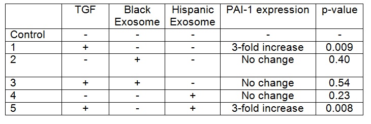
Institutional Review Board approval and patient or parental/guardian consent was obtained. HepG2 cells were grown to 60-70% confluence and then co-cultured with TGFΒ-1 for 48 hours. VAT exosomes from one Black (BMI 51) and one Hispanic (BMI 46) female patient were applied to HepG2 cells in culture with and without TGFΒ-1 exposure. PAI-1 protein expression was analyzed with ELISA, and compared via t-test. Experiments were performed in duplicate and repeated once for each donor.

Results:

TGFB-1 exposure to HepG2 cells increased PAI-1 expression when compared to control cells (p= <0.05). Addition of exosomes from the Black patient decreased PAI-1 expression in HepG2 cells exposed to TGFB-1 (p=0.034), with expression levels that were similar to baseline (Table). In contrast, there was no difference in PAI-1 expression in HepG2 cells exposed to TGFB-1 and exosomes from the Hispanic patient when compared to cells co-cultured with TGFB-1 alone. Exosomes from neither donor altered PAI-1 expression in the absence of TGFB (p= >0.05).

Conclusion:
HepG2 cells co-cultured with TGFΒ-1 had an increase in PAI-1 protein expression. VAT exosomes from a Black patient inhibited TGFB-1 induced production of PAI-1 in hepatocytes. However, exosomes from a Hispanic patient did not inhibit this TGFB-1 induced upregulation of PAI-1. Racial differences in exosomal content and function likely exist, and may be the mechanism behind the known ethnic and racial differences in NAFLD prevalence.

01.06 A Large Animal Model of Liver Injury Induced by Retrorsine and Partial Hepatectomy

K. Inomata1, H. Yagi1, K. Tajima1, H. Shimoda1, T. Hibi1, Y. Abe1, M. Kitago1, M. Shinoda1, O. Itano1, Y. Kitagawa1 1Keio Universiry,Surgery,Shinanomachi, Shinjukuku, TOKYO, Japan

Introduction: Recently, regenerative medicine approaches have been made great advantage, therefore evaluation of its feasibility in animal model is absolutely necessary for clinical application. However, pre-clinical model for the study of liver regeneration is yet to be established. Regeneration process of the liver requires continuous regenerative signals induced by hepatic injury, on the other hand, transplanted cells require functional advantage compared to the remnant cells to repopulate efficiently in recipient’s body. One of the sufficient models for these demands is clearly retrorsine with partial heaptectomy (PH) model. However, no study has been reported using large animal including porcine. Here we demonstrate the first established porcine model with retrorsine and PH.

Methods: Göttingen miniature pigs were treated with 2 intra-peritoneal administrations of retrorsine in different doses; 30mg, 50mg and 100mg/kg respectively, or saline as a control, 2 weeks apart. Four weeks after the second administration of retrorsine, animals underwent PH in different volumes; 50%, 60% and 85%. The animals were sacrificed on 10, 14 and 28 days after the PH and the resected liver volume and histological alterations were evaluated. Blood samples were obtained every 7 days until and every 3 days after the PH to analyze hepatobilially enzymes and synthesized proteins. The quantity of serum and liver tissue retrorsine concentration was determined by LC-MS/MS. Hematoxylin and eosin staining and immunohistochemical staining for Ki-67 and EpCAM were performed on the liver tissue.

Results: The Animals injected 100 mg/kg retrorsine or performed 85% PH were resulted in dead with severe liver injury. Blood test demonstrate that distinct liver disorder was sustained after PH. Liver regeneration was inhibited in animals as 20% volume reduction at day 10 of 60% PH animals with pre-surgical administration of 50mg/kg retrorsine. The histological study demonstrated the explosive puff and cytoplasmic vacuoles were remained in retrorsine treated animals at day 10. The immunohistochemical staining showed the expression of Ki-67 in hepatocytes and EpCAM in biliary epithelium were suppressed in retrorsine treated animals. LC-MS/MS determined the quantity of accumulated retrorsine in the liver tissue while retrorsine was not detected in the blood.

Conclusion: We could successfully demonstrate the first large animal model of retrorsine and 60% PH characterized by sustained liver injury with suppression of hepatic regeneration. We believe that it is a promising model for improving preclinical study of liver regenerative medicine.

01.07 Establishment of a Novel Murine Model of Obstructive Jaundice

H. Aoki1,2, M. Aoki1,2, E. Katsuta1,2, J. Yong3, X. Wang3, H. Zhou3, S. Spiegel2, K. Takabe1,2 1Virginia Commonwealth University,Division Of Surgical Oncology, Department Of Surgery,Richmond, VA, USA 2Virginia Commonwealth University,Department Of Biochemistry & Molecular Biology,Richmond, VA, USA 3Virginia Commonwealth University,Department Of Microbiology And Immunology,Richmond, VA, USA

Introduction: Obstructive jaundice is one of the classic signs of pancreatic head mass, or tumor in the biliary tree. Although there are numerous publications on clinical management of obstructive jaundice, there are very few reports regarding the effects of it on cancer biology itself partly due to lack of stable model. The standard murine model for cholestasis is the partial ligation of the common bile duct (pBDL), which is known to have very short survival, usually less than 3-4 days. Recently, a long term survival cholestasis model that totally ligate hepatic bile duct (tHBDL) was reported. Here we compared tHBDL model with standard pBDL model on their characteristics.

Methods: C57Black6 mice were subjected to sham, pBDL and tHBDL operations. For the tHBDL model, total ligation of the hepatic bile duct was performed at the level where left and median lobe bile ducts, as well as the duct from upper right lobe were completely obstructed, whereas right lower lobe bile duct was spared. Of note, cholecystectomy was added to all tHBDL model. For pBDL model, the suture was tied down tightly at the common bile duct with surgical needle placed by the side, and then the needle was removed leaving a defined lumen within the ligation. Survival was determined by humane endpoints and assessed by Kaplan-Meier method. Weights of body, liver and spleen were measured on the indicated days. Intraoperative complications were also recorded.

Results: 24 mice that received pBDL mice died between 3-4 days after operation. On the other hand, mice that underwent tHBDL were observed to survive over 14 days, longest survivor was over 2 months. After 2 days, pBDL model showed the significant body weight loss compared with sham and tHBDL. An increase of liver weight was observed in both pBDL and tHBDL groups, where it was much heavier in tHBDL group than pBDL group. Spleen of tHBDL model was heavier than other groups, on the other hand, weight of spleen in pBDL group was lighter than sham surgery group. In tHBDL model, enlargement of right lobe was observed. Most common complication during the procedure was bleeding from portal vein or liver and technique for tHBDL model was stabilized due to good exposure of procedure field with assistance.

Conclusion: Total hepatic bile duct ligation model was clearly more stable model than standard partial bile duct ligation model, which is most commonly used. Given the longer survival, total hepatic bile duct ligation model provide us tool to investigate the biological impact of obstructive jaundice.

01.08 Potential of Liver Organoids as Implantation Therapy for Liver Insufficiency

V. X. Zhou1, M. Lolas1, T. T. Chang1 1University Of California – San Francisco,Surgery,San Francisco, CA, USA

Introduction: Liver transplantation is currently the only treatment for end-stage liver disease (ESLD) and is limited by the critical shortage of donor organs. Implantation of liver organoids generated ex vivo by three-dimensional (3D) culture techniques may serve as an adjunct or alternative to liver transplantation. Efficient high-engraftment methods of introducing organoids into the liver parenchyma have not been established. In this study, we aim to develop a reliable surgical technique to implant liver organoids into the liver and assess their ability to engraft and function.

Methods: Liver organoids composed of either hepatocytes alone or hepatocytes co-cultured with stellate cells were generated by 3D culture in rotating wall vessel bioreactors. Primary hepatocytes and stellate cells were isolated from ROSA26 C57BL/6 mice, in which β-galactosidase was expressed under a ubiquitous promoter, so that engrafted cells could be easily identified by X-gal staining when implanted into wild-type mice. Some recipient mice underwent simultaneous two-thirds partial hepatectomy to determine whether hepatocytes within implanted organoids proliferated in response to acute liver insufficiency. As control, freshly isolated primary hepatocytes were transplanted as single-cell suspensions. At various time-points after implantation, engraftment of liver organoids was qualitatively and quantitatively determined.

Results: Confocal microscopy analysis demonstrated that hepatocytes within liver organoids had cortical actin organization and produced extracellular matrix. Organoids maintained their 3D structure up to 7 days after implantation within recipient livers and engraftment efficiency was superior to that of single cells. Generating liver organoids with stellate cell co-culture and performing two-thirds partial hepatectomy on recipient mice did not appear to improve engraftment efficiency.

Conclusion: Implantation of liver organoids generated by 3D tissue culture is promising for development as a novel surgical therapy for ESLD. Further optimization of 3D culture and implantation techniques are required to increase engraftment efficiency.

01.04 SDF-1α Decreases Inflammation in Diabetic Dermal Fibroblasts by Upregulating miR-146a Expression

M. M. Hodges1, C. Zgheib1, J. Hu1, J. Xu1, K. W. Liechty1 1University Of Colorado Denver,Laboratory For Fetal And Regenerative Biology, Department Of Surgery,Aurora, CO, USA

Introduction: In 2014, the United States Center for Disease Control and Prevention (CDC) estimated that 29.1 million Americans (9.3% of Americans) were living with diabetes, with the economic burden of diabetes related wound care estimated as $38.6 billion in 2007. With the global prevalence of diabetes expected to double between 2000 and 2030, there will be an increasing need for innovative technologies to address the commensurate rise in chronic wounds. We have previously demonstrated that diabetic wounds exhibit markedly decreased levels of the anti-inflammatory microRNA miR-146a. Furthermore, we have shown improved healing of diabetic wounds after treatment with a lentiviral construct expressing stromal derived growth factor (SDF-1α). We hypothesize that the improved healing observed in diabetic wounds after SDF-1α treatment is partly due to increased miR-146a expression and the subsequent decrease in inflammation.

Methods: To test our hypothesis we isolated dermal fibroblasts from the skin of 10 week old diabetic (Db/Db) and non-diabetic (Db/+) mice. These fibroblasts were then cultured and transfected with a lentivirus expressing either SDF-1α or green fluorescent protein (GFP) as a control. RNA was extracted from these fibroblasts, and real time PCR analysis was utilized to quantify the gene expression of NFkB, TRAF6, IRAK1, IL-6, MIP2 (IL-8), and miR-146a.

Results: When compared to fibroblasts from the skin of non-diabetic mice, fibroblasts isolated from the skin of diabetic mice demonstrated significantly decreased levels of miR-146a and increased levels of NFkB, TRAF6, IRAK1, IL-6, and MIP2 (IL-8). When transfected with a lentivirus expressing SDF-1α, fibroblasts from the skin of diabetic mice demonstrated significantly increased expression of miR-146a, and significantly decreased expression of NFkB, TRAF6, IRAK1, IL-6, and MIP2 (IL-8).

Conclusions: The significantly increased expression of inflammatory mediators NFkB, TRAF6, IRAK1, IL-6, and MIP2 (IL-8) observed in diabetic fibroblasts is nearly reversed with lenti-SDF-1α transfection. Similarly, the decreased level of miR-146a observed in diabetic fibroblasts is completely reversed when diabetic fibroblasts are transfected with lenti-SDF-1α. The accelerated wound healing previously observed in diabetic skin after lenti-SDF-1α treatment may be due in part to the increased expression of miR-146a and decreased expression of inflammatory mediators NFkB, TRAF6, IRAK1, IL-6, and MIP2 (IL-8). These results support further investigation of the role of SDF-1α in the pathogenesis of diabetic wound healing impairment and as a possible therapeutic target in the treatment of diabetic wounds.

01.05 Mechanistic Analysis: Alcohol Induces Apoptosis and Autophagy in the Liver of Hypercholesterolemic Swine

I. J. Lawandy1, B. A. Potz1, N. Y. Elmadhun1, A. D. Lassaletta1, J. A. Feng1, F. W. Sellke1 1Brown University,Surgery/Cardiothoracic Surgery/ Warren Alpert Medical School,Providence, RHODE ISLAND, USA

Introduction: Autophagy serves as a cellular protective mechanism against alcohol induced tissue injury but can also be detrimental leading to apoptosis. Our lab has previously shown moderate alcohol consumption in a swine model of metabolic syndrome worsens glucose metabolism by altering activation of the insulin signaling pathway in the liver. We examined the effect of alcohol consumption on apoptosis and autophagy signaling in the liver in our clinically relevant animal model of chronic myocardial ischemia and hypercholesterolemia.

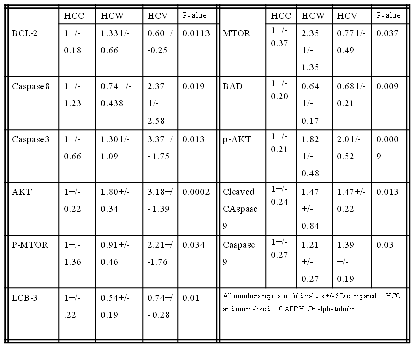
Methods: Twenty-six Yorkshire swine were fed a hypercaloric, high-fat diet for 4 weeks then split into 3 groups: hypercholesterolemic diet alone (HCC, n= 9), hypercholesterolemic diet with vodka (HCV, n= 9), and hypercholesterolemic diet with wine (HCW,n = 8) for 7 weeks. Animals underwent euthanasia and liver tissue samples were harvested for analysis.

Results: There were significant increases in the pro-apoptotic proteins caspase 3, caspase 8, caspase 9 and cleaved caspase 9 in the HCV group and a trend increase in the HCW group compared to control. There was a significant decrease in pro-apoptotic protein Bad in the HCW and HCV groups compared to control. There was a significant increase in anti-apoptoic signal BCL-2 in the HCW group compared to the HCV group and trend increase in the HCW group compared to control. There were significant increases in pro-survival proteins AKT and p-AKT in the HCW and the HCV group compared to control. There was significant increase in MTOR in HCW compared to HCV and a trend increase in compared to control. There was a significant increase in p-MTOR in the HCV group compared to the control. There was a significant decrease in autophagy protein LCB-3 in the HCW and a trend decrease in HCV compared to the control. (See table)

There was a trend increase in anti-apoptotic signal p-BCL-2 in the HCW and HCV groups (p=0.151) compared to control. There were trend increases in the HCV group in AMPKA (p= 0.071) and TNF-alpha (p=0.071) compared to control. There was a trend increase pro- autophagy protein Beclin 1 in the HCW group (p=0.431) compared to control. There were trend increases in pro-autophagy proteins Lamp-1 and Lamp-2 in the HCW group and a trend decrease in the HCV group (p=0.120) compared to control. There were no significant differences in cleaved Caspase 3 (p=0.296), Bax (p= 0.169), p-Bad (p =0.675), LKB-1 (p=0.523), P-AMPKA (p=0.758) or ATG5 (p=0.319) compared to the control.

Conclusions: Moderate Alcohol consumption altered cell survival protein expression related to apoptosis and autophage in pig liver in the setting of hypercholesterolemia. Vodka may induce more pro apoptotic pathways and wine may induce more pro survival pathways in pig liver tissue.

01.03 Tumor-Targeting Nanotheranostic Micelles for Neuroendocrine Cancer Therapy

R. Jaskula Sztul1,3, G. Chen2, A. Harrison1, S. Gong2, H. Chen1,3 1University Of Wisconsin,Surgery,Madison, WI, USA 2University Of Wisconsin,Wisconsin Institutes For Discovery,Madison, WI, USA 3University Of Alabama,Surgery,Birmingham, Alabama, USA

Introduction: Although neuroendocrine tumors (NETs) are slow growing, they are frequently metastatic at the time of their discovery and no longer amenable to curative surgery. Therefore, there is a great need to develop novel therapeutic strategies both to reduce tumor burden and control the release of hormones. To address this need, we developed and optimized a family of novel multifunctional upconversion nanoparticle (UCNP)-based theranostic unimolecular micelles capable of delivering a newly reported anticancer drug AB3 for NET-targeted combination chemotherapy, photodynamic therapy (PDT), and bioimaging. These UCNP-based micelles conjugated with photosensitizer (Rose Bengal, RB) specifically target NET cells using somatostatin analog (KE108). In the current study we assessed the antitumor effects of the nanotheranostic micelles both in vitro and in vivo.

Methods: Stable UCNP-based micelles were prepared in an aqueous solution using multi-arm star amphiphilic block copolymer. KE108 was conjugated for active tumor-targeting. AB3 was loaded into the photosensitive hydrophobic core of the resulting micelles. The effect of 980nm on singlet oxygen generation and in vitro drug release of the UCNP-based micelles were studied. Cell proliferation was assessed by MTT assay in human medullary thyroid cancer cell line (MZ-CRC-1) treated with a family of AB3-loaded micelles (AB3 mM) for 48h with or without 980nm irradiation. The effect of the KE108 targeting ligands on the cellular uptake of the nanotheranostic micelles was measured by flow cytometry and confocal laser scanning microscope (CLSM). The antitumor efficacy of AB3-loaded micelles was determined in GI neuroendocrine (BON) xenografts after two intravenous injections performed with 7 day interval with a dose of 20 mg/kgBW. The group treated with UCNP-based micelles containing RB was irradiated for 15 min with 980nm laser at 4h post injection.

Results: The family of UCNP-based NET-targeting unimolecular micelles was developed for targeted delivery of AB3 and RB to NETs. UCNP-based micelles for targeted and combined chemotherapy with PDT exhibited the strongest antiproliferative effect. Moreover, the targeted micelles exhibited a much higher cellular uptake than non-targeted micelles based on flow cytometry and CLSM analyses. Additionally, AB3 loaded micelles conjugated with RB and KE108 (i.e., T-RB-AB3), enabling combined chemo-therapy and PDT, induced the best antitumor efficacy (82% reduction in tumor volume) and did not cause any significant changes in body weight or survival. Pathological assessment of H&E-stained sections of different organs of mice treated with T-RB-AB3 micelles did not indicate any signs of inflammation or necrotic regions.

Conclusion:The AB3-loaded UCNP-based micelles conjugated with both RB and KE108, offering combination chemotherapy and PDT, are more effective at suppressing NET cell growth while having minimal toxicity to non-NET cells.

09.21 Surgical Quality in Low- and Middle-Income Countries: a Systematic Literature Review

S. Saluja4,5,6, S. Mukhopadhyay4,6,8, A. Silverstein4,6,10, Y. Lin4,6,9, R. Sood4,6,10, N. Raykar3,6, P. Moberger12, J. Von Schreeb11, J. G. Meara4,6, M. G. Shrime6,7 3Beth Israel Deaconess Medical Center,Department Of Surgery,Boston, MA, USA 4Children’s Hospital Boston,Department Of Plastic Surgery,Boston, MA, USA 5Weill Cornell Medical College,Department Of Surgery,New York, NY, USA 6Harvard Medical School,Program In Global Surgery And Social Change,Boston, MA, USA 7Massachusetts Eye And Ear Infirmary,Boston, MA, USA 8University Of Connecticut,Department Of Surgery,Storrs, CT, USA 9University Of Colorado Denver,Department Of Surgery,Aurora, CO, USA 10University Of Miami,Miami, FL, USA 11Karolinska Institutet,Health System And Policy Research,Stockholm, , Sweden 12Uppsala University,Department Of Surgery,Uppsala, , Sweden

Introduction:

In recent years, there has been a large and successful movement towards putting surgery on the global health agenda. Thus far much work has focused on improving access to care. The study of surgical quality, however, remains a nascent field, especially in low- and middle-income countries. We systematically evaluate the literature assessing surgical quality in LMIC and discuss possible areas for further investigation.

Methods:
We conducted a search of the English language literature published after January 1, 2000 using Medline and Embase. MeSH, Emtree, and individual word searches were used to capture three domains: quality of care, surgical procedure, and low- and middle-income country. We included studies evaluating surgeries that comprise the DCP-3 Essential Surgery Package for first level hospitals, excluding studies of thoracostomy, colposcopy, and vacuum extraction/forceps delivery. Furthermore, we excluded any study where all patients did not undergo surgical intervention. The data extracted included: sample size, study design, type of surgery performed, and whether processes or outcomes were evaluated. We further determined whether authors evaluated checklist utilization, mortality, morbidity, or health-related quality of life (HRQL). Our search and extraction followed PRISMA guidelines.

Results:
Our search identified a total of 4885 articles; 301 studies remained for data extraction after title and abstract screening. The mean sample size of the studies was 1841 patients (SD 9359.7) with a median of 104 patients. 78.2% of studies were observational, of which 55.6% were retrospective. 28.9% of studies evaluated multiple surgical procedures. Studies of operative fracture reduction (11.9%), hernia surgery (11.2%), Cesarean sections (11.2%), and laparotomy for perforated viscus (10.9%) were the most common of the DCP-3 procedures evaluated. There were no studies evaluating quality for patients specifically undergoing escharotomy/fasciotomy and only one study each for dilation & curettage, skin graft, vasectomy, and debridement of osteomyelitis. We found process-based measures reported in 10.3% of studies; 99% of studies reported outcomes-based measures. Amongst studies reporting processes, 22.6% were of checklists. Amongst studies reporting outcomes, mortality was reported in 47.2%, morbidity in 88.0% and HRQL in 16.3%.

Conclusion:

As LMICs expand access to surgical care, there must be a directed effort to monitor the care delivered. Studies of surgical quality vary greatly in size, but many are small. The vast majority of these studies are observational in design. Further, HRQL and process-based measures are frequently overlooked dimensions of quality, while numerous essential surgeries are underrepresented in the literature. Future studies should expand on these parameters to close gaps in the literature.