03.08 Sepsis Induces Alterations of PD-1/PD-L1 Expression on Peritoneal Neutrophils.

E. A. Fallon1, A. Rossi1, W. G. Cioffi1, A. Ayala1, D. S. Heffernan1  1Brown University / Rhode Island Hospital,Department Of Surgery,Providence, RI, USA

Introduction:  Sepsis remains a leading cause of death, with much of the morbidity and mortality deriving from immune dysfunction. Specifically, sepsis induces profound immunoparalysis. We have shown a role for the checkpoint protein Programmed cell death receptor-1 (PD-1) in regulating the immune response to sepsis. Although considerable data has emerged regarding the role and function of PD-1 and its primary ligand PD-L1 upon lymphocytes, little information is available regarding the role of PD-1/PD-L1 upon neutrophils in response to intra-abdominal murine sepsis.

Methods: Cecal ligation and puncture (CLP) versus sham laparotomy was undertaken in 8 to 12 week old male C57BL/6 mice. At 24 hours, mice were euthanized and peritoneal cells were obtained by lavage. Samples were analyzed by flow cytometry with monoclonal antibodies. Neutrophils were identified as CD11b+/Ly6G+ cells. These cells were then further analyzed for PD-1 or PD-L1 expression. Chi-squared and Mann-Whitney-U tests were used for statistical analysis.

Results: Sepsis was noted to induce an increase in both absolute number of neutrophils into the peritoneal cavity, as well as neutrophils as a percentage of peritoneal cells (21% versus 43%; p<0.05). PD-1 expression upon peritoneal neutrophils was unchanged following sepsis (52% versus 59%; p=0.1), whereas PD-L1 expression was increased upon peritoneal neutrophils 22% vs 51%; p=0.02).

Conclusion: Neutrophils play a critical role in the immune response to polymicrobial sepsis. Although expression of PD-1, a key checkpoint protein of sepsis pathophysiology, upon peritoneal neutrophils is unchanged in response to sepsis, PD-L1 expression is elevated. This is akin to previous findings of the PD-1/PD-L1 relationship on lymphocytes. Our data support the concept that upregulation of PD-L1 is associated with migration into the peritoneal cavity in response to sepsis.

 

03.07 Targeting of Epigenetic Regulators from Sepsis Dervied Microvesicles Prevents Immunosupression

J. Wisler1, D. Dakhlallah2, T. Eubank2, C. Marsh2  1Ohio State University,Surgery,Columbus, OH, USA 2West Virginia University Robert C Byrd Health Sciences Center,Microbiology, Immunology And Cell Biology,Morgantown, WV, USA

Introduction: Systemic immunosuppression associated with severe sepsis is a poor prognostic marker and predisposes survivors to increased infection risk, morbidity and mortality. Immunosuppression can persist for years following the initial insult. We have previously presented this immunosuppression is related to epigentic regulators that can be transferred via circulating microvesicles (MV).  Epigenetic events occur at the promoter sites of several key proinflmmatory genes including IL-1[beta] and TNF-[alpha].  We hypothesized that these circulating MVs can be targeted for therapy to prevent these epigenetic events from occuring and maintain a functional host immune response.

Methods: Naïve human monocytes were treated with MVs from patients with septic shock, sepsis, critical illness without sepsis, and non-septic normal controls. After incubation with MV, monocytes were stimulated with LPS and TNF-a expression measured and DNA methylation was assessed at the TNF-[alpha] promoter.  Using a competitive inhibition model, dose response curves were generated to assess inhibition of uptake.  Cells were also treated with a methylation inhibitor, 5'-Azacitidine, to prevent methylation events from occuring.

Results: Naïve monocytes treated with MVs from patients with sepsis demonstrated increased expression of DNMTs at 24 hours. Promoter methylation at TNF-[alpha] was increased significantly in recipient monocytes and these cells demonstrated a dramatically reduced TNF-[alpha] expression in response to LPS challenge (all p < 0.04, Fig 1).  Treatment of naive cells with healthy donor derived MVs significantly reduced TNF-[alpha] promoter methylation.  Additionally, treatment of naive cells with demethylating agents significantly reduced TNF-[alpha] promoter methylation events and these cells maintained their ability to generate TNF-[alpha] in response to LPS stimulation.

Conclusion: These data demonstrate that targeting of sepsis derived MV may prevent transfer of methylation events to naive recipient cells and prevent immunosuppressive consequences.  The use of healthy donor derived MVs or demethylating agents significantly reduced the transfer of epigenetic elements and allowed naive monocytes to maintain their immunocompetence.  This has potential as a novel therapeutic target during the septic process

03.06 The Role of the Innate and Adaptive Immune Systems in the Pathogenesis of Traumatic Brain Injury in Mice

J. C. Alonso-Escalante1, D. F. Hamade2, C. P. Sodhi1, W. Fulton1, D. J. Hackam1, I. W. Nasr1  1Johns Hopkins University School Of Medicine,Pediatric Surgery,Baltimore, MD, USA 2American University Of Beirut School Of Medicine,Beirut, BEIRUT, Lebanon

Introduction:
The morbidity of traumatic brain injury (TBI) develops after the convergence of acute and chronic neuroinflammatory pathways that cause axonal injury. The role of the innate and adaptive immune systems in these processes remains incompletely understood. We now hypothesize that innate toll-like receptor 4 (TLR4), and adaptive immune cells are decisive in the development of TBI in mice, and sought to address this by development of an experimental model of TBI in 4-8 week-old male mice.

Methods:
A Controlled Cortical Impact (CCI) murine TBI model using C57BL/6 wild-type (WT), adaptive immune deficient Rag1 KO, and innate immune receptor TLR4 knockout (KO) mice, was performed by shooting a 3-mm diameter piston over intact dura mater with a speed of 2m/s, and a depth of 1.5mm. The volume of brain injury was assessed using H&E stained sections and quantitative microscopy, and MRI of total cerebral cortex. Using confocal immunofluorescence (IF) stained for CD3, TUNEL and NeuN, sections of injured cortex were labeled to determine the extent of CD3+ cell infiltration and neuronal apoptosis. All analyses were performed at 1 day and 7 days after CCI.

Results:
The Volume of brain injury, as measured by MRI, showed a mean lesion volume of 81±2mm3 in WT C57BL/6 mice (n=2), 140±7mm3 in Rag1 KO mice (n=3), and 156±2mm3 in TLR4 KO mice (n=3) 1 day after cortical impact. After 7 days, the mean lesion volumes were 50±3mm3 in WT C57BL/6 mice (n=2), 132±3mm3 in Rag1 KO mice (n=2), and 36±1mm3 in TLR4 KO mice (n=2). H&E results were positively correlated with MRI results and demonstrated a volume of 90±5mm3 and 56±8mm3 in C57BL/6 WT, 148±4mm3 and 160±5mm3 in Rag1 KO, 180±6mm3 and 28±4mm3 in TLR4 KO mice, all at 1 day and 1 week post-TBI respectively.  TLR4 KO mice showed a statistically significant increase in the number of apoptotic neurons (208±26cells/hpf) compared to WT C57BL/6 (87±6cells/hpf) 1 day after CCI. After 1 week of trauma, TLR4 KO mice showed a significant decrease in apoptotic neurons (126±1cells/hpf) when compared to WT C57BL/6 (210±7cells/hpf) (p<0.05). CD3+ cell infiltration was significantly greater at 1 day after CCI in TLR4 KO mice (14±1cells/hpf) vs WT C57/BL6 mice (8±2 cells/hpf) (p<0.05). Strikingly, after 1 week of CCI, CD3+ cell infiltration was significantly decreased in TLR4 KO mice (6.3±1cells/hpf) vs WT C57BL/6 mice (12±2cells/hpf) (p<0.05) suggesting a significant role for the innate immune system in the pathogenesis of this disease.

Conclusion:
We now conclude that in accordance with our hypothesis, the development of TBI in mice requires the input of the innate and adaptive immune system, and that these findings are time dependent. These results raise the possibility that strategies that modulate the innate and adaptive immune systems may have a role in reducing the severity of brain injury in post-TBI patients.
 

03.05 Integrinβ3 Positively Regulates TLR-induced Inflammation by Targeting CD14 Expression in Septic Mice

Z. Chen1, Z. Shao1, Q. Li1  1East Hospital,Tongji University School Of Medicine,Shanghai, SHANGHAI, China

Introduction:

Sepsis is one of the life-threatening diseases worldwide. It is characterized by inappropriate amplified systemic inflammatory response that may promote multiple organ dysfunction and mortality. TLR activation plays a key role in sepsis.CD14 is necessary for TLRs dependent induction of pro-inflammatory cytokines. Recent studies indicated that integrinβ3 involved in TLR-triggered innate immunity. Our previously study found that integrin inhibitor RGDs alleviated sepsis induced lung injury and improved survival rate. However, the interaction between integrinβ3 and TLR activation in sepsis remains largely unknown. This study aims to uncover the mechanism of integrinβ3 and TLR activation in in vivo and in vitro study.

Methods:
In vivo: WT, TLR4 and integirnβ3-/-mice undergoing cecal ligation and puncture (CLP) induced sepsis were sacrificed at indicated time. Serum was collected to detected inflammatory cytokines via ELISA. Tissue was fixed with 4% paraformaldehyde (PFA) for HE stain or immunohistochemical. Tissue lysate for CD14 expression was detected by Western blot.
In vitro: Peritoneal macrophages isolated from WT , TLR4 and integirnβ3-/-mice were stimulated with LPS at different concentration or indicated time, then assayed CD14 expression by Wb and flow cytometry. Medium was collected for TNF-α and IL-6 release via ELISA. Pretreated WT macrophages with TAK or anti-β3 Ab then stimulated with LPS and detected CD14 expression by Wb and flow cytometry.  293T cells were cotransfected with Flag-TLR4 and HA-integrinβ3 for 24h and stimulated with LPS for 30 and 60min then assayed by co-immunoprecipitation.

Results:

Integrinβ3−/− mice are resistant to CLP induced sepsis. Sepsis induced CD14 up-regulation of the tissue was suppressed in TLR4-/- and integrinβ3−/− mice. LPS induced CD14 expression was blocked in PMB or TAK pretreated WT macrophages and TLR4 mutant macrophages. LPS increased CD14 expression was suppressed in integrinβ3−/− macrophages. Integrinβ3 inhbition via anti-β3 or P11did not affect CD14 expression in LPS stimulated macrophages. Activation integrinβ3 by vitronectin also had no effect in CD14 expression in macrophages. Both integrinβ3 inhibition and integrinβ3 depletion attenuated LPS induced pro-inflammatory cytokines production. TLR4 and integrinβ3 interaction at rest condition and increased under LPS stimulation.

Conclusion:
Integrinβ3 positively regulates TLR4-triggered inflammatory responses through two different ways. Integrinβ3 signaling activation involved in LPS induced inflammatory cytokines production. Integrinβ3 interacted with TLR4 contributed to LPS induced CD14 expression. Specifically targeting integrinβ3/TLR4-CD14 signaling may represent a novel strategy in polymicrobial sepsis.

03.04 Pancreatic Cancer Reverses the Gut-protective Effects of EGF Overexpression in Sepsis

J. D. Lyons1, Z. Liang1, N. Klingensmith1, K. Fay1, C. Chen1, C. Coopersmith1  1Emory University,Surgery,Atlanta, GA, USA

Introduction:
Sepsis increases intestinal epithelial cell (IEC) death, decreases IEC proliferation, and worsens gut barrier function. The trophic hormone epidermal growth factor (EGF), given either systemically or by local tissue over-expression, has been repeatedly shown in mouse models to limit gut damage during sepsis and improve survival. Pre-existing malignancy, however, is known to worsen sepsis survival and alter patterns of gut damage in septic shock, possibly by inducing changes in circulating lymphocyte populations. We sought to determine if EGF remained beneficial in septic mice with pre-existing cancer.

Methods:

Experiments were performed on age-matched fabpi-EGF mice (animals with enterocyte-specific expression of EGF), wild-type (WT) mice, and RAG1-/- mice (animals that lack mature lymphocytes). Mice were given subcutaneous injections of a pancreatic cancer cell line (Pan02), observed for 3 weeks to allow tumor growth, and then received intra-tracheal injections of Pseudomonas aeruginosa to model sepsis from bacterial pneumonia. Mice were either sacrificed at 24 hours or followed for 7-day survival. IEC death by apoptosis was quantified histologically, counting morphologically apoptotic cells on hematoxylin-eosin staining. IEC proliferation was quantified by injecting mice with the thymidine analogue bromodeoxyuridine (BRDU) prior to sacrifice and staining for BRDU-positive cells in intestinal crypts. Gut permeability was assayed by detecting serum concentrations of fluorescent dextran (FD4), which was orally gavaged prior to sacrifice. Means from each group were compared via T-test with Welch’s correction, and survival studies were compared via Log-Rank test. P < 0.05 determined significant throughout.

Results:
Although gut EGF expression has been shown to be enterocyte-protective in healhy animals subjected to sepsis modles, Fabpi-EGF mice with cancer paradoxically had increased rates of septic IEC death compared to mice with cancer alone (29.6±15.8 apoptotic cells/100 intestinal crypts v. 14.6±6.3, n=7-8, p=0.049). Gut permeability was also surprisingly significantly increased in septic cancer mice that expressed EGF as compared to septic mice with only cancer (5990±3500 ng FD4/ml serum v. 2840±1230, n=6-10, p=0.02). Rates of IEC proliferation did not differ between groups (258.3±36.5 BRDU-positive cells/100 crypts v. 249.9±43.3, n=6-9, p=0.69). Furthermore, there was no difference in 7-day survival between fabpi-EGF mice with cancer and mice with cancer alone (35% vs 17%, n=35-36, p=0.09) though EGF therapy has previously exerted a strong survival benefit. RAG1-/- mice with cancer had significantly worsened gut cell death after sepsis than RAG1-/- mice without cancer (38.8±25.5 apoptotic cells/100 crypts v. 7.3±5.4, n=4-5, p=0.049), suggesting cancer may impact IEC death processes in sepsis independent of lymphocytes.

Conclusion:
Gut-specific expression of EGF, while protective in healthy animals, loses its ability to improve survival and appears to actually worsen septic gut damage when mice have pre-existing cancer. How this reversal comes about is unclear, but our data suggest cancer’s impacts on IEC death after sepsis may be independent of cancer’s known effects on immune cell populations. 
 

01.01 Ex-situ Hepatic Transection with Use of Electrocautery

K. Bittner1, M. C. Mora1, K. Douglas1, K. Wong1, D. Tashjian2, K. Moriarty2, M. Tirabassi2  1Baystate Medical Center,Springfield, MA, USA 2Baystate Children’s Hospital,Springfield, MA, USA

Introduction:
Blood loss is an important factor affecting perioperative outcomes in patients undergoing ex-situ split liver transplantation. The purpose of this study was to determine if monopolar electrocautery can be used to achieve hemostasis in an ex-situ liver transection model. 

Methods:

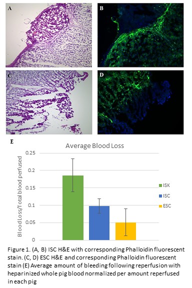
Following euthanasia of Yorkshire swine utilized from an IACUC-approved study, a midline laparotomy was performed. The left hepatic lobe was incised twice to length 6cm and depth 1cm utilizing either monopolar electrocautery (in-situ cautery, ISC) or a 10 blade (in-situ knife, ISK). For ISC, the animal was grounded using a standard adhesive grounding pad. Vascular inflow and outflow to the liver was controlled. The liver was then perfused with heparinized ice-cold lactated ringers.  Next, the liver was harvested, transferred to a sterile back table, and placed into a container containing slush solution. The container was set on a draped reusable non-contact grounding electrode pad.  Ex-situ, the liver was incised with monopolar electrocautery (ex-situ cautery, ESC) and was then re-perfused with heparinized whole pig blood (203±129mL/pig). After re-perfusion, total blood loss from each liver parenchymal incision was quantified by weight via capture into absorptive gauze. Hematoxylin and eosin (H&E), and fluorescent staining with Phalloidin (F-actin) on liver samples were performed to compare the effects of ISC and ESC. 

Results:
In all three re-perfused pig livers, ISK demonstrated the greatest blood loss. In 2/3 pigs, ESC resulted in less bleeding versus ISC.  As each pig had a different amount of blood reperfused, data was normalized as a percentage of reperfused blood for each pig (Figure 1E). In ESC and ISC, H&E sections of livers revealed fragmented margins, cell distortion, and elongated nuclei consistent with electrocautery injury (Figure 1A&C). Staining with Phalloidin in both groups revealed a zone of denatured actin corresponding to areas of electrocautery use (Figure 1B&D). 

Conclusion:
Monopolar electrocautery can effectively be performed in ex-situ liver transections using a commercially available non-contact grounding electrode pad.  Incorporation of this technique in ex-situ liver procedures can potentially result in reduced blood loss after transplantation.

02.14 Xanthohumol Increases DR5 Expression and Enhances Growth Suppression with TRAIL in Neuroblastoma

S. Engelsgjerd1, S. Kunnimalaiyaan1, T. Gamblin1, M. Kunnimalaiyaan1  1Medical College Of Wisconsin,Surgical Oncology/Surgery,Milwaukee, WI, USA

Introduction:  High-risk neuroblastoma (NB) is a lethal childhood cancer. Published data have reported the anti-proliferative effect of Xanthohumol (XN), a prenylated chalcone, in various cancer types suggesting that XN could be a useful small molecule compound against cancer.  The effect and mechanism of XN on NB cell proliferation is unknown. This study hypothesizes that XN will inhibit NB growth, and the effects of XN on cellular proliferation as well as the mechanism of action in NB cell lines were examined. The TNF-Related Apoptosis Inducing Ligand (TRAIL) is an endogenous ligand that is expressed in monocytes, macrophages, dendritic cells, natural killer (NK) cells, and activated T cells. TRAIL mediates apoptosis through binding of transmembrane receptors, death receptor 4 (DR4) and/or death receptor 5 (DR5). Cancer cells are frequently resistant to TRAIL-mediated apoptosis, and the cause of this may be decreased expression of death receptors. This study hypothesizes that XN increases DR5 expression in NB cells thus sensitizing them to TRAIL.

Methods:  The effect of XN in human NB cell lines NGP, SH-SY-5Y, and SK-N-AS was determined via MTT assay. Cell confluency assay by cell live imaging was also carried out for SK-N-AS and NGP cell lines after XN treatment. Cell lysates were analyzed through Western blotting for pro- and anti-apoptotic markers as well as death receptor (DR5). Synergistic analysis of XN and TRAIL in SK-N-AS cells was performed via MTT assay.

Results: XN treatment causes a statistically significant decrease in the viability of NB cells with IC50 values of approximately 12 µM for all three cell lines. Inhibition of cell proliferation via apoptosis was evidenced by an increase in pro-apoptotic markers, including cleaved PARP, cleaved caspase-3, and Bax, and a reduction in anti-apoptotic markers BcL-2 and survivin. Importantly, XN treatment increased expression of DR5. Furthermore, statistically significant synergistic reduction was observed following pretreatment with XN (41%) compared to either TRAIL or XN alone (10%) in SK-N-AS cells.

Conclusion: XN treatment reduces NB cell growth via apoptosis in a dose-dependent manner. Treatment is associated with an increase in DR5 expression. Most importantly, enhanced growth reduction was observed in combination with TRAIL. This is the first study to demonstrate that XN alters the expression of DR5 as well as the synergistic effect of XN on TRAIL in NB. This study provides a strong rationale for further preclinical in vitro and in vivo analysis of XN in combination with TRAIL. 


02.16 Tumor Derived Microparticles and Exosomes Induce Invasion in Pancreatic Cancer

I. A. Naqvi1, R. Gunaratne1, J. Yeh4, D. Pisetsky5, B. Sullenger2, R. White3  1Duke University Medical Center,School Of Medicine,Durham, NC, USA 2Duke University Medical Center,Department Of Surgery,Durham, NC, USA 3University Of California – San Diego,Department Of Surgery,San Diego, CA, USA 4University Of North Carolina At Chapel Hill,School Of Medicine,Chapel Hill, NC, USA 5Duke University Medical Center,Department Of Medicine,Durham, NC, USA

Introduction:  Pancreatic cancer (PC) has the worst prognosis of the major cancers. Metastatic progression is one of the main reasons PC is so deadly. Microparticles (MPs) and exosomes (EXOs) are cell-derived lipid particles derived from the plasma membrane and endoplasmic reticulum, respectively. Recent work has shown that intercellular communication via MPs and EXOs may play a role in tumor progression. We investigated the functional effects of tumor-derived MPs and EXOs on a pancreatic cancer cell line derived from a genetically engineered mouse model (KPC).

Methods:  KPC cells were cultured in EXO-free media at 100% confluence for 72 hours after which cell supernatant was collected and centrifuged at 20,000g to isolate MPs and further at 120,000g to isolate EXO. Effects of MPs and EXO on cellular invasion were quantified by Transwell Matrigel-Invasion assay. Briefly, cells were plated in the upper chamber of a Transwell chamber with either MPs, EXO, or vehicle control and allowed to invade for 24 hours. The Transwell membrane was then fixed and stained with crystal violet. Invaded cells were quantified using Image-J software. The effects of MPs and EXO on intracellular signaling were investigated via western blot.

Results: We observed that treating KPC cells with tumor cell-derived MPs and EXOs significantly increased their invasiveness in the Transwell Matrigel-Invasion Assay Figure 1A and 1B). We also observed that treating with MPs and EXOs caused increased phosphorylation of both p65 NFkB and p-38 MAPK, both known to be important mediators of tumor progression. 

Conclusion: Our results are consistent with recent work showing that MPs and EXOs may be playing a critical role in cancer progression and metastasis. Ongoing work is focused on inhibiting the effects of MPs and EXOs on cancer cells and understanding the molecular underpinnings of the effects induced by MP and EXO treatment. 


02.17 Examining the Role of Telomere Length and Inflammation in Glioma Oncogenesis with an Agent-Based Model

D. Kokosi2, G. An1  1University Of Chicago,Surgery,Chicago, IL, USA 2National And Kapodistrian University Of Athens,School Of Medicine,Athens, ATHENS, Greece

Introduction:  Gliomagenesis is a phenomenon of complex molecular etiology, with recent genomic screening supporting the importance of telomere biology. There is a suggestion of a the correlation between long telomere length predisposition and increased risk of glioma. The hypothesis that longer telomere length enhances the proliferative capacity of each cell and therefore increases the possibility of malignant transformation is backed by various studies related to different types of carcinogenesis; however, a systemic approach to understanding the dynamics of this correlation is difficult to accomplish in vivo due to the complexity of the constituting factors. Agent-based modeling, a computational modeling method, can be applied to examine hypotheses related to specific elements of gliomagenesis in a dynamic and systemic fashion, and evaluate macroscopic evidence through cell-level systems components and functions. Consequently, we have developed an abstract agent-based model (ABM) that encompasses the fundamental aspects of neural tissue function in healthy and injured states, and incorporated a possible role of telomere length and inflammation in glioma oncogenesis. 

Methods:  The ABM that was developed includes agents representing neurons, astrocytes, microglia and stem cells. The inter-agent interaction via signaling molecules in both healthy and injured states is based on published mechanisms. The model was calibrated to simulate the generation and accumulation of mutations in astrocytes in relation to their degree of activation during injury response. Important parameters that were incorporated and examined during experimental runs include: activation of glial cells, astrocyte proliferation, lifespan of cells, telomere length and mutation levels of astrocytes, neuron damage and degeneration, inflammation and chemotaxis. Stem cell differentiation was triggered via a counter or due to local injury. 

Results: The ABM identified that the ability of astrocytes to acquire, accumulate, and pass on mutations was enhanced during an activated state due to elevated levels of stress and proliferation caused by inflammation-induced injury. The proliferative capacity of astrocytes was increased by longer telomere length as a larger number of cell divisions were permitted before cell senescence. Simulations of 80-year trials suggest that this mechanism is plausible in explaining the role of longer telomere length in gliomagenesis.

Conclusion: Gliomagenesis is comprised of an intricate network of pathways that are the subject of ongoing research. The ABM effectively represents fundamental responses of neural tissue to inflammation-induced injury and indicates a plausible explanation for the role of longer telomere length in glioma oncogenesis. ABMs provide a means of defining a basic biological framework into which detailed elements can be dynamically incorporated in order to visualize, evaluate, and reshape hypotheses.


02.18 HDAC Inhibitors Modulate Notch3 Expression though a Unique Transcriptional Motif in NE Cancer Cells

S. Jang1, H. Jin1, R.Jaskula-Sztul1, H. Chen1  1University Of Alabama At Birmingham,Surgery,Birmingham,, AL, USA

Introduction: It is known that Notch signaling is minimally active in neuroendocrine (NE) cancer cells and the induction of Notch isoforms alter the malignant neuroendocrine phenotype. The induction of Notch3 by Histone Deacetylase (HDAC) Inhibitors appears to be the result of increased mRNA expression at the transcriptional level, which leads to suppression of cancer cell proliferation and to apoptosis. The aim of our study is to investigate the molecular mechanism of HDAC inhibitor activation on the Notch3 pathway.

Methods: We functionally characterized the Notch3 promoter using deletion mapping. The mapping started with the truncated genomic DNA fragment fused with a luciferase reporter in plasmid vector, transfected into BON cell, a carcinoid cell line, screened for luciferase activity. Protein-DNA binding was then performed by electrophoretic mobility shift assay (EMSA).

Results: One HDAC inhibotor, Thailandepsin-A, was shown to induce luciferase activity controlled by a small distal region of Notch3 promoter, from -120 to +1 of the start codon ATG.  Further, we identified a functional DNA motif that is responsible for HDAC induction located at -120 to -100 of the Notch3 promoter region. Thus, an in vitro assay, EMSA revealed the transcription factor-DNA complex formed in the flanking sequence.

Conclusion: We have identified the DNA motif located in the Notch3 promoter region that is responsive to HDAC inhibitor. This understanding of how HDAC inhibitor act on the Notch3 promoter may lead to the development of novel therapies for neuroendocrine cancers.


02.19 Sphingosine-1-phosphate in the Lymphatic Fluid Determined by Novel Methods

M. Nagahashi1, A. Yamada2, T. Aoyagi2, J. Allegood3, T. Wakai1, S. Spiegel3, K. Takabe4  1Niigata University Graduate School Of Medical And Dental Sciences,Division Of Digestive And General Surgery,Niigata, NIIGATA, Japan 2Virginia Commonwealth University School Of Medicine,Division Of Surgical Oncology And The Massey Cancer Center,Richmond, VA, USA 3Virginia Commonwealth University School Of Medicine,Department Of Biochemistry And Molecular Biology And The Massey Cancer Center,Richmond, VA, USA 4Roswell Park Cancer Institute,Breast Surgery, Department Of Surgical Oncology,Buffalo, NY, USA

Introduction:

Sphingosine-1-phosphate (S1P) is a pleiotropic bioactive lipid mediator that regulates many physiological and pathological processes. S1P produced by sphingosine kinases (SphK1 and SphK2) is secreted from cells and signals in autocrine and/or paracrine manners by binding to its specific cell surface receptors. We have recently reported that S1P is strongly associated with lymphatic network development (FASEB J 2013) and lymphatic metastasis in cancer patients (Cancer Research 2012, JSR 2016). Further, we have shown that S1P links inflammation and cancer in colitis-associated colon cancer (Cancer Cell 2013). It has been suggested that S1P gradient with high concentrations in the blood and lymphatic fluid and low concentrations in the peripheral tissue plays important roles in immune cell trafficking and potentially cancer progression. Although S1P levels in blood have been published to be associated with lymphatic metastasis by our group and others, only a few reports have assessed its levels in lymphatic fluid due to lack of established method in experimental setting. Here, we report simple technique for collection of lymphatic fluid to measure sphingolipids in murine models.

Methods:

The lymphatic fluid was collected directly with a catheter needle (classical method) or was absorbed onto filter paper after incision of cisterna chyli (new method). Whole blood, serum, lymphatic fluid and mesenteric lymph nodes were corrected from WT and SphK2 knockout mice to determine levels of sphingolipids including S1P by mass spectrometry.

Results:

S1P levels were measured in the lymphatic fluid collected either by classical and new methods. The volume of the lymphatic fluid collected by the new method was at least three times greater than those collected by the old one. S1P levels in lymphatic fluid corrected by both classical and new methods showed consistent results with minimal variation. In 8 weeks old mice, S1P concentrations in lymphatic fluid were in the range of 100 to 400 nM, compared to more than 1500 nM in whole blood and more than 600 nM in serum, and less than 50 nM in the mesenteric lymph node tissue. SphK2 knockout mice showed higher levels of S1P in whole blood and serum than WT mice, partially due to overexpression of SphK1 in the blood endothelial cells. Interestingly, S1P levels in lymphatic fluid from SphK2 knockout mice were also significantly higher than those in the WT mice, suggesting an important role of SphK2 and/or SphK1 in the lymphatic endothelial cells to provide S1P into the lymphatic fluid.

Conclusion:

We determined the levels of S1P in lymphatic fluid, which is lower than blood and higher than lymph nodes. In agreement with the previous theory, our results confirm the “S1P gradient” among blood, lymphatic fluid and the peripheral lymphatic tissues. Convenient methods for collection and measurement of sphingolipids in lymphatic fluid are expected to provide new insights on functions of bioactive sphingolipids.


02.20 A Pre-Metastatic Niche in the Omentum of Human Pancreatic Cancer Patients

J. C. King1, X. Jung1, M. Xu1, A. Schmidt1, C. Chou1, O. J. Hines1, G. Eibl1  1University Of California – Los Angeles,Surgery,Los Angeles, CA, USA

Introduction: Pancreatic cancer (PC) is almost universally metastatic to the liver and / or peritoneum and current methods for detection and prognostication like serum tumor markers or peritoneal cytology are poor predictors of outcome following curative resection. Recent animal research demonstrates extracellular matrix protein deposition and cellular changes such as stellate cell activation in organs at risk for metastatic spread may predict metastasis-termed the pre-metastatic niche. We sought to identify features of the pre-metastatic niche in human PC patients.

Methods: Histologically verified non-tumor tissue samples from 9 human PC patients were stratified by the distribution of metastases: no metastases (n=3), liver only metastases (n=3), liver and peritoneal metastases (n=3). We identified pre-metastatic niche changes by staining uninvolved omentum and liver by immunofluorescence for fibronectin (FN) and smooth muscle actin (SMA). Samples were scored by fluorescence signal intensity in three independent high power fields and normalized to total cell count by DAPI staining.

Results:FN expression was increased in the uninvolved omentum of patients with metastatic disease (33.2±16.7 vs 71.7±23.6; p=0.04). Omentum SMA expression was similar between local only and metastatic patients (56.8±27.1 vs 60.4±36.0; p=0.81). There was a trend toward increased expression of FN (11.3±4.1 vs 67.6±46.3; p=0.10) and SMA (20.0±20.4 vs 50.9±43.4; p=0.10) in uninvolved liver of patients with metastatic disease compared to those without metastases.

Conclusion:We found the presence of metastatic disease was associated with increased FN expression in uninvolved tissues of the omentum. There appeared to be a trend towards increased FN and SMA expression in the livers of patients with metastatic disease with no association between metastatic disease and SMA expression in the omentum. These findings typify the pre-metastatic niche found in animal models. FN expression in the omentum may be an early indicator metastagenesis in patients with pancreatic cancer.


03.01 Heart-rate Variability Predicts Mortality in a Hypothermic Coagulopathic Swine Hemorrhage Model

K. R. Koko1, J. P. Gaughan1, B. McCauley1, M. W. Fromer1, A. L. Hagaman1, N. S. Ryan1, S. S. Brown1, J. P. Hazelton1  1Cooper University Hospital,Trauma,Camden, NJ, USA

Introduction:  Spectral analysis of continuous blood pressure and heart-rate variability provides a quantitative assessment of autonomic response to hemorrhage. This may reveal markers of mortality as well as endpoints of resuscitation.

Methods:  28 Yorkshire swine, ranging in weight from 35kg-40kg were included in the analysis. Four groups of animals were compared. Normothermic pigs who survived [S] and did not survive [NS], and coagulopathic pigs who survived [C-S] and did not survive [C-NS.] Hypothermia and coagulopathy was achieved by exchanging 20% of estimated blood volume with refrigerated normal saline solution. All pigs underwent a laparotomy and then sustained a standardized retrohepatic IVC injury Animals were then allowed to progress to class III hemorrhagic shock and where then treated with abdominal sponge packing followed by 6 hours of crystalloid resuscitation. Fast Fourier transformation calculations were used to convert the components of blood pressure and heart-rate variability into corresponding frequency classifications. Autonomic tones are represented as the following: HF=parasympathetic tone, LF=Sympathetic, VLF=Renin-angiotensin aldosterone system. The relative sympathetic to parasympathetic tone was expressed as LF/HF ratio.

Results: Baseline hemodynamic parameters were equal for the Survival [S](n=11), Non-Survival [NS](n=3), coagulopathic survival [C-S](n=11), and coagulopathic non-survival [C-NS](n=3) groups. LF/HF ratio decreased during initial laparotomy and bowel manipulation in the non-survival group ([S]1.3 vs [NS]0.3 p<0.05). LF/HF ratio increased significantly before death compared to corresponding times in survival groups (e.g. [S]2.3 vs [NS]0.8 p<0.05). Baseline sympathetic tone was increased during induction of coagulopathy and hypothermia compared to non-coagulopathic pigs; however, pigs who were able to recover to a lower sympathetic to parasympathetic ratio survived to complete the 6-hour resuscitation.  Lastly, the non-survival group had significantly lower VLF signal during the hemorrhage and resuscitation period (e.g. [S]29.8±2.5 vs [NS]5.6±3.9 p<0.05).

Conclusion: An increased LF/HF ratio, indicative of sympathetic predominance following injury and during resuscitation was a marker of impending death. Spectral analysis of heart rate variability can also identify autonomic lability in hypothermic coagulopathic pigs, with implications for first responder triage. Furthermore, a decreased VLF signal during resuscitation indicates an additional marker of hemodynamic instability and death. These data indicate that continuous quantitative assessment of autonomic response can be a predictor of mortality and potentially guide resuscitation of patients in hemorrhagic shock.


03.02 Expression and Activation of Matrix Metalloproteinase-2 and -9 in Filter-implanted Vena Cava

Z. Wang1, D. W. Ashley1, L. Kong1, J. Kang1, D. K. Nakayama1, P. S. Dale1  1Mercer University School Of Medicine, The Medical Center Navicent Health, Macon,Department Of Surgery,Macon, GA, USA

Introduction: Insertion of a vena cava filter (VCF) is a therapy for preventing patients from fatal pulmonary embolism. But long-term filter placement may cause significant complications including thrombosis, occlusion and dense adherence of the device to the vessel wall. Retrieval, while effective for reducing risk of long-term complications, is limited by intimal hyperplasia and fibrosis in the caval wall. Matrix metalloproteinases (MMPs), especially active MMP-2 and latent MMP-9, have been found and considered to play a key role in smooth muscle cells (SMCs)-enriched neointima and fibrosis caused by balloon angioplasty. However, it is unknown regarding molecular and cellular mechanisms underlying intimal development after VCF insertion. We sought to investigate if VCF insertion would cause elevation and activation of MMP-2 and -9, and if so, if the proteases would be associated with intimal SMCs. 

Methods: Filters were placed in pig infrarenal vena cava (VC) for 30 days and then removed. Tissues of normal VC segments and neointimal tissues adherent to the filter struts were collected. Gelatin zymography was used to examine MMP content and activity. Localization of MMP-2, MMP-9 and SMCs in the intimal tissues was analyzed with immunohistology with antibodies to the proteases and smooth muscle alpha-actin. 

Results: MMP-2 was found in normal vena cava tissues. The intimal tissues contained higher amount of latent and active MMP-2 than the normal tissues (p<0.05). Moreover, both latent and active forms of MMP-9 were found in the intimal tissues but not in the normal tissues.  The intima was composed predominantly of SMCs. In the intimal tissues, significant immunoreactivities of both MMP-2 and -9 were detected and found to be remarkably associated with SMCs.  

Conclusion: The present study demonstrates for the first time that VCF implantation causes elevation and activation of MMP-2 and -9, and the proteases are associated with SMC hyperplasia in neointima.  Our data suggest that the MMP activity would significantly contribute to development of intimal hyperplasia and matrix remodeling in filter-inserted vena caval walls. MMP-2 and 9 might be therapeutic targets for inhibiting filter-caused intimal overgrowth and improving filter retrieval. 


03.03 Sphingosine Treatment Rescues Susceptible Mice from Pulmonary Pseudomonas aeruginosa Infection

A. M. Pugh1, T. C. Rice1, A. P. Seitz1, B. E. Whitacre1, M. J. Edwards1, E. Gulbins1,2, C. C. Caldwell1  1University Of Cincinnati,Division Of Research, Department Of Surgery,Cincinnati, OH, USA 2University Of Duisburg-Essen,Department Of Molecular Biology,Essen, , Germany

Introduction:
Pulmonary infection by Pseudomonas aeruginosa (PA) occurs in a high percentage of injured, septic, and aged patients with increased mortality rates.  Due to increased susceptibility of this population, there is a need to identify innate defense mechanisms against bacterial infection that may be defective and evaluate for possible novel therapies.  Microparticles (MPs) are small vesicles derived from the cell membrane and are known to play a significant role in the immune response.  MPs are also a source of sphingosine (SPH), a long chain base that has previously been demonstrated to have antimicrobial properties.  However, the role of SPH-containing MPs in the defense against pulmonary infection in patients has not been elucidated.  We hypothesize that 1) decreased MP SPH content increases susceptibility to infection and 2) MP treatment will prevent pulmonary infection with PA.

Methods:
Similar to what is observed clinically, we demonstrate that injured, septic, or aged mice (susceptible mice) with pulmonary PA infection have increased mortality as compared to infected sham mice.  To determine underlying mechanisms of this mortality, we observed a reduction in bronchoalveolar lavage (BAL) fluid MP SPH content in each of the susceptible mouse models.  To test our hypothesis, SPH, MPs, and SPH enriched MPs were used as treatments against PA in vitro and in vivo bacterial killing assays.

Results:
When SPH, MPs, and SPH enriched MPs were applied to PA in vitro cultures, each were shown to directly reduce bacterial growth.  Next, susceptible mice were given exogenous MPs from healthy donors, which significantly improved survival and/or bacterial load compared to untreated infected mice.  Upon testing the impact of SPH treatment alone, we also observed significant improvement when susceptible mice were treated with aerosolized SPH.  Overall, we demonstrated that treatment with SPH or SPH containing MPs improves survival and rescues susceptible mice from pulmonary PA infection.

Conclusion:
The data indicates that MPs and SPH are important in the innate antimicrobial defense of the pulmonary system, specifically the upper airways.  We suggest that healthy tracheal epithelial cells release SPH-containing MPs that eliminate invading bacteria and pathogens.  Although the mechanism is unknown, we hypothesize that the release of these MPs decreases in injured, septic, and aged patients therefore increasing their susceptibility to PA infection.  Potential treatment with SPH or SPH-enriched MPs could restore the innate immune response to PA and decrease the mortality in susceptible patients.

02.04 Roles of Sphingosine-1-Phosphate Produced by Sphingosine Kinases in Pancreatic Cancer Progression

M. Nakajima1, Y. Miura1, T. Ando1, K. Yuza1, J. Tsuchida1, Y. Tajima1, M. Abe2, K. Sakimura2, T. Wakai1, M. Nagahashi1  1Niigata University Graduate School Of Medical And Dental Sciences,Division Of Digestive And General Surgery,Niigata City, , Japan 2Brain Research Institute, Niigata University,Department Of Cellular Neurobiology,Niigata City, , Japan

Introduction: Pancreatic cancer is one of the most lethal diseases known, and it is important to develop new therapeutic agents. Sphingosine-1-phosphate (S1P) is a pleiotropic lipid mediator that regulates cell survival, migration, immmune cell recruitment, angiogenesis and lymphangiogenesis, which are all factors involved in cancer progression. S1P, which functions intra- and extracellularly, is generated inside the cell by two sphingosine kinases (SphK1 and SphK2). We have reported that SphK1 plays an important role in S1P secretion (J Biol Chem 2010) and cancer progression (Cancer Res 2012, J Surg Res 2016), and that SphK2 has a unique role in regulating cellular functions in the liver (Hepatology 2015). Little is known, however, about the role of SphK1 and SphK2 in pancreatic cancer progression. The aim of this study is to investigate the role of SphK1 and SphK2 in pancreatic cancer progression using SphK-knockout (KO) cells generated by CRISPR/Cas9 technology.

Methods: We generated Pan02 murine pancreatic cancer cell lines with a CRISPR/Cas9 mediated targeted deletion of the SphK1 or SphK2 gene. Western blotting and RT-qPCR determined the expression levels of SphKs in these cell lines. To investigate the role of SphK1 or SphK2 in cellular proliferation, we assessed cell growth by a spectrophotometric technique using the water-soluble tetrazolium salt, WST-8. Cell migration was measured by an in vitro scratch assay.

Results: We confirmed the knockout of SphK1 or SphK2 in Pan02 pancreatic cancer cells by western blotting and RT-qPCR. SphK2 KO cells were significantly less proliferative than wild type (WT) cells. Unexpectedly, SphK1 KO cells were significantly more proliferative than WT cells. The in vitro scratch assay indicated that SphK2 KO cells were less migratory than WT cells, and that SphK1 KO cells had greater migratory ability than WT cells. These results indicate that S1P produced by SphK2, rather than by SphK1, may have important roles in proliferation and migratory behavior in pancreatic cancer cell lines.

Conclusion: Our findings indicate that S1P produced by SphK2, rather than by SphK1, promotes pancreatic cancer cell proliferation and migration. Targeting SphK2 may be a potential strategy for pancreatic cancer treatment. Further studies that include in vivo models are needed to explore this therapeutic possibility.
 

02.05 COMP Gene Is Over-expressed in Early-Onset Colon Cancer and Associated with Poor Survival.

V. N. Nfonsam1, J. Koblinski1, J. Jandova1  1University Of Arizona,Surgery,Tucson, AZ, USA

Introduction: Although the overall incidence of colon cancer (CC) has steadily declined over the last three decades, the incidence has increased in patients younger than 50. The etiology of early-onset (EO) CC is not fully understood. COMP gene has been shown to confer tumor aggressiveness in pancreatic cancer. The aim of this study was to elucidate gene expression patterns in EOCC and show its uniqueness compared to late-onset (LO) disease.

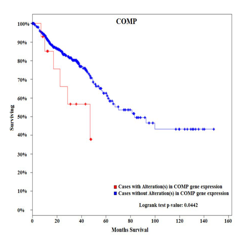
Methods: Two cohorts of patients with sporadic CC were identified. Tumors and matching noninvolved tissues from six EOCC patients (<50) and six late-onset colon cancers (LOCC) patients (>65) were obtained from pathology archives. De-paraffinized tissues were macro-dissected from FFPE sections, RNA isolated, and used for expression profiling of 770 cancer-related genes representing 13 canonical pathways. Survival analysis was performed using the cBioPortal for cancer genomics using 382 CRC patients from the TCGA Provisional database.

Results:Among 770 genes assayed, changes in expression levels of 93 genes were statistically significant between EOCC and matching noninvolved tissues. There were also significant differences in expression levels of 118 genes between LOCC and matching noninvolved tissues. Detailed comparative gene expression analysis between EOCC and LOCC normalized to their matching noninvolved tissues revealed that changes in expression of 88 genes were unique to EOCC using the cutoff criteria of expression levels difference >2 fold and P value <0.01. From these differentially expressed genes specific to EOCC, 28 genes were upregulated and 60 genes downregulated. At the pathway level, PI3K-AKT signaling was the most deregulated pathway in EOCC and cell cycle in LOCC. COMP glycoprotein was one of the genes that were uniquely over-expressed in EOCC. Survival analysis of 382 patients with CRC tumors using the cBioPortal for cancer genomics showed that CRC patients with alterations (all of them with up-regulated COMP) in COMP expression present with poorer overall survival compared to patients without alterations in COMP expression.

Conclusion:
These results suggest that sporadic EOCC is characterized by distinct molecular events compared to LOCC. In addition, COMP glycoprotein is associated with poor overall survival. COMP gene can potentially serve as a novel biomarker associated with EOCC as COMP glycoproteins could be detected in serum and urine.  
 

02.06 Obesity-induced Sphingosine-1-phosphate in Breast Cancer Interstitial Fluid Promotes Angiogenesis

J. Tsuchida1, M. Nakajima1, K. Moro1, A. Ohtani1, M. Endo1, T. Niwano1, K. Tatsuda1, C. Toshikawa1, M. Hasegawa1, M. Ikarashi1, Y. Koyama1, J. Sakata1, T. Kobayashi1, H. Kameyama1, T. Wakai1, K. Takabe2, M. Nagahashi1  1Niigata University Graduate School Of Medical And Dental Sciences,Division Of Digestive And General Surgery,Niigata, NIIGATA, Japan 2Roswell Park Cancer Institute,Breast Surgery, Department Of Surgical Oncology,Baffalo, NY, USA

Introduction: It is well established that obesity evokes chronic inflammation, which aggravates cancer progression by facilitating interactions between cancer cells and stromal cells including blood endothelial cells. Sphingosine-1-phosphate (S1P) is a bioactive lipid mediator produced by sphingosine kinases (SphKs) that plays critical roles in inflammation and cancer progression. We have recently discovered that the SphK1/S1P/S1PR1 axis play critical roles in inflammation-associated cancer progression, and that the S1PR1 functional antagonist, FTY720, suppresses cancer progression by inhibition of the S1P axis during chronic inflammation. We have recently developed a method to measure S1P levels in tumor interstitial fluid (IF), which is a component of the tumor microenvironment that bathes cancer cells in the tumor. Our findings suggested the possibility that S1P secreted from tumor cells to IF may be important for metastasis. We hypothesized that obesity and its related inflammation up-regulate SphK1, which produces more S1P, and the elevated levels of S1P in both tumor and tumor IF stimulate angiogenesis and progression of breast cancer. In this study, we test this hypothesis utilizing animal models with obesity.

Methods: Orthotopically-implanted E0771 syngeneic breast cancer mouse models were used. Mice were fed with normal or high-fat diet (HFD). FTY720 was administered orally (1 mg/kg/day). Angiogenesis in the primary tumor was determined by measurement of microvessel density. S1P levels in tumor and tumor IF were measured by mass spectrometry.

Results: HFD induced obesity significantly worsened the cancer progression. Obesity elevated inflammatory cytokines such as IL-6 and TNF-α in both circulation and in the tumor. Mass spectrometry analysis revealed that while S1P levels in the normal breast mammary fat pad were increased with HFD feeding, S1P levels were even higher in breast tumors. Consistent with increased SphK1 and S1P in tumors, S1P was also significantly increased in the tumor IF. Further, microvessel density analysis revealed that significantly more angiogenesis in peri-tumor area in mice fed with HFD than in mice fed with normal diet. Importantly, FTY720 dramatically reduced the levels of cytokines as well as SphK1, S1PR1 and S1P in primary tumor. Further, FTY720 suppressed not only primary tumor growth, but also angiogenesis in the primary tumor with suppression of aggregation of tumor-associated macrophages.

Conclusion: Our results indicate an important role of S1P in tumor microenvironment, which is affected by obesity, for obesity-related angiogenesis and breast cancer progression. S1P will be one of the promising targets for breast cancer patients with obesity.


02.07 Peri-tumor Sphingosine-1-phosphate Levels Are Affected by the Tumor

J. Tsuchida1, K. Moro1, A. Ohtani1, M. Endo1, T. Niwano1, K. Tatsuda1, C. Toshikawa1, M. Hasegawa1, M. Ikarashi1, M. Nakajima1, Y. Koyama1, J. Sakata1, T. Kobayashi1, H. Kameyama1, K. Takabe2, T. Wakai1, M. Nagahashi1  1Niigata University Graduate School Of Medical And Dental Sciences,Division Of Digestive And General Surgery,Niigata, NIIGATA, Japan 2Roswell Park Cancer Institute,Breast Surgery, Department Of Surgical Oncology,Buffalo, NEW YORK, USA

Introduction: Sphingosine-1-phosphate (S1P), a pleiotropic bioactive lipid mediator, has been implicated as a key regulatory molecule in cancer through its ability to promote cell proliferation, migration, angiogenesis and lymphangiogenesis. Although many researchers including us have reported important roles of S1P in breast cancer progression utilizing in vitro and in vivo models, there have been very few data on human patients due to the difficulty in accurate measurements of S1P levels in the clinical samples. Recently, we have reported that high S1P levels in the tumor determined by mass spectrometry are associated with lymph node metastasis in breast cancer patients. In this study, to reveal further roles of sphingolipids in the patients, we measured the levels of sphingolipids including S1P in plasma and breast tissue including peri-tumor tissue.

Methods: Forty-nine serum prior to operation and 20 tumors from invasive cancer larger than 1.5cm were collected from breast cancer patients who underwent operation from November 2015 to March 2016 in Niigata University Medical and Dental Hospital. The levels of sphingolipids were determined by mass spectrometry.

Results: The levels of sphingolipids including sphingosine, dihydro-sphingosine, S1P, dihydro-S1P were detected successfully in the plasma from 49 breast cancer patients and in the tissue samples from 20 patients. Despite our expectation, no significant differences in plasma S1P levels were identified by hormone receptors (ER, PgR) and HER2 status, Ki-67 index, nuclear grade, lymphatic and vascular invasion of the tumor, pT, pN, and pStage. The levels of sphingosine in patients with lymph node metastasis (pN1,2) were significantly higher than those in patients without metastasis (pN0) (P = 0.023). Interestingly, dihydro-S1P levels in patients with high nuclear grade (NG2,3) were significantly lower than those in patients with low nuclear grade (NG1) (P = 0.025). Moreover, there were significant differences among S1P levels in tumor, peri-tumor, and normal breast tissue (P = 0.0004). Significant differences were also seen among sphingosine (P = 0.0007) and dihydro-sphingosine levels (P = 0.0236) in the three different tissue types.

Conclusion: This is the first report to measure the S1P levels in peri-tumor tissue compared to the tumor and normal breast tissue. This is in agreement with our notion that S1P that is generated in cancer is secreted out to peri-tumoral microenvironment where it aggravates cancer progression, such as lymphangiogenesis. Further study will be needed to reveal the role of S1P in breast cancer patients.

02.08 Paradoxical Association of Postoperative Sphingosine-1 Phosphate and Breast Cancer Aggressiveness

R. Ramanathan1, A. Raza1, J. Young2, J. Sturgill1, D. Lyon1, K. Takabe2  1Virginia Commonwealth University,Richmond, VA, USA 2Roswell Park Cancer Institute,Breast Surgery,Buffalo, NY, USA

Introduction: Sphingosine-1 phosphate (S1P) is bioactive lipid mediator that has been shown to serve an important regulatory function in breast cancer progression. The dynamics of the circulating S1P levels during the postoperative period, however, have yet to be delineated. In this study, we analyze plasma S1P levels in breast cancer patients undergoing adjuvant therapy as compared to control healthy volunteers.

Methods: With institutional review board approval, plasma S1P was measured in 20 healthy women without breast cancer (control) as well as 158 patients with breast cancer who underwent adjuvant chemotherapy with or without irradiation after surgical resection. Among patients with breast cancer, postoperative plasma S1P was measured two weeks prior to adjuvant therapy (baseline), prior to the 4th cycle of chemotherapy (midpoint) and two weeks after completion of adjuvant therapy (completion). Correlations with demographics, treatments and inflammatory markers were analyzed with the use of chi-square tests, paired and non-paired t-tests and ANOVA analyses.

Results: 452 plasma S1P samples among 158 breast cancer patients were analyzed, along with 20 control healthy volunteers. Mean S1P levels did not differ between cancer patients and controls (1221.7 vs. 1139 pmol/mL, p=NS). S1P levels similarly did not associate with age or race in our cohort. Smoking was associated with higher S1P levels (1445.4 vs. 1163.3 pmol/mL, p<0.05). Midpoint S1P levels during adjuvant therapy were lower than baseline (1088.8 vs. 1221.8, p<0.05), with near return to baseline after completion (1121.5 vs. 1221.8, p=0.06), indicating a relationship between chemotherapy and circulating S1P. Furthermore, while stage of disease did not correlate with plasma S1P levels, they were lower among patients with Her2 enriched and triple negative breast cancer as compared to luminal type cancer (1119.2 and 1167.1 vs. 1280.8, p<0.05). Additionally, patients with intermediate and high grade tumors similarly had lower S1P than those with low grade tumors (1230.0 and 1176.5 vs. 1570.8 pmol/mL, p<0.05), further suggesting an inverse relationship between plasma S1P levels and tumor aggressiveness. There were no significant differences based on chemotherapy regimen and radiation.

Conclusions: We found that plasma S1P levels are paradoxically suppressed in aggressive breast cancer and during adjuvant chemotherapy, which raises the possibility that plasma S1P levels do not reflect S1P secretion from cancer cells.